Orthopaedic quiz.by.yapa wijeratne
•Descargar como PPSX, PDF•
69 recomendaciones•18,131 vistas

Quiz on Orthopaedics for medicine undergraduates..

Recomendados
Más contenido relacionado
La actualidad más candente
La actualidad más candente (20)
Destacado
Destacado (20)
Más de Yapa
Más de Yapa (20)
Último
Último (20)
Orthopaedic quiz.by.yapa wijeratne
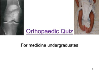
- 1. Orthopaedic Quiz For medicine undergraduates 1
- 2. 1.What is the diagnosis? Q:1 2.Name 2 complications associated with this fracture. 2
- 3. Q:2 1.Identify this clinical deformity 3
- 4. Q:3 1.Describe the mechanism of injury? 2.Name 2 serious complications of this injury? 4
- 5. 1.Identify the prosthesis Q:4 2.What is the indication for its use? 5
- 6. 1.Identify this equipment Q:5 2. Name 2 indications for its use. 6
- 7. 1.Identify this fracture. Q:6 2.What is the best way to manage this fracture? 7
- 8. 1.Identify this injury? 2. What is the mechanism of injury. Q:7 8
- 9. 1.Identify the type of fracture Q:8 2.What is the clinical condition associated with this fracture? 9
- 10. 1.Identify this clinical deformity Q:9 2.How do you manage this child during first six months? 10
- 11. 1.Describe this injury. Q:10 2.Name two complications associated with this injury 11
- 12. 1.What is the injury? Q:11 2. What is the best way to manage this injury 12
- 13. 1.What is the mechanism of injury Q:12 2.How do you manage this injury? 13
- 14. 1.Describe the type of injury Q:13 2.How do you manage this type of injury? 14
- 15. 1.What is the diagnosis Q:14 2.How do manage this problem? 15
- 16. 1.What is the diagnosis Q:15 2.name 2 radiological features of this condition 16
- 17. 1.What is the diagnosis Q:16 2. How do you manage this problem? 17
- 18. 1.What is the diagnosis? Q:17 2.Name 2 radiological features of this condition 18
- 19. 1.What is the radiological feature seen n this x-ray? Q:18 2. Name 2 tumour markers associated with this condition 19
- 20. 1.What is the diagnosis 2.Name 2 long term complications associated with this condition Q:19 20
- 21. 1.What is the diagnosis Q:20 2. How do you manage this problem? 21
- 22. 1.What is the diagnosis Q:21 2. How do you manage this problem? 22
- 23. 1.What is the diagnosis Q:22 2. How do you manage this problem? 23
- 24. 1.What is the diagnosis? Q:23 2.Name 2 complications associated with this fracture. 24
- 26. 1. Identify the deformity. Q:25 2. How do you manage this problem? 26
- 27. 27
- 28. 1.What is the diagnosis? Q:1 2.Name 2 complications associated with this fracture. • 1. – Colles fracture • 2. – Avascular necrosis of Scaphoid – Osteoarthritis – Non-union – Delayed -union 28
- 29. Q:2 1.Identify this clinical deformity • Positive Trendelenburg’s sign on left due to the weakness of left hip abductors (gluteus medius/minimus) 29
- 30. Q:3 1.Describe the mechanism of injury? 2.Name 2 serious complications of this injury? • 1. – Falling on outstretched hand (straight elbow) • 2. – Brachial artery damage – Compartment syndrome 30
- 31. 1.Identify the prosthesis Q:4 2.What is the indication for its use? • 1. – Austin Moore Hemiprosthesis • 2. – Intracapsular neck of the femur fracture 31
- 32. 1.Identify this equipment Q:5 2. Name 2 indications for its use. • 1. • C-arm image intensifier • 2. • Manipulation • Checking progress of surgery/ intraoperative x-rays • Cardiac catheterization • DHS 32
- 33. 1.Identify this fracture. Q:6 2.What is the best way to manage this fracture? • 1. – Forearm fracture of the proximal radius & ulna • 2. – Open reduction & internal fixation (OR+IF) 33
- 34. 1.Identify this injury? 2. What is the mechanism of injury. Q:7 • Transverse patellar fracture 34 • Avulsion fracture/ violent contraction of quadriceps tendon.
- 35. 1.Identify the type of fracture Q:8 2.What is the clinical condition associated with this fracture? • 1. – Wedge fracture of the vertebral body • 2. – Osteoporosis 35
- 36. 1.Identify this clinical deformity Q:9 2.How do you manage this child during first six months? • 1. CTEV • 2. serial plaster casts as soon as identify 36
- 37. 1.Describe this injury. Q:10 2.Name two complications associated with this injury • 1. – Posterior dislocation of hip • 2. – Sciatic nerve injury – Avascular necrosis of femoral head due to twisting & rupture of retinacular arteries. 37
- 38. 1.What is the injury? Q:11 2. What is the best way to manage this injury • 1. – Fracture of lateral malleolus • 2. – OR+IF (as it is a unstable #) 38
- 39. 1.What is the mechanism of injury Q:12 2.How do you manage this injury? • 1. – Sudden inversion of the foot – (violent contraction of peroneus brevis) • 2. – Internal fixation (with K-wire) 39
- 40. 1.Describe the type of injury Q:13 2.How do you manage this type of injury? 1. Transient facet dislocation 2. Stabilize with cervical collar & if B/L, fixation 40
- 41. 1.What is the diagnosis Q:14 2.How do manage this problem? • 1.Giant cell tumor • 2. • Curettage, bone graft /cementing 41
- 42. 1.What is the diagnosis Q:15 2.name 2 radiological features of this condition • 1.Ewing’s sarcoma • 2. • periosteal reaction • Onion peel appearance 42
- 43. 1.What is the diagnosis Q:16 2. How do you manage this problem? • 1. Osteochondroma • 2.Excision of the tumor + symptomatic treatment 43
- 44. 1.What is the diagnosis? Q:17 2.Name 2 radiological features of this condition • 1.osteosarcoma • 2. sun ray specules, Codman’s triangle 44
- 45. 1.What is the radiological feature seen n this x-ray? Q:18 2. Name 2 tumour markers associated with this condition • 1.osteosclerosis of the pubis & ischium • 2. – PSA – Prostatic acid phosphatase 45
- 46. 1.What is the diagnosis 2.Name 2 long term complications associated with this condition Q:19 • 1. – Perthes’ disease • 2. – Secondary osteoarthritis – Persistent limp – Pain – Dislocation – Deformity – Shortening 46
- 47. 1.What is the diagnosis Q:20 2. How do you manage this problem? • 1.Chronic osteomyelitis • 2 • Pus drainage • Antibiotics • Excision of sequestrum 47
- 48. 1.What is the diagnosis Q:21 2. How do you manage this problem? • 1.Fracture of the clavicle (Displaced fracture of the middle third of the clavicle) • 2. “8” figure brace 48
- 49. 1.What is the diagnosis Q:22 2. How do you manage this problem? • 1. • Myositis ossificans • 2. • DO NOT heat or forcefully move • Indomethacin 49
- 50. 1.What is the diagnosis? Q:23 2.Name 2 complications associated with this fracture. • 1. – Scaphoid fracture • 2. – Avascular necrosis of (proximal) Scaphoid – Osteoarthritis – Non-union – Delayed -union 50
- 51. Q:24 1.Identify the followings 1. Flexible Intramedullary nails 2. Locked Intramedullary nails 3. Plate & screw 4. External fixator 51
- 52. 1. Identify the deformity. Q:25 2. How do you manage this problem? • 1.Cubitus varus • 2.Corrective (wedge shape) osteotomy for correcting carrying angle 52
- 53. Reference • Apleys System of Orthopaedics and Fractures 9 ed • Prepared as a slideshow – by Yapa Wijeratne 53