Short case...Neuromyelitis optica
•
2 recomendaciones•206 vistas

Short case...Neuromyelitis optica http://yassermetwally.com http://yassermetwally.net

Recomendados
Recomendados
Más contenido relacionado
Destacado
Destacado (18)
Similar a Short case...Neuromyelitis optica
Similar a Short case...Neuromyelitis optica (20)
Más de Professor Yasser Metwally
Más de Professor Yasser Metwally (20)
Short case...Neuromyelitis optica
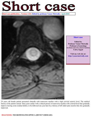
- 1. Short case publication... Version 3.18 | Edited by professor Yasser Metwally | April 2010 Short case Edited by Professor Yasser Metwally Professor of neurology Ain Shams university school of medicine Cairo, Egypt Visit my web site at: http://yassermetwally.com 29 years old female patient presented clinically with transverse myelitis with a high cervical sensory level. The medical history of the patient started three years earlier with a clinical picture of transverse myelitis with a dorsal level that gradually improve leaving some residual deficit, to be followed one year later by a history of left sided optic neuritis that also gradually improved. DIAGNOSIS: NEUROMYELITIS OPTICA (DEVIC'S DISEASE)
- 2. Figure 1. MRI T2 images showing a longitudinally extensive T2 hyperintensities involving the whole cervical spinal cord and the the upper four segments of the dorsal spine. Figure 2. Precontrast MRI T1 images showing a longitudinally extensive T1 hypointensities involving the whole cervical spinal cord and the the upper four segments of the dorsal spine.
- 3. Figure 3. Precontrast MRI T1 image (A), and MRI T2 images (B,C) showing central hypointensity (A) and central hyperintensity involving more than 2/3 of the spinal cord in cross section (B,C). Notice the central dot sign. The MRI signal changes represent central cord vasogenic edema and the central dot sign represents compressed gray matter. Figure 4. Precontrast MRI 1 images showing optic nerve enlargement on the left side.
- 4. References 1. Metwally, MYM: Textbook of neurimaging, A CD-ROM publication, (Metwally, MYM editor) WEB-CD agency for electronic publishing, version 11.2a April 2010 Addendum A new version of short case is uploaded in my web site every week (every Saturday and remains available till Friday.) To download the current version follow the link "http://pdf.yassermetwally.com/short.pdf". You can download the long case version of this short case during the same week from: http://pdf.yassermetwally.com/case.pdf or visit web site: http://pdf.yassermetwally.com To download the software version of the publication (crow.exe) follow the link: http://neurology.yassermetwally.com/crow.zip At the end of each year, all the publications are compiled on a single CD-ROM, please contact the author to know more details. Also to view a list of the previously published case records follow the following link: (http://wordpress.com/tag/case-record/) or click on it if it appears as a link in your PDF reader To inspect the patient's full radiological study, click on the attachment icon (the paper clip icon in the left pane) of the acrobat reader then double click on the attached file Click here to download the long case version of this short case in PDF format