SlideShare una empresa de Scribd logo
1 de 4
Descargar para leer sin conexión
Short case publication... Version 3.2| Edited by professor Yasser Metwally | October 2009


                                                                                                   Short case

                                                                                                  Edited by
                                                                                          Professor Yasser Metwally
                                                                                            Professor of neurology
                                                                                        Ain Shams university school of medicine
                                                                                                   Cairo, Egypt

                                                                                             Visit my web site at:
                                                                                          http://yassermetwally.com


A 35 years old female patient presented clinically with a gradual progressive paraplegia of 5 years duration and with a
sensory level at D2. Urge inconvenience started to occur 3 years before clinical presentation.

DIAGNOSIS: SPINAL SYNCYTIAL MENINGIOMA
Figure 1. MRI T2 images showing a dorsal cord syncytial meningioma. The meningioma is hyperintense relative to
the signal intensity of the spinal cord, with wide- base attachment. A hyperintense CSF cleft is present between the
tumor and the spinal cord. The tumor is retromedullary, pushing the spinal cord anteriorly.


Figure 2. MRI T2 images showing wedging of the vertebral body of L2 by a hyperinetse osteolytic lesion with
preservation of the intervertebral discs above and below. The osteolytic lesion is seen extending posteriorly to the
epidural spaces and encroaching upon the cauda roots.
Figure 3. Postcontrast MRI T1 image showing a dorsal cord
                                              syncytial meningioma. Notice the dense contrast enhancement and
                                              the dural tail.


MR IMAGING OF MENINGIOMAS

Precontrast and postcontrast MR imaging studies can easily diagnose meningioma as well as CT. MR
imaging can also predict histologic subtypes of meningioma.

Diagnosis of meningiomas using MR imaging is made by demonstrating the extra-axial nature of the mass.
Several key MR imaging signs aid in this distinction including: (1) the CSF cleft sign (a cleft of CSF
between the lesion and the brain); (2) direct visualization of displaced or involved dura; (3) demonstration
of displaced pial vessels, which lie between the brain and the extra-axial mass; and (4) buckling of the gray-
white matter junction. Meningiomas are thus characterized by the existence of a hypointense cleft between
the tumour and the brain that probably represents blood vessels or a CSF interface.Anther characteristic
feature is the existence meningeal tail on the enhanced T1 images. The tail extends to a variable degree
away from the meningioma site. This tail does not represent neoplastic infiltration and may instead reflect
fibrovascular proliferation in reaction to the tumour.

   The dural tail or "dural flair"

The dural tail is a curvilinear region of dural enhancement adjacent to the bulky hemispheric tumor. The
finding was originally thought to represent dural infiltration by tumor, and resection of all enhancing dura
mater was thought to be appropriate. However, later studies helped confirm that most of the linear dural
enhancement, especially when it was more than a centimeter away from the tumor bulk, was probably
caused by a reactive process. This reactive process includes both vasocongestion and accumulation of
interstitial edema, both of which increase the thickness of the dura mater. Because the dural capillaries are
"nonneural," they do not form a blood-brain barrier, and, with accumulation of water within the dura
mater, contrast material enhancement occurs.

Grossly meningiomas are characterized, by the existence of a vascular rim that surrounds the meningioma
and appears signal void on both T1,T2 MRI images, this finding is consistent with the overall blood supply
of meningiomas (the peripheries of meningiomas are supplied by branches from the anterior or middle
cerebral arteries that encircle the tumour and form the characteristic vascular rim). Meningiomas encase,
narrow and parasitize pial and meningeal vessels. Vascular rim is common in syncytial and angioblastic
types and much less commonly seen in transitional meningiomas.

  Heterogeneous appearance of meningiomas in T2-weighted pulse sequence can be due to tumor vascularity,
  calcifications, and cystic foci. MR imaging has also been found to be superior to CT in evaluating
  meningiomas for venous sinus invasion or internal carotid artery encasement. Brain edema is observed in
  about 50% of meningiomas, with severe edema occurring with syncytial and angioblastic types. [1]

  Metwally [1] reported a strong correlation between tumor histology and tumor intensity on T2-weighted
  images compared with those of the cortex. Meningiomas are classified into four basic subtypes: fibroblastic,
  transitional, syncytial, and angioblastic. [1] Metwally [1] have stated that meningiomas significantly
  hyperintense to cortex tend to be primarily of syncytial or angioblastic type, whereas meningiomas
  hypointense to cortex tend to be primarily of fibrous or transitional type.


References

1. Metwally, MYM: Textbook of neurimaging, A CD-ROM publication, (Metwally, MYM editor) WEB-CD agency for
electronic publishing, version 10.4a October 2009


Addendum

   A new version of short case is uploaded in my web site every week (every Saturday and remains available till Friday.)
   To download the current version follow the link "http://pdf.yassermetwally.com/short.pdf".
   You can download the long case version of this short case during the same week from: http://pdf.yassermetwally.com/case.pdf or visit web
   site: http://pdf.yassermetwally.com
   To download the software version of the publication (crow.exe) follow the link: http://neurology.yassermetwally.com/crow.zip
   At the end of each year, all the publications are compiled on a single CD-ROM, please contact the author to know more
   details.
   Screen resolution is better set at 1024*768 pixel screen area for optimum display
   For an archive of the previously reported cases go to www.yassermetwally.net, then under pages in the right panel,
   scroll down and click on the text entry "downloadable short cases in PDF format"
   Also to view a list of the previously published case records follow the following link (http://wordpress.com/tag/case-
   record/) or click on it if it appears as a link in your PDF reader

Más contenido relacionado

La actualidad más candente

Presentation2.pptx. posterior fossa tumour
Presentation2.pptx. posterior fossa tumourPresentation2.pptx. posterior fossa tumour
Presentation2.pptx. posterior fossa tumour
Abdellah Nazeer
 
Presentation1.pptx radio;ogical imaging of benign and malignant soft tissue t...
Presentation1.pptx radio;ogical imaging of benign and malignant soft tissue t...Presentation1.pptx radio;ogical imaging of benign and malignant soft tissue t...
Presentation1.pptx radio;ogical imaging of benign and malignant soft tissue t...
Abdellah Nazeer
 
Presentation1, radiological imaging of cavernous sinus lesions.
Presentation1, radiological imaging of cavernous sinus lesions.Presentation1, radiological imaging of cavernous sinus lesions.
Presentation1, radiological imaging of cavernous sinus lesions.
Abdellah Nazeer
 
Radiological pathology of butterfly brain tumors
Radiological pathology of butterfly brain tumorsRadiological pathology of butterfly brain tumors
Radiological pathology of butterfly brain tumors
Professor Yasser Metwally
 
Presentation1, radiological imaging of trigeminal schwanoma.
Presentation1, radiological imaging of trigeminal schwanoma.Presentation1, radiological imaging of trigeminal schwanoma.
Presentation1, radiological imaging of trigeminal schwanoma.
Abdellah Nazeer
 

La actualidad más candente (20)

Embryology of human eye
Embryology of human eyeEmbryology of human eye
Embryology of human eye
 
Case record...Multiple meningiomas
Case record...Multiple meningiomasCase record...Multiple meningiomas
Case record...Multiple meningiomas
 
Short case...Multiple sclerosis
Short case...Multiple sclerosisShort case...Multiple sclerosis
Short case...Multiple sclerosis
 
Presentation2.pptx. posterior fossa tumour
Presentation2.pptx. posterior fossa tumourPresentation2.pptx. posterior fossa tumour
Presentation2.pptx. posterior fossa tumour
 
ECR2013_C-1604
ECR2013_C-1604ECR2013_C-1604
ECR2013_C-1604
 
Foramen Magnum Meningioma
Foramen Magnum MeningiomaForamen Magnum Meningioma
Foramen Magnum Meningioma
 
Spinal neoplasms
Spinal neoplasmsSpinal neoplasms
Spinal neoplasms
 
Surgical Anatomy of The Foramen Magnum_Crimson Publishers
Surgical Anatomy of The Foramen Magnum_Crimson PublishersSurgical Anatomy of The Foramen Magnum_Crimson Publishers
Surgical Anatomy of The Foramen Magnum_Crimson Publishers
 
Spinal tumors
Spinal tumorsSpinal tumors
Spinal tumors
 
Presentation1.pptx radio;ogical imaging of benign and malignant soft tissue t...
Presentation1.pptx radio;ogical imaging of benign and malignant soft tissue t...Presentation1.pptx radio;ogical imaging of benign and malignant soft tissue t...
Presentation1.pptx radio;ogical imaging of benign and malignant soft tissue t...
 
Foramen Magnum Meningioma
Foramen Magnum MeningiomaForamen Magnum Meningioma
Foramen Magnum Meningioma
 
Presentation1, radiological imaging of cavernous sinus lesions.
Presentation1, radiological imaging of cavernous sinus lesions.Presentation1, radiological imaging of cavernous sinus lesions.
Presentation1, radiological imaging of cavernous sinus lesions.
 
Radiological pathology of butterfly brain tumors
Radiological pathology of butterfly brain tumorsRadiological pathology of butterfly brain tumors
Radiological pathology of butterfly brain tumors
 
Intramedullary tumors
Intramedullary tumorsIntramedullary tumors
Intramedullary tumors
 
Neuroradiology primary spinal cord tumours
Neuroradiology   primary spinal cord tumoursNeuroradiology   primary spinal cord tumours
Neuroradiology primary spinal cord tumours
 
Surgical Anatomy of the Clivus_Crimson Publishers
Surgical Anatomy of the Clivus_Crimson PublishersSurgical Anatomy of the Clivus_Crimson Publishers
Surgical Anatomy of the Clivus_Crimson Publishers
 
Presentation1, radiological imaging of trigeminal schwanoma.
Presentation1, radiological imaging of trigeminal schwanoma.Presentation1, radiological imaging of trigeminal schwanoma.
Presentation1, radiological imaging of trigeminal schwanoma.
 
Diagnostic Imaging of Brain Tumors
Diagnostic Imaging of Brain TumorsDiagnostic Imaging of Brain Tumors
Diagnostic Imaging of Brain Tumors
 
Spinal tumors- Imaging
Spinal tumors- ImagingSpinal tumors- Imaging
Spinal tumors- Imaging
 
Spinal cord tumors
Spinal cord tumorsSpinal cord tumors
Spinal cord tumors
 

Destacado

Destacado (20)

Issues in brainmapping...EEG in the evaluation of focal cerebral dysfunction
Issues in brainmapping...EEG in the evaluation of focal cerebral dysfunctionIssues in brainmapping...EEG in the evaluation of focal cerebral dysfunction
Issues in brainmapping...EEG in the evaluation of focal cerebral dysfunction
 
Short case...Multiple dural arteriovenous fistulas
Short case...Multiple dural arteriovenous fistulasShort case...Multiple dural arteriovenous fistulas
Short case...Multiple dural arteriovenous fistulas
 
Short case...Spinal dural arteriovenous fistula
Short case...Spinal dural arteriovenous fistulaShort case...Spinal dural arteriovenous fistula
Short case...Spinal dural arteriovenous fistula
 
Case record...Spinal meningioma
Case record...Spinal meningiomaCase record...Spinal meningioma
Case record...Spinal meningioma
 
Case record...brain stem glioma
Case record...brain stem gliomaCase record...brain stem glioma
Case record...brain stem glioma
 
Topic of the month.... Endothelial nitric oxide synthase (eNOS) And Stroke: P...
Topic of the month.... Endothelial nitric oxide synthase (eNOS) And Stroke: P...Topic of the month.... Endothelial nitric oxide synthase (eNOS) And Stroke: P...
Topic of the month.... Endothelial nitric oxide synthase (eNOS) And Stroke: P...
 
Topic of the month.... The role of serotonin in attention deficit/hyperactivi...
Topic of the month.... The role of serotonin in attention deficit/hyperactivi...Topic of the month.... The role of serotonin in attention deficit/hyperactivi...
Topic of the month.... The role of serotonin in attention deficit/hyperactivi...
 
Talking psychiatry...The neurology of schizophrenia
Talking psychiatry...The neurology of schizophreniaTalking psychiatry...The neurology of schizophrenia
Talking psychiatry...The neurology of schizophrenia
 
Topic of the month...Neuroimaging of smelling
Topic of the month...Neuroimaging of smellingTopic of the month...Neuroimaging of smelling
Topic of the month...Neuroimaging of smelling
 
Issues in brainmapping...Neonatal EEG
Issues in brainmapping...Neonatal EEGIssues in brainmapping...Neonatal EEG
Issues in brainmapping...Neonatal EEG
 
Short case...Spinal metastasis
Short case...Spinal metastasisShort case...Spinal metastasis
Short case...Spinal metastasis
 
Short case...Neuromyelitis optica
Short case...Neuromyelitis opticaShort case...Neuromyelitis optica
Short case...Neuromyelitis optica
 
Short case...Cerebral palsy
Short case...Cerebral palsyShort case...Cerebral palsy
Short case...Cerebral palsy
 
case record...Heavily calcified dorsal disc herniation at D11,D2 disc level
case record...Heavily calcified dorsal disc herniation at D11,D2 disc levelcase record...Heavily calcified dorsal disc herniation at D11,D2 disc level
case record...Heavily calcified dorsal disc herniation at D11,D2 disc level
 
Short case...Neuro-Behcet
Short case...Neuro-BehcetShort case...Neuro-Behcet
Short case...Neuro-Behcet
 
Short case...Myxopapillary ependymoma
Short case...Myxopapillary ependymomaShort case...Myxopapillary ependymoma
Short case...Myxopapillary ependymoma
 
Undergraduate downloads...Clinical neurological examination
Undergraduate downloads...Clinical neurological examinationUndergraduate downloads...Clinical neurological examination
Undergraduate downloads...Clinical neurological examination
 
Topic of the month.... Antiangiogenic Therapy in Brain Tumors
Topic of the month.... Antiangiogenic Therapy in Brain TumorsTopic of the month.... Antiangiogenic Therapy in Brain Tumors
Topic of the month.... Antiangiogenic Therapy in Brain Tumors
 
Talking psychiatry...Functional neuroimaging of schizophrenia
Talking psychiatry...Functional neuroimaging of schizophreniaTalking psychiatry...Functional neuroimaging of schizophrenia
Talking psychiatry...Functional neuroimaging of schizophrenia
 
Case record...Epidural secondary CNS lymphoma
Case record...Epidural secondary CNS lymphomaCase record...Epidural secondary CNS lymphoma
Case record...Epidural secondary CNS lymphoma
 

Similar a Short case...Spinal meningioma

Similar a Short case...Spinal meningioma (20)

Short case...brain stem glioma
Short case...brain stem gliomaShort case...brain stem glioma
Short case...brain stem glioma
 
Short case...Cortical dysplasia
Short case...Cortical dysplasiaShort case...Cortical dysplasia
Short case...Cortical dysplasia
 
Case record...Cerebellopontine angle meningioma
Case record...Cerebellopontine angle meningiomaCase record...Cerebellopontine angle meningioma
Case record...Cerebellopontine angle meningioma
 
Short case...tectal plate glioma
Short case...tectal plate gliomaShort case...tectal plate glioma
Short case...tectal plate glioma
 
Short case...Occipital lobe cavernoma
Short case...Occipital lobe cavernomaShort case...Occipital lobe cavernoma
Short case...Occipital lobe cavernoma
 
Short case...Epidural secondary CNS lymphoma
Short case...Epidural secondary CNS lymphomaShort case...Epidural secondary CNS lymphoma
Short case...Epidural secondary CNS lymphoma
 
Short case...Spinal multiple sclerosis
Short case...Spinal multiple sclerosisShort case...Spinal multiple sclerosis
Short case...Spinal multiple sclerosis
 
Short case...Neuromyelitis optica
Short case...Neuromyelitis opticaShort case...Neuromyelitis optica
Short case...Neuromyelitis optica
 
Short case...Acute postinfectious transverse myelitis
Short case...Acute postinfectious transverse myelitisShort case...Acute postinfectious transverse myelitis
Short case...Acute postinfectious transverse myelitis
 
Short case...Friedreich ataxia
Short case...Friedreich ataxiaShort case...Friedreich ataxia
Short case...Friedreich ataxia
 
Short case...Subependymal primary CNS lymphoma
Short case...Subependymal primary CNS lymphomaShort case...Subependymal primary CNS lymphoma
Short case...Subependymal primary CNS lymphoma
 
Short case...Cervical vascular spondylotic myelopathy
Short case...Cervical vascular spondylotic myelopathyShort case...Cervical vascular spondylotic myelopathy
Short case...Cervical vascular spondylotic myelopathy
 
Short case...Spinal dural arteriovenous fistula withn congestive myelopathy
Short case...Spinal dural arteriovenous fistula withn congestive myelopathyShort case...Spinal dural arteriovenous fistula withn congestive myelopathy
Short case...Spinal dural arteriovenous fistula withn congestive myelopathy
 
Short case...Parasagittal meningioma
Short case...Parasagittal meningiomaShort case...Parasagittal meningioma
Short case...Parasagittal meningioma
 
Short case...Neurofibromatosis type 2
Short case...Neurofibromatosis type 2Short case...Neurofibromatosis type 2
Short case...Neurofibromatosis type 2
 
Short case...Multiple craniocervical anomalies
Short case...Multiple craniocervical anomaliesShort case...Multiple craniocervical anomalies
Short case...Multiple craniocervical anomalies
 
Short case...Lymphomatous leptomeningitis
Short case...Lymphomatous leptomeningitisShort case...Lymphomatous leptomeningitis
Short case...Lymphomatous leptomeningitis
 
Short case...Parasellar craniopharyngioma
Short case...Parasellar craniopharyngiomaShort case...Parasellar craniopharyngioma
Short case...Parasellar craniopharyngioma
 
Short case...Intramedullary cystic spinal cord metastasis
Short case...Intramedullary cystic spinal cord metastasisShort case...Intramedullary cystic spinal cord metastasis
Short case...Intramedullary cystic spinal cord metastasis
 
Short case..Subacute cerebral hemorrhage
Short case..Subacute cerebral hemorrhageShort case..Subacute cerebral hemorrhage
Short case..Subacute cerebral hemorrhage
 

Más de Professor Yasser Metwally

Más de Professor Yasser Metwally (20)

The Egyptian Zoo in Cairo 2015
The Egyptian Zoo in Cairo 2015The Egyptian Zoo in Cairo 2015
The Egyptian Zoo in Cairo 2015
 
End of the great nile river in Ras Elbar
End of the great nile river in Ras ElbarEnd of the great nile river in Ras Elbar
End of the great nile river in Ras Elbar
 
The Lion and The tiger in Egypt
The Lion and The tiger in EgyptThe Lion and The tiger in Egypt
The Lion and The tiger in Egypt
 
The monkeys in Egypt
The monkeys in EgyptThe monkeys in Egypt
The monkeys in Egypt
 
The Snake, the Scorpion, the turtle in Egypt
The Snake, the Scorpion, the turtle in EgyptThe Snake, the Scorpion, the turtle in Egypt
The Snake, the Scorpion, the turtle in Egypt
 
The Egyptian Parrot
The Egyptian ParrotThe Egyptian Parrot
The Egyptian Parrot
 
The Egyptian Deer
The Egyptian DeerThe Egyptian Deer
The Egyptian Deer
 
The Egyptian Pelican
The Egyptian PelicanThe Egyptian Pelican
The Egyptian Pelican
 
The Flamingo bird in Egypt
The Flamingo bird in EgyptThe Flamingo bird in Egypt
The Flamingo bird in Egypt
 
Egyptian Cats
Egyptian CatsEgyptian Cats
Egyptian Cats
 
Radiological pathology of epileptic disorders
Radiological pathology of epileptic disordersRadiological pathology of epileptic disorders
Radiological pathology of epileptic disorders
 
Radiological pathology of cerebrovascular disorders
Radiological pathology of cerebrovascular disordersRadiological pathology of cerebrovascular disorders
Radiological pathology of cerebrovascular disorders
 
Radiological pathology of spontaneous cerebral hemorrhage
Radiological pathology of spontaneous cerebral hemorrhageRadiological pathology of spontaneous cerebral hemorrhage
Radiological pathology of spontaneous cerebral hemorrhage
 
Radiological pathology of cerebral amyloid angiography
Radiological pathology of cerebral amyloid angiographyRadiological pathology of cerebral amyloid angiography
Radiological pathology of cerebral amyloid angiography
 
Radiological pathology of cerebral microbleeds
Radiological pathology of cerebral microbleedsRadiological pathology of cerebral microbleeds
Radiological pathology of cerebral microbleeds
 
The Egyptian Zoo in Cairo
The Egyptian Zoo in CairoThe Egyptian Zoo in Cairo
The Egyptian Zoo in Cairo
 
Progressive multifocal leukoencephalopathy
Progressive multifocal leukoencephalopathyProgressive multifocal leukoencephalopathy
Progressive multifocal leukoencephalopathy
 
Progressive multifocal leukoencephalopathy
Progressive multifocal leukoencephalopathyProgressive multifocal leukoencephalopathy
Progressive multifocal leukoencephalopathy
 
Issues in radiological pathology: Radiological pathology of watershed infarct...
Issues in radiological pathology: Radiological pathology of watershed infarct...Issues in radiological pathology: Radiological pathology of watershed infarct...
Issues in radiological pathology: Radiological pathology of watershed infarct...
 
Radiological pathology of cortical laminar necrosis
Radiological pathology of cortical laminar necrosisRadiological pathology of cortical laminar necrosis
Radiological pathology of cortical laminar necrosis
 

Último

Call Girl In Indore 📞9235973566📞 Just📲 Call Inaaya Indore Call Girls Service ...
Call Girl In Indore 📞9235973566📞 Just📲 Call Inaaya Indore Call Girls Service ...Call Girl In Indore 📞9235973566📞 Just📲 Call Inaaya Indore Call Girls Service ...
Call Girl In Indore 📞9235973566📞 Just📲 Call Inaaya Indore Call Girls Service ...
Sheetaleventcompany
 
Premium Call Girls Nagpur {9xx000xx09} ❤️VVIP POOJA Call Girls in Nagpur Maha...
Premium Call Girls Nagpur {9xx000xx09} ❤️VVIP POOJA Call Girls in Nagpur Maha...Premium Call Girls Nagpur {9xx000xx09} ❤️VVIP POOJA Call Girls in Nagpur Maha...
Premium Call Girls Nagpur {9xx000xx09} ❤️VVIP POOJA Call Girls in Nagpur Maha...
Sheetaleventcompany
 
Pune Call Girl Service 📞9xx000xx09📞Just Call Divya📲 Call Girl In Pune No💰Adva...
Pune Call Girl Service 📞9xx000xx09📞Just Call Divya📲 Call Girl In Pune No💰Adva...Pune Call Girl Service 📞9xx000xx09📞Just Call Divya📲 Call Girl In Pune No💰Adva...
Pune Call Girl Service 📞9xx000xx09📞Just Call Divya📲 Call Girl In Pune No💰Adva...
Sheetaleventcompany
 
❤️Amritsar Escorts Service☎️9815674956☎️ Call Girl service in Amritsar☎️ Amri...
❤️Amritsar Escorts Service☎️9815674956☎️ Call Girl service in Amritsar☎️ Amri...❤️Amritsar Escorts Service☎️9815674956☎️ Call Girl service in Amritsar☎️ Amri...
❤️Amritsar Escorts Service☎️9815674956☎️ Call Girl service in Amritsar☎️ Amri...
Sheetaleventcompany
 
Difference Between Skeletal Smooth and Cardiac Muscles
Difference Between Skeletal Smooth and Cardiac MusclesDifference Between Skeletal Smooth and Cardiac Muscles
Difference Between Skeletal Smooth and Cardiac Muscles
MedicoseAcademics
 
💚Chandigarh Call Girls Service 💯Piya 📲🔝8868886958🔝Call Girls In Chandigarh No...
💚Chandigarh Call Girls Service 💯Piya 📲🔝8868886958🔝Call Girls In Chandigarh No...💚Chandigarh Call Girls Service 💯Piya 📲🔝8868886958🔝Call Girls In Chandigarh No...
💚Chandigarh Call Girls Service 💯Piya 📲🔝8868886958🔝Call Girls In Chandigarh No...
Sheetaleventcompany
 
Call Girl in Chennai | Whatsapp No 📞 7427069034 📞 VIP Escorts Service Availab...
Call Girl in Chennai | Whatsapp No 📞 7427069034 📞 VIP Escorts Service Availab...Call Girl in Chennai | Whatsapp No 📞 7427069034 📞 VIP Escorts Service Availab...
Call Girl in Chennai | Whatsapp No 📞 7427069034 📞 VIP Escorts Service Availab...
amritaverma53
 
Kolkata Call Girls Service ❤️🍑 9xx000xx09 👄🫦 Independent Escort Service Kolka...
Kolkata Call Girls Service ❤️🍑 9xx000xx09 👄🫦 Independent Escort Service Kolka...Kolkata Call Girls Service ❤️🍑 9xx000xx09 👄🫦 Independent Escort Service Kolka...
Kolkata Call Girls Service ❤️🍑 9xx000xx09 👄🫦 Independent Escort Service Kolka...
Sheetaleventcompany
 

Último (20)

Call Girl In Indore 📞9235973566📞 Just📲 Call Inaaya Indore Call Girls Service ...
Call Girl In Indore 📞9235973566📞 Just📲 Call Inaaya Indore Call Girls Service ...Call Girl In Indore 📞9235973566📞 Just📲 Call Inaaya Indore Call Girls Service ...
Call Girl In Indore 📞9235973566📞 Just📲 Call Inaaya Indore Call Girls Service ...
 
Premium Call Girls Nagpur {9xx000xx09} ❤️VVIP POOJA Call Girls in Nagpur Maha...
Premium Call Girls Nagpur {9xx000xx09} ❤️VVIP POOJA Call Girls in Nagpur Maha...Premium Call Girls Nagpur {9xx000xx09} ❤️VVIP POOJA Call Girls in Nagpur Maha...
Premium Call Girls Nagpur {9xx000xx09} ❤️VVIP POOJA Call Girls in Nagpur Maha...
 
Pune Call Girl Service 📞9xx000xx09📞Just Call Divya📲 Call Girl In Pune No💰Adva...
Pune Call Girl Service 📞9xx000xx09📞Just Call Divya📲 Call Girl In Pune No💰Adva...Pune Call Girl Service 📞9xx000xx09📞Just Call Divya📲 Call Girl In Pune No💰Adva...
Pune Call Girl Service 📞9xx000xx09📞Just Call Divya📲 Call Girl In Pune No💰Adva...
 
Cardiac Output, Venous Return, and Their Regulation
Cardiac Output, Venous Return, and Their RegulationCardiac Output, Venous Return, and Their Regulation
Cardiac Output, Venous Return, and Their Regulation
 
Kolkata Call Girls Shobhabazar 💯Call Us 🔝 8005736733 🔝 💃 Top Class Call Gir...
Kolkata Call Girls Shobhabazar  💯Call Us 🔝 8005736733 🔝 💃  Top Class Call Gir...Kolkata Call Girls Shobhabazar  💯Call Us 🔝 8005736733 🔝 💃  Top Class Call Gir...
Kolkata Call Girls Shobhabazar 💯Call Us 🔝 8005736733 🔝 💃 Top Class Call Gir...
 
(RIYA)🎄Airhostess Call Girl Jaipur Call Now 8445551418 Premium Collection Of ...
(RIYA)🎄Airhostess Call Girl Jaipur Call Now 8445551418 Premium Collection Of ...(RIYA)🎄Airhostess Call Girl Jaipur Call Now 8445551418 Premium Collection Of ...
(RIYA)🎄Airhostess Call Girl Jaipur Call Now 8445551418 Premium Collection Of ...
 
❤️Amritsar Escorts Service☎️9815674956☎️ Call Girl service in Amritsar☎️ Amri...
❤️Amritsar Escorts Service☎️9815674956☎️ Call Girl service in Amritsar☎️ Amri...❤️Amritsar Escorts Service☎️9815674956☎️ Call Girl service in Amritsar☎️ Amri...
❤️Amritsar Escorts Service☎️9815674956☎️ Call Girl service in Amritsar☎️ Amri...
 
Difference Between Skeletal Smooth and Cardiac Muscles
Difference Between Skeletal Smooth and Cardiac MusclesDifference Between Skeletal Smooth and Cardiac Muscles
Difference Between Skeletal Smooth and Cardiac Muscles
 
💚Chandigarh Call Girls Service 💯Piya 📲🔝8868886958🔝Call Girls In Chandigarh No...
💚Chandigarh Call Girls Service 💯Piya 📲🔝8868886958🔝Call Girls In Chandigarh No...💚Chandigarh Call Girls Service 💯Piya 📲🔝8868886958🔝Call Girls In Chandigarh No...
💚Chandigarh Call Girls Service 💯Piya 📲🔝8868886958🔝Call Girls In Chandigarh No...
 
Chandigarh Call Girls Service ❤️🍑 9809698092 👄🫦Independent Escort Service Cha...
Chandigarh Call Girls Service ❤️🍑 9809698092 👄🫦Independent Escort Service Cha...Chandigarh Call Girls Service ❤️🍑 9809698092 👄🫦Independent Escort Service Cha...
Chandigarh Call Girls Service ❤️🍑 9809698092 👄🫦Independent Escort Service Cha...
 
Chennai ❣️ Call Girl 6378878445 Call Girls in Chennai Escort service book now
Chennai ❣️ Call Girl 6378878445 Call Girls in Chennai Escort service book nowChennai ❣️ Call Girl 6378878445 Call Girls in Chennai Escort service book now
Chennai ❣️ Call Girl 6378878445 Call Girls in Chennai Escort service book now
 
Call Girls in Lucknow Just Call 👉👉8630512678 Top Class Call Girl Service Avai...
Call Girls in Lucknow Just Call 👉👉8630512678 Top Class Call Girl Service Avai...Call Girls in Lucknow Just Call 👉👉8630512678 Top Class Call Girl Service Avai...
Call Girls in Lucknow Just Call 👉👉8630512678 Top Class Call Girl Service Avai...
 
Call Girl in Chennai | Whatsapp No 📞 7427069034 📞 VIP Escorts Service Availab...
Call Girl in Chennai | Whatsapp No 📞 7427069034 📞 VIP Escorts Service Availab...Call Girl in Chennai | Whatsapp No 📞 7427069034 📞 VIP Escorts Service Availab...
Call Girl in Chennai | Whatsapp No 📞 7427069034 📞 VIP Escorts Service Availab...
 
Kolkata Call Girls Service ❤️🍑 9xx000xx09 👄🫦 Independent Escort Service Kolka...
Kolkata Call Girls Service ❤️🍑 9xx000xx09 👄🫦 Independent Escort Service Kolka...Kolkata Call Girls Service ❤️🍑 9xx000xx09 👄🫦 Independent Escort Service Kolka...
Kolkata Call Girls Service ❤️🍑 9xx000xx09 👄🫦 Independent Escort Service Kolka...
 
Call Girls Bangalore - 450+ Call Girl Cash Payment 💯Call Us 🔝 6378878445 🔝 💃 ...
Call Girls Bangalore - 450+ Call Girl Cash Payment 💯Call Us 🔝 6378878445 🔝 💃 ...Call Girls Bangalore - 450+ Call Girl Cash Payment 💯Call Us 🔝 6378878445 🔝 💃 ...
Call Girls Bangalore - 450+ Call Girl Cash Payment 💯Call Us 🔝 6378878445 🔝 💃 ...
 
ANATOMY AND PHYSIOLOGY OF REPRODUCTIVE SYSTEM.pptx
ANATOMY AND PHYSIOLOGY OF REPRODUCTIVE SYSTEM.pptxANATOMY AND PHYSIOLOGY OF REPRODUCTIVE SYSTEM.pptx
ANATOMY AND PHYSIOLOGY OF REPRODUCTIVE SYSTEM.pptx
 
Low Cost Call Girls Bangalore {9179660964} ❤️VVIP NISHA Call Girls in Bangalo...
Low Cost Call Girls Bangalore {9179660964} ❤️VVIP NISHA Call Girls in Bangalo...Low Cost Call Girls Bangalore {9179660964} ❤️VVIP NISHA Call Girls in Bangalo...
Low Cost Call Girls Bangalore {9179660964} ❤️VVIP NISHA Call Girls in Bangalo...
 
7 steps How to prevent Thalassemia : Dr Sharda Jain & Vandana Gupta
7 steps How to prevent Thalassemia : Dr Sharda Jain & Vandana Gupta7 steps How to prevent Thalassemia : Dr Sharda Jain & Vandana Gupta
7 steps How to prevent Thalassemia : Dr Sharda Jain & Vandana Gupta
 
Circulatory Shock, types and stages, compensatory mechanisms
Circulatory Shock, types and stages, compensatory mechanismsCirculatory Shock, types and stages, compensatory mechanisms
Circulatory Shock, types and stages, compensatory mechanisms
 
Race Course Road } Book Call Girls in Bangalore | Whatsapp No 6378878445 VIP ...
Race Course Road } Book Call Girls in Bangalore | Whatsapp No 6378878445 VIP ...Race Course Road } Book Call Girls in Bangalore | Whatsapp No 6378878445 VIP ...
Race Course Road } Book Call Girls in Bangalore | Whatsapp No 6378878445 VIP ...
 

Short case...Spinal meningioma

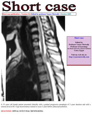
  • 1. Short case publication... Version 3.2| Edited by professor Yasser Metwally | October 2009 Short case Edited by Professor Yasser Metwally Professor of neurology Ain Shams university school of medicine Cairo, Egypt Visit my web site at: http://yassermetwally.com A 35 years old female patient presented clinically with a gradual progressive paraplegia of 5 years duration and with a sensory level at D2. Urge inconvenience started to occur 3 years before clinical presentation. DIAGNOSIS: SPINAL SYNCYTIAL MENINGIOMA
  • 2. Figure 1. MRI T2 images showing a dorsal cord syncytial meningioma. The meningioma is hyperintense relative to the signal intensity of the spinal cord, with wide- base attachment. A hyperintense CSF cleft is present between the tumor and the spinal cord. The tumor is retromedullary, pushing the spinal cord anteriorly. Figure 2. MRI T2 images showing wedging of the vertebral body of L2 by a hyperinetse osteolytic lesion with preservation of the intervertebral discs above and below. The osteolytic lesion is seen extending posteriorly to the epidural spaces and encroaching upon the cauda roots.
  • 3. Figure 3. Postcontrast MRI T1 image showing a dorsal cord syncytial meningioma. Notice the dense contrast enhancement and the dural tail. MR IMAGING OF MENINGIOMAS Precontrast and postcontrast MR imaging studies can easily diagnose meningioma as well as CT. MR imaging can also predict histologic subtypes of meningioma. Diagnosis of meningiomas using MR imaging is made by demonstrating the extra-axial nature of the mass. Several key MR imaging signs aid in this distinction including: (1) the CSF cleft sign (a cleft of CSF between the lesion and the brain); (2) direct visualization of displaced or involved dura; (3) demonstration of displaced pial vessels, which lie between the brain and the extra-axial mass; and (4) buckling of the gray- white matter junction. Meningiomas are thus characterized by the existence of a hypointense cleft between the tumour and the brain that probably represents blood vessels or a CSF interface.Anther characteristic feature is the existence meningeal tail on the enhanced T1 images. The tail extends to a variable degree away from the meningioma site. This tail does not represent neoplastic infiltration and may instead reflect fibrovascular proliferation in reaction to the tumour. The dural tail or "dural flair" The dural tail is a curvilinear region of dural enhancement adjacent to the bulky hemispheric tumor. The finding was originally thought to represent dural infiltration by tumor, and resection of all enhancing dura mater was thought to be appropriate. However, later studies helped confirm that most of the linear dural enhancement, especially when it was more than a centimeter away from the tumor bulk, was probably caused by a reactive process. This reactive process includes both vasocongestion and accumulation of interstitial edema, both of which increase the thickness of the dura mater. Because the dural capillaries are "nonneural," they do not form a blood-brain barrier, and, with accumulation of water within the dura mater, contrast material enhancement occurs. Grossly meningiomas are characterized, by the existence of a vascular rim that surrounds the meningioma and appears signal void on both T1,T2 MRI images, this finding is consistent with the overall blood supply of meningiomas (the peripheries of meningiomas are supplied by branches from the anterior or middle cerebral arteries that encircle the tumour and form the characteristic vascular rim). Meningiomas encase, narrow and parasitize pial and meningeal vessels. Vascular rim is common in syncytial and angioblastic
  • 4. types and much less commonly seen in transitional meningiomas. Heterogeneous appearance of meningiomas in T2-weighted pulse sequence can be due to tumor vascularity, calcifications, and cystic foci. MR imaging has also been found to be superior to CT in evaluating meningiomas for venous sinus invasion or internal carotid artery encasement. Brain edema is observed in about 50% of meningiomas, with severe edema occurring with syncytial and angioblastic types. [1] Metwally [1] reported a strong correlation between tumor histology and tumor intensity on T2-weighted images compared with those of the cortex. Meningiomas are classified into four basic subtypes: fibroblastic, transitional, syncytial, and angioblastic. [1] Metwally [1] have stated that meningiomas significantly hyperintense to cortex tend to be primarily of syncytial or angioblastic type, whereas meningiomas hypointense to cortex tend to be primarily of fibrous or transitional type. References 1. Metwally, MYM: Textbook of neurimaging, A CD-ROM publication, (Metwally, MYM editor) WEB-CD agency for electronic publishing, version 10.4a October 2009 Addendum A new version of short case is uploaded in my web site every week (every Saturday and remains available till Friday.) To download the current version follow the link "http://pdf.yassermetwally.com/short.pdf". You can download the long case version of this short case during the same week from: http://pdf.yassermetwally.com/case.pdf or visit web site: http://pdf.yassermetwally.com To download the software version of the publication (crow.exe) follow the link: http://neurology.yassermetwally.com/crow.zip At the end of each year, all the publications are compiled on a single CD-ROM, please contact the author to know more details. Screen resolution is better set at 1024*768 pixel screen area for optimum display For an archive of the previously reported cases go to www.yassermetwally.net, then under pages in the right panel, scroll down and click on the text entry "downloadable short cases in PDF format" Also to view a list of the previously published case records follow the following link (http://wordpress.com/tag/case- record/) or click on it if it appears as a link in your PDF reader