2. outline
• Relevant anatomy
• Clinical approach to hand injury
–History
–Examination
–Imaging
• Principles of treatment
• Common Traumatic Hand Injuries
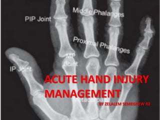
3. SKELETAL ANATOMY
• Hand consist of 27 bones:
– 14 Phalangeal
• Five proximal
• Four middle
• Five distal
– 5 metacarpals
– 8 carpal bones
10. MUSCLE ANATO…………
• Intrinsic muscle of the hand
• Have their origins and
insertions within the
hand.
• Consist the following:
– Thenar muscles
– Hypothenar muscles
– the interossei and the
lumbricals
– adductor pollicies
13. Motor supply to hand –
Ulnar nerve.
• the medial two lumbricals
• interossei
• the adductor pollicis, the deep head of FPB
• Muscles of hypothenar eminence
Median nerve
LOAF" for Lumbricals 1 & 2, Opponens pollicis,
Abductor pollicis brevis and Flexor pollicis brevis
15. Approach to Hand …………
1. History:
General
◦ Age
◦ Hand dominance and occupation
◦ Other past medical history, especially diabetes, vascular
problems
◦ Smoking history
◦ History of previous hand problems
When and where did this injury take place?
How was the trauma sustained?
What was the posture of the hand at the time of the
injury?
16. 2. Physical examination
A. General considerations
– Examine the patient while sitting,
– explain each step of the examination
– Encourage the patient to look at the injured hand, to
better participate in planning treatment
B. Skin
– Note location, and estimate area and depth of
lacerations or burns.
– Note color and temperature of the injured extremity.
17. 2. Physical examin…………..
C. Circulation
– Look for color changes
– capillary refill (< 1 second),
– radial pulse
– Allen test
– Perform a Doppler examination, if pulses are not
palpable.
18. 2. Physical examin…………..
• D. Neurologic assessment:
• fingers: assess digital nerves at distal tips with two-
point discrimination on radial and ulnar aspects (> 6
mm indicates axonal loss)
• immersion test
• skin moisture
• pinch and grip strength
• hand
23. 2. Physical examin…………..
F. Bone and joints
– Palpate and look for deformities
– Passive and active range of motion
– Stability
– Open vs closed dislocations or fractures
24. 3. Diagnostic tests
• X rays- AP, lateral &oblique views
Ultra sound
◦ Has a growing role in locating foreign bodies and in
evaluating soft tissues
◦ Can detect ruptured tendons and assess dynamic function
of tendons non-invasively.
MRI
◦ Highly sensitive but not have a role in management of hand
wounds.
◦ Angiography or Doppler examination to assess arterial
integrity
25. PRINCIPLES OF TREATMENT
• goals of treatment
– to maintain or restore distal circulation,
– obtain a healed wound,
– preserve motion, and retain distal sensation
• The majority of hand injuries will not require antibiotic
therapy. Important exceptions include
• animal/human bites,
• grossly contaminated wounds,
• contaminated penetrating trauma,
• high-pressure injection injuries, and
• amputations.
26. PRINCIPLES OF TREA………..
• Appropriate treatment requires thorough
knowledge of
– local and regional anesthesia,
– use of a tourniquet
– correct placement of incisions
– appropriate use of dressings and splints
27. General Operative Principles
• Local And Regional Anesthesia
– The classic methods for obtaining hand anesthesia
in the ED are
• local anesthetic infiltration,
• digital nerve blocks, and
• anatomic forearm nerve blocks
30. General Operative…………..
• Tourniquet Application
– used to provide a bloodless field so that clear
visualization of all structures.
– standard pressures: 100-150 mmHg >SBP.
– cuff is deflated every 2 hrs for 15-20 min to revascularize
distal tissues and to relieve pressure on nerves locally.
32. General Operative…………..
• Wound Closure
– Incisions and wounds of the hand can generally be
closed with a single layer of interrupted sutures.
– Vertical mattress sutures are needed if the skin
edges tend to invert..
– Wound closure for children with 5-0 or 6-0 plain
– sutures are removed after 7 to 10 days to
minimize suture marks
33. POSTOPERATIVE PRINCIPLES
• Dressings and Splints
– purposes: to protect wounds, absorb drainage, & help
splint repaired structures
– splints: to protect only the part necessary to be
immobilized and not the remainder extremity.
34. POSTOPERATIVE PRINC………
• Protective Position
– When immobilization is
essential, it should be in
the “protective” or “safe
position
– There are just three
essential elements of the
protective position:
– interphalangeal (IP)
joint extension,
– Metacarpophalangeal
(MCP) joint flexion, and
– palmar abduction of the
thumb
35. POSTOPERATIVE PRINC………
• Elevation
– An accidentally or surgically injured limb should
be continuously elevated until effective pumping
activity is again functioning to propel venous
blood and lymph back to the body.
37. Anatomy
– Fingertip
• The portion of the digit distal to the insertion of the
flexor and extensor tendons on the distal phalanx
– subcutaneous soft tissue
– nail
– nail bed
– distal end of the
terminal phalanx
Finger tip Injuries
43. Tendon injuries
• Tendon injuries are classified as
• partial lacerations,
• complete lacerations, and
• tendon avulsion injuries
• Tendon lacerations of less than 50% of the diameter of
the tendon are usually asymptomatic and do not need
repair
• Prerequisites for tendon repair include
• a clean wound debrided of all devitalized tissue,
• bony stabilization, and
• adequate stable soft-tissue coverage.
44. Tendon injur………..
• Flexor Tendon Injury
– Flexor tendon injuries most commonly result from
lacerations or puncture wounds on the palmar
surface of the hand
– They are best treated by a surgeon experienced in
the management of these injuries
– Flexor tendon injuries are divided into five zones
46. Flexor Tendon Inju……
• Timing of Flexor Tendon
Repair
– primary tendon repair :
< 12 hrs ( 24 hrs )
– delayed primary repair :
24 hrs ~ 10 days
– early secondary repair :
10 days ~ 4 weeks
– late secondary repair : > 4
weeks
• Relative contraindications to
immediate tendon repair
– Injuries more than 12 hours old
– Crush wounds with poor skin
coverage
– Contaminated wounds, especially
human bites
– Tendon loss greater than 1 cm
– Injury at multiple sites along the
tendon
– Destruction of the pulley system
48. Post OP Protocols
• Kleinert :
– Active extension,
Passive flexion by rubber
bands
• Duran :
– controlled passive
motion
• Strickland : early active
ROM
49. Flexor Tendon Inju……
49
Zone Presentation Management
I
Loss of active flexion at
DIP joint
Hyperextension of DIP
joint
(Jersey finger )
•Primary or Secondary tendon repair
•Careful suturing prevent post-op
adhesions.
II
Loss of active flexion
at MCP joint
•Skin closure then secondary repair by
tendon grafting
•Primary repair performed by skilled
hand surgeon to minimize post-op
adhesions.
III, IV
Thumb
Same
•Primary or secondary tendon repair
•Examine carefully for thenar muscle
injury and recurrent branches of
median nerve.
50. Extensor tendon Injury:
– subcutaneous location of extensor tendons makes them
susceptible to crush, laceration, and avulsion injuries
– all lacerations are repaired if ≥50% of the tendon is
divided
– Divided into Zones according to anatomical
location of injury
– In the hand and wrist there are 7 extensor
tendon zones
50
Tendon inju………..
52. Extensor tendon In……
Zone Presentation Management
I Mallet Deformity
•Closed: splinting 6-8 weeks
• At the end of the six-week course,
gentle active flexion with night splinting
should be done for 2 to 4 weeks
•Open: suture repair for fixation. Soft
tissue reconstruction
III
Boutonniere’s
Deformity
•Closed: splinting PIP in hyperextension
for 6 weeks
•Open: suture repair (figure of 8 suture)
V Fixed flexion of MCP
•Closed: splinting ,45 extension at wrist
and 20 flexion at MCP
•Open: suture repair. 52
53. Nerve injuries
Effect of injury: “Seddon’s Classification”
◦ Neuropraxia:
conduction block that occurs without axonal disruption
Recovery is usually complete within days to a few months
◦ Axonotmesis:
Injury to both Schwann sheath and axon.
Distal part undergoes Wallerian degeneration.
Stimulation of nerve 72 hours after injury does not elicit response.
Regeneration occurs with the average rate of 1-2 mm/day.
Neorutmesis:
• Injury to all anatomical components, myelin sheath, axons and the
surrounding connective tissue.
• This total nerve disruption makes regeneration impossible.
• Surgical intervention is necessary.
54. Bone injury
Scaphoid fractures:
– >50% of all carpal fractures
– Common in young adults.
– Signs.
• Tenderness in the anatomic snuff box•.
– Treatment.
– Immobilize in short arm cast or thumb spica splint until
radiologically healed, usually 6 to 12 weeks.
– Displaced or unstable fractures may require primary surgery.
55. Bone injury
• Metacarpal neck fractures
– they are among the most common fractures of the hand
– Etiology
• Direct axial force or compressive force
• Fractures of the 5th metacarpal = Boxer’s Fracture
– Management
• stable fractures can be treated with closed manipulation and
plaster immobilization
• Unstable fractures of the II and III metacarpals generally require
surgical intervention
• Unstable fractures in the IV and/or V metacarpals, also known as a
boxer’s fracture, can be reduced in the ED after adequate
anesthesia
56. Bone injuri…….
• Thumb Metacarpal Fractures
– Bennett's fracture
• intraarticular fracture of the first CMC
joint.
• it is unstable and often requires surgical
fixation after reduction.
• treat with percutaneous pinning, thumb
spica x 6 weeks
– Rolando's fracture
• T or Y-shaped intra-articular fracture of
the base of the thumb metacarpal
• treat with open reduction and internal
fixation (ORIF
57. Bone injuri…….
• Phalangeal bone fracture
– Nondisplaced, stable fractures are buddy taped or splinted
for 3 weeks.
– Unstable fractures are treated with closed reduction and
K-wire fixation, if possible, or with open reduction and
internal fixation through plate or K-wires.
– Intraarticular fractures must be reduced to minimize the
risk for posttraumatic arthritis.
58. Amputation and replantation
• Hand or Finger
–emergency management:
• injured patient and amputated part require
attention
–patient: radiographs, clean wound and irrigate with NS,
dress stump with non adherent, cover with dry sterile
compression dressing, tetanus and antibiotic prophylaxis
– care of the amputated part:
» Irrigate with Hartmann
» Wrap in wet swab
» Place in bag and place in ice
59. Amputation and rep………..
• Indications for replantation
– amputations of the thumb,
– trans metacarpal hand amputations
– multiple digit amputations, and
– amputations in children.
• contraindications to replantation
– severely crushed or mangled parts;
– multilevel amputation;
– amputations in patients with arteriosclerotic vessels;
– amputations in patients with other serious injuries or diseases
61. The normal sequence of the operative procedure
• Debridement
• Identification and/or tagging of vital structures
• Skeletal stabilization
• Extensor tenorrhaphy
• Placing sutures within flexor tendon ends
• Digital artery repair
• Neurorrhaphy of digital nerve
• Repair of flexor tendons
• Venous repair
• Skin closure
• Dressing
62. Amputation and rep………..
• Post operative monitoring
– check any venous congestion
– Check any arterial insufficiency
– Keep warm
– Anticoagulant
– Rest and no smoking
Notas del editor
1. Each digit, except the thumb, is composed of three bones extending from their respective metacarpal (proximal, middle, and distal phalanx).
The proximal carpal row of bones articulates with the distal radius and ulna, providing the ability to flex and extend the hand and
perform radial and ulnar deviation.
The distal row bones articulate with the metacarpals: the trapezium with the thumb, the trapezoid with the index, the capitate with the middle, and the hamate with the ring and smal
The capitate, the largest carpal bone and first to ossify in a child, lies between the lunate and the middle finger MC
the extrinsic muscles have their muscle bellies in the forearm and their tendon insertions in the hand
Intrinsic muscles of the hand are those that have their origins and insertions in the hand
There are two flexor tendons for each digit and one for the thumb
The long flexors of the fingers all originate from the medial epicondyle of the humerus.
The flexor digitorum profundus (FDP) inserts on the base of the distal phalanx and primarily flexes the DIP joint. The four profundus tendons have a common muscle belly
The flexor digitorum superficialis (FDS) inserts on the base of the middle phalanx of each finger and primarily flexes the PIP joint.
The flexor pollicis longus (FPL) originates more distally. It inserts on the base of the distal phalanx of the thumb and primarily flexes the IP joint.
As the flexor tendons pass distal to the metacarpal neck, they enter the fibroosseous tunnel, or digital flexor sheath.
The fibroosseous tunnel extends distally to the proximal aspect of the distal phalanx.
The tendinous sheath consists of annular pulleys, which provide mechanical stability, and cruciate pulleys, which provide flexibility
Each finger has five annular and three cruciate pulleys The second and fourth (A2 and A4) pulleys are the critical structures that prevent bowstringing of the finger.
The remaining pulleys can be divided as needed for surgical exposure or to relieve a stricture area.
The thumb has two annular pulleys (at the proximal and distal phalanx) and an oblique pulley between them that is important and needs to be preserved.
The four fingers are extended by EDC;. The index and little fingers also have independent extensor muscles—EIP and EDM,. These tendons usually lie ulnar and deep to the communis tendons to these two fingers.
The extensor digitorum communis tendons of the middle, ring, and little fingers are tethered together by juncturae tendinum over the dorsum of the hand proximal to the metacarpophalangeal joint .
lacerations proximal to the juncturae may not impair digit extension due to the connection to the adjacent digits
The thumb is extended by three tendons: the first metacarpal by APL, the proximal phalanx by EPB, and the distal phalanx by EPL.
the MP and IP joints of the thumb can both be extended by EPL due to the attachments of the dorsal apparatus.
The dorsal hood apparatus, frequently referred to as the extensor mechanism, is the confluence of intrinsic and extrinsic tendon insertions on the dorsal aspect of the finger.
The tendons insert proximally into the MP joint volar plate (via the transverse metacarpal ligament) through attachments known as the sagittal bands.
Distal to the MP joint, the extensor tendons divide into one central and two lateral slips. The central slip inserts into the middle phalanx and extends the PIP joint. The lateral slips reunite distally and attach to the distal phalanx, extending the DIP joint
The insertions of the interossei and lumbricals enter into the extensor hood as the lateral bands
The thenar muscles originate from the volar radial surface of the scaphoid and trapezium and the flexor retinaculum. The abductor pollicis brevis The opponens pollicis (OP) , The flexor pollicis brevis (FPB)
The hypothenar muscles originate from the pisiform, hamate, and flexor retinaculum and insert on theulnar base of the small finger proximal phalanx.
The lumbrical muscles are unique in the body in that they originate from a tendon. Each finger's lumbrical originates from the FDP tendon in the palm. The lumbrical tendon passes along the radial aspect of the digit to flex the MP and extend the IP joints
The interosseous muscles occupy the space between the MC bones. Their tendons insert on the bases of the proximal phalanges. All act to flex the MP joints and extend the IP joints.
The three palmar interosseous muscles adduct the fingers. The four dorsal interosseous muscles abduct the finger
The adductor pollicis originates from the middle finger MC and inserts on the ulnar base of the thumb proximal phalanx
The ulnar artery is larger than the radial artery and provides the primary arterial contribution to the hand)
In the anatomic snuffbox, the RA gives off the dorsal radial carpal branch and the dorsal digital vessels and the first dorsal metacarpal artery, which supplies the skin over the dorsum of the thumb.
The superficial palmar arch is located distal to the deep palmar arch. The arterial arch is complete, with total communication between the radial and ulnar arteries in 34% of hands and incomplete communication in 20%.
Within the finger, the proper digital arteries travel lateral to the bones and tendons, just palmar to the midaxis of the digit, but dorsal to the proper digital nerves
The major veins and lymphatics lie on the dorsum of the forearm and hand. The lymphatics follow the distribution of the major vein with
the radial half of the hand draining into the cephalic vein and the ulnar side of the hand into the basilic vein to the epitrochlear nodes and then
To the axillary nodes.
The median nerve supplies sensation to the radial side of the palm and the palmar surface of the thumb, index finger, long finger, and the radial side of the ring finger.
It also supplies sensation to the dorsal surfaces of these fingers distal to the distal interphalangeal (DIP) joint.
The ulnar nerve supplies sensation to the hypothenar eminence, the palmar and dorsal surfaces of the little finger, and the ulnar side of the ring finger.
The radial nerve innervates the dorsal surface of the palm and fingers, from the thumb to the radial side of the ring finger, proximal to the DIP joints.
The deep branch of the ulnar nerve supplies the hypothenar muscles, the medial two lumbricals, the adductor pollicis, the deep head of FPB and all the interossei
The ulnar nerve is referred to as the nerve of fine movements because it innervates muscles that are concerned with intricate hand movements
important questions from past medical history include previous injuries or surgeries to the affected limb, current medications , medication allergies, and whether the patient is immunocompromised.
Smoking and diabetes are particularly important, as they slow wound healing
When and where did this injury take place?
Determine the likelihood of severe injury and probability of contamination with foreign matter.
How was the trauma sustained?
This gives clues to the most likely injury
The position of the hand at the time of the injury may give some insight into the position of the distal tendon stump
Injuries that occur with the fingers extended will result in the distal end of the tendon being located close to the wound.
injury to a flexed finger will result in the distal tendon retracting away from the wound as the finger is straightened
Finally, If the skin is broken, history should include tetanus immunization status
expose entire upper extremity
❏compare with unaffected region/hand
The physical examination of an isolated hand injury should begin by assessing the general appearance of the hand for gross deformity, active bleeding, and amputations or avulsions as well as how the patient holds the limb at rest. Check skin integrity by examining for any lacerations, swelling/edema, or scars.
The color of the digits should be observed, and any pallor, hyperemia or cyanosis noted. Capillary refill greater than 2 seconds is not normal. Cold, blue digits with poor (> 2 sec) capillary refill are concerning for arterial injury
Arterial inflow to the hand is determined by palpating the radial and ulnar artery pulses
The Allen test has variable sensitivity, but it may be used to help assess perfusion to the hand.
Compress radial and ulnar arteries at the wrist.
Instruct the patient to open and close the fist to exsanguinate the hand.
Have the patient open the hand.
Release the radial artery. The examiner observes how long it takes for each of the fingers to regain its pink color
If the hand fills with blood within 5 seconds, the radial artery is patent. Repeat the test for the ulnar artery.
in cases where vascular injury is in doubt, Doppler ultrasound as well as pulse oximetry can be particularly useful tools in confirming diagnosis
Neurologic testing of the hand includes motor and sensory function of 3 nerve distributions: ulnar, radial, and median. Testing
should be performed before local anesthetics or regional nerve blocks are performed
Two-point sensory discrimination is the most sensitive method for testing for sensory loss and is easily done by using a bent paperclip .Two-point discrimination should be 2-3 mm on the pulp of the fingers.
A measurement of greater than 5 mm is abnormal in most people
in children two point discrimination may not be practical
immersion test – immerse hand in water for 5-10 minutes (skin on palmar surface of hand should wrinkle)
skin moisture (skin becomes dry with loss of sympathetic innervation)
Testing motor function of the median, ulnar and radial nerves.
(A and B) The median nerve: (A) abducting the thumb; (B) testing opposition.
(C-E) The ulnar nerve: (C) testing the interossei; (D) testing the first interosseus; (E) testing adductor pollicis.
(F) The radial nerve: testing the extensors of the wrist and fingers
The hand should be observed in the resting position.
The resting hand has a normal cascade of the fingers, with the small finger flexed most and the index finger least
If a finger is out of sequence there may be a tendon injury (less flexed suggests flexor injury, more flexed an extensor injury).
If the patient is unable to cooperate, extension of the wrist will produce passive flexion of the fingers and also demonstrate a deficit. This is referred to as the tenodesis maneuver.
Likewise, on palmar flexion, the fingers extend in the uninjured hand
extensor digitorum communis tendons. Evaluate by asking the patient to extend the fingers.
When testing for extensor function, remember to place the MP joints in extension as the intrinsics can provide extension with the MPs flexed
The FDS is evaluated by immobilizing the surrounding fingers in extension and having the patient flex the finger at the PIP joint
Flexor digitorum profundus: While holding the PIP joint in extension, instruct the patient to bend the tip of the finger.
.
Examine the bones for proper anatomic alignment, tenderness, and active/passive range of motion
Examine for ligamentous injury by placing varus and valgus stress on injured joints, especially the (DIP), (PIP), and l (MCP) joints
Inspect the digits for rotational variation. If all fingers are not pointing in the same direction when the fist is closed, there is likely a spiral fracture
Check each joint for active and passive ROM (2):
IP thumb joint: Normal ROM, 0–80°
MCP thumb joint: Normal ROM, 0–50°
DIP and MCP finger joints: Normal ROM, 0–90°
PIP finger joints: Normal ROM, 0–100°
Wrist: Normal values, 80° of flexion and 70° of extension
Almost every hand evaluation should include plain x-rays of the injured/affected part.
A standard, anteroposterior, lateral, and oblique view of the hand or wrist (as appropriate) usually provides sufficient information about the bony structures to achieve a diagnosis in conjunction with the symptoms and findings
Bilateral views in children to avoid confounding epiphyseal plates with fractures
MRI provides the best noninvasive visualization of the soft tissue structures MRI can demonstrate soft tissue injuries such as cartilage or ligament tears or tendonitis
It can demonstrate occult fractures that are not sufficiently displaced to be seen on x-ray or CT (again, by demonstrating edema)
Tetanus prophylaxis should be considered for every patient with a wound.
For clean wounds, tetanus toxoid should be administered if the patient has not been immunized within 10 years or has had fewer than the usual series of immunization doses.
For highly contaminated or extensive wounds, tetanus toxoid usually should be administered, and if the patient has not been immunized within 5 years, tetanus immune globulin is recommended.
Appropriate treatment of upper extremity problems requires a thorough knowledge of
local and regional anesthesia,
use of a tourniquet to provide a bloodless field,
correct placement of incisions to minimize later scar contracture, and
appropriate use of dressings and splints to reduce edema and maintain a functional position, and
above all, a clear knowledge of the unique anatomy of the hand and upper extremity
Anesthetic blockade can be administered at the wrist level, digital level, or with local infiltration, as needed using lidocaine without epinephrine
When longer anesthesia is desired, bupivacaine or other longer-acting local anesthetics without epinephrine may be substituted
the past 10 years, several studies have shown that the true incidence of epinephrineinduced ischemia is extremely low Only 17 cases have ever been reported in worldwide literature.Furthermore, phentolamine, the injectable antidote, is readily available in hospitals. Based on this recent data, epinephrine use is likely very safe for use in digital nerve blocks . It should still be avoided in injuries with suspected vascular damage and in patients with known digital vasospasm (such as Raynaud’s) or peripheral vascular disease
Digital blocks are the preferred type of anesthesia for procedures done distal to the PIP join
There are two principal ways to anesthetize a digit .
The flexor sheath technique introduces the needle in the volar skin at the MC head level;
Blocking of the digital nerves at the MC head level is useful for volar injuries distal to this point and for dorsal
injuries beyond the midpoint of the middle phalanx
the intermetacarpal technique introduces the needle in the web space skin, but requires two injections for a single digit..
that is quite painful to patients
Four major nerves cross the wrist: the median nerve, SRN, ulnar nerve, and dorsal sensory branch of the ulnar nerve
For a complete block of the hand, inject 5 to 7 mL of the local anesthetic to each of the four injection sites.
Block the ulnar nerve at the level of the proximal wrist crease, just radial to the flexor carpi ulnaris.
The sensory branch can be anesthetized at the level of the ulnar styloid process on the dorsum of the wrist.
Radial nerve: The sensory branch of this nerve can be anesthetized by injecting local anesthetic over the dorsum of the forearm 2 cm proximal to the radial styloid process.
Median nerve: Block the median nerve between the palmaris longus and flexor carpi radialis tendon. Local anesthetic injected at the junction of the vertical mid-palmar and distal wrist creases will achieve anesthesia of this nerve.
The pressure must exceed systolic pressure by about 100 mm Hg to maintain a bloodless field, but excessive pressure can cause direct injury to tissues under the tourniquet, especially the nerves
We use 250 mmHg in adults and 200 mmHg in children
The definitive limits of tourniquet time have not been conclusively defined. The most commonly accepted guidelines advocate 2 h maximum time.
If a tourniquet must be reinflated during a prolonged procedure, the tourniquet should be deflated for a period of 5 minutes for every 30 minutes that the tourniquet was inflated to revascularize distal tissues and to relieve pressure on nerves locally
In general, it is safer to release the tourniquet before skin closure and to check hemostasis
Incisions in the digits should be based on the laceration pattern.
The two most useful incisions in the finger are the midaxial and the Bruner, or zigzag,incisions.
For lacerations that are oblique on the digit, Brunner-style diagonal incisions between interphalangeal joints should be utilized.
For transverse lacerations, axial incisions should be performed.
Palmar incisions follow the pattern of skin creases.
The neurovascular bundles of the digits lie along the lateral volar surface of the digit and should be protected at all
Lacerated wounds that cross flexion creases need reorientation by Z-plasties
sutures should be removed in 10-14 days except for those on the palm, which require 14-21 days
Protect wound,Promote healing and decrease pain,Prevent stiffness
The basic hand dressing consists of three layers.
The first layer is flat, with slightly moist gauze applied directly to thewounds.
The second layer is fluffed gauze placed between the fingers to prevent skin maceration and to cover potential pressure points, such as the ulnar head.
The third layer is a 2- or 3-inch roll gauze applied circumferentially to hold the other layers in place but with diminishing pressure from distal to proximal to promote venous return.
After evaluation and/or treatment, patients should be splinted to protect the injured digits and keep the collateral
ligaments of the injured joints on tension
When immobilization is essential, it should be in the “protective” or “safe position.” This keeps ligaments under maximum stretch to prevent their shortening and subsequent restriction of joint mobility.
Maintaining the interphalangeal joint in extension prevents shortening of the volar plate and flexor tendon sheath
keeping the MCP joints in flexion prevents shortening of their collateral ligaments and loss of jointmobility during immobilizatio
Holding the wrist in slight extension to make it easier to keep the MCP joints in flexion.
Finally, palmar abduction of the thumb simply keeps the soft tissues of the first web space stretched to prevent an adduction contracture
Common splints used for hand injuries/surgeries
A. Ulnar gutter splint. for digits IV and V. C. Thumb spica splint. for thumb injuries, B. Dorsal four-finger splint., finger MCP joints are flexed to 90° with IP joints kept fully extended
Postoperatively, hand elevation is important to reduce edema
The heart is the point of reference for elevation. While walking, the patient should hold the hand over the contralateral shoulder or even rest it on the head.
Slings tend to promote edema by holding the hand at the level of the umbilicus, which is lower than the heart.
Ideally the limb should be elevated from the moment the tourniquet is released, while the dressings are applied, through into the recovery ward, and for the first 24 to 72 hours, until the acute phase begins to settle
The fingertip is the most frequently injured part of the hand, and the middle finger is most vulnerable because it is the most distal and therefore the last to be withdrawn
Lunula—the white line in the nail bed that demarcates the germinal matrix from the sterile matrix
If sufficient local tissue is present, homodigital V-Y flaps can be considered. If volar skin is in excess, a volar V-Y flap can be raised and advanced distally.
If not, bilateral V -Y flaps can be raised and advanced distally to meet in the midline
For the thumb only, the entire volar skin including both neurovascular bundles can be raised and advanced distally up to 1.5 cm
For wounds too large to cover with homodigital tissue, regional flaps can be considered
The skin from the distal radial thenar eminence can be raised as a random pattern flap
Alternatively, the skin from the dorsum of the middle phalanx of an adjacent digit can be raised as a flap to cover the volar
for transverse midnail or dorsally directed fingertip amputations
V-Yadvancementflap.
V” incision is made adjacent to the defect. The skin and subcutaneous tissues are advanced forward, and the proximal defect is closed
end-to-end. After closure, the proximal portion of the wound forms the vertical line of the “Y.”
The Kutler V-Y flap is also useful for transverse amputations (13,22,35). The design of this flap is similar to the V-Y advancement flap
The entire volar surface of an injured digit can be advanced distally as a neurovascular flap to reconstruct an amputated
Although this procedure has been described for all digits of the hand, the Moberg flap is best suited for transverse thumb tip amputations
Use of moberg flap, may lead to dorsal skin necrosis. The dorsal skin of the thumb however, is protected against necrosis by dorsal arterial branches from the radial artery
Two parallel incisions to create the flap are made just dorsal to the neurovascular bundles of the thumb. The flap is elevated from the flexor tendon sheath with the neurovascular bundles included in the flap.
The base of the flap is usually placed at the metacarpophalangeal joint flexion crease and the flap advanced to cover the tip defect.
The standard cross-finger flap is best performed for cases of volar fingertip pulp amputation.
The flap is designed over the dorsal surface of the middle phalanx of an adjoining digit and elevated superficial to the extensor peritenon.
After turning the flap over like the page of a book, the injured finger is flexed and the flap sutured over the volar tip defect.
The donor site is skin grafted and the digits immobilized. Because adipose tissue is absent, revascularization is rapid and the pedicle may be divided on the eighth or ninth postoperative day
the procedures are discouraged in patients predisposed to finger stiffness. This includes patients older than 50 years of age, patients with rheumatoid arthritis, and patients with multiple injured digits
The thenar flapis most often used to cover the index and long finger tip amputations.
The donor site is found by placing the injured finger tip(s) over the thenar eminence, and an H-shaped incision is made to bury the stump in the thenar pad.
The flap is separated after about 2 weeks
Ihenar flap.
(A)Design the flap. Proximal and ulnar to the proximal thumb crease.
(B) Apply a skin graft to the secondary defect. Do not use a tie over dressing. Do not suture the graft to the pedicle of the flap.
(C)Suture the skin graft to the proximal margin of [he primary defect. (D)Suture the flap to the primary defect
The finger is maintained in flexion for 14 to 21 days until division of the flap pedicle and inset of the flap
In general, tendon injuries are classified as partial lacerations, complete lacerations, and tendon avulsion injuries
Injuries to hand tendons most often occur due to laceration, crush, or forceful hyperextension/hyperflexion injuries
Zone I extends from the insertion of the profundus on the distal phalanx to the insertion of the flexor digitorum superficialis on the middle phalanx
Zone II, which extends from the proximal portion of the A1 pulley to the insertion of the superficialis tendon, is the most problematic region of injury because it contains both the profundus and superficialis tendons in a relatively avascular region
Zone III injuries are located between the proximal edge of the A1 pulley and the distal edge of the transverse carpal ligament
In zone IV injuries, the area beneath the transverse carpal ligament
Tendon zones to the thumb are T1 through T3
The repair of flexor tendons can be classified as primary,delayed primary, or secondary (early and late). “
Primary”indicates repair is performed within 24 hours after injury. Contraindications to primary repair are high-grade contaminations,
Late secondary” repairs are performed 5 weeks or more after injury and may require tendon substitution procedures rather than a direct repair..
The ideal suture material for tendon repairs has not been found. A 4-0 coated polyester or braided nylon is the best material for the core suture
The preferred end-to-end tendon repair is the Tajima modification of the four-corner grabbing stitch of Kessler
Use two double-armed 3-0 or 4-0 monofilament polypropylene sutures
Pass the suture transversely through the tendon about 1 cm behind the cut edge
Then reinsert each needle just behind the transverse suture and direct it on its side to the open end
Once the sutures have been placed, cut the needles off
A running, epitendinous suture line of 6-0 permanent monofilament suture provides both strength and a smooth gliding surface.
Postoperative care: • Therapy is the corner stone to a functional outcome. • Early mobilization improves tendon healing,reduces scarring, and improves overall functional outcome
Kleinert technique uses elastic band traction, which passively holds the finger in flexion but allows active extension within the limits of a dorsal blocking splint. Beginning on the first day after surgery Instruct the patient to actively extend the IPJ fully 5 to 10 times every hour
After 3 weeks, the dorsal splint is removed and a wrist band with a hook for the rubber band is used for an additional 3 weeks . No passive extension or active flexion is permitted
The wrist band splint is discontinued between 6 and 8 weeks, and dynamic extension splinting is used to prevent contractures of the proximal interphalangeal join Between 8 and 10 weeks, strengthening exercises are permitted
Duran technique allows for controlled passive mobilization. A dorsal blocking splint is used to hold . Passive flexion of interphalangeal joints, which is done several times each day for 4 to 5 weeks
In properly motivated and cooperative patients, an active hold program is begun during the first week. The therapist passively brings the hand into flexion and the patient is asked to maintain the position.
most commonly in the ring finger. The injury occurs most often during a football game when,, the fingers grasp onto the jersey of an opposing player. As the opposing player runs away, the FDP is extended forcibly while in maximal contraction, resulting in an avulsion of the tendon insertion off the distal phalanx
Zone I injuries usually require percutaneous button suturing of the proximal tendon to the distal phalanx due to the frequent shortage of distal tendon available
Zone II injuries (within the flexor sheath) are the most difficult cases and should only be attempted by a surgeon experienced with these injuries. Treatment of Zone II was associated with increased incidence of post operative cross-adhesions.
Both the flexor digitorum superficialis and profundus tendons should be repaired.
Zone III injuries (in the palm) are generally more straightforward and heal well. Zone IV injuries are rare and are frequently associated with injuries to the median nerve. Zone V injuries (in the forearm) can be complicated if the injury occurs at the musculotendinous junction, as muscle does not hold sutures securely
The thumb has its own classification system:
• T1—IP joint
• T2—dorsum of the proximal phalanx
• T3—MCP joint
• T4—first metacarpal
• T5—CMC joint and radial styloid process
Proximal to the CMC joint—same as for the other fingers.
Zone 1 and 2 injuries (DIP and middle phalanx) can produce a mallet finger deformity. A mallet finger is a very common injury of the extensor tendon insertion into the distal phalanx, usually caused by forced flexion of the DIP joint
A mallet finger is loss of active extension of the DIP joint. Rx continuous splinting of the DIP joint in full extension to hyperextension for at least 6 weeks If the wound is open, most would treat with direct suture of the tendon, and K-wire fixation of the DIP joint in extension for 6 weeks
Zone 3 and 4 injuries (PIP and proximal phalanx) can produce the boutonniere deformity more commonly thought of as a complication of rheumatoid arthritis, but can occur as a result of acute injury to the central slip
is loss of active extension of the PIP joint with subsequent hyperextension of the DIP joint
Extensor tendon lacerations over the MP joints (zone V) are often due to human bites (either biting or striking an open mouth) Such wounds are at very high risk of infection and the tendon should never be repaired primarily. It can be repaired a week later after the wound is no longer contaminated.
In extensor tendon injuries, the wrist and fingers are immobilized in extension.
Classification of nerve injuries to guide treatment and prognosis.
Neurapraxia is usually caused by blunt trauma or pressure (e.g., “Saturday night palsyâ€). Axons are intact and full recovery is the rule, usually within days to months. Support with splint as indicated.
Axonotmesis is usually caused by crush or stretch injury. Epineurium is intact, but the axons are disrupted. Because axonal growth occurs at approximately 1 mm/day, recovery is good but slow
Neurotmesis involves a severed nerve. Requires surgical repair for optimal recovery, which rarely is complete.
All nerve injuries should be splinted to prevent further nerve damage
They usually occur in young adult men following falls on their outstretched palms
Anatomically, the scaphoid may be divided into proximal, middle, and distal thirds. The middle third is termed the waist
Middle third fractures account for approximately 70% of scaphoid fractures, proximal pole fractures for 20%, and distal pole 10
Most of the blood supply to the scaphoid enters distally. Almost 100% of proximal pole fractures develop ischemic or aseptic necrosis
Patients usually present with radial wrist pain. There is often no visible deformity, and swelling may be minimal. Tenderness is elicited in the anatomic snuffbox
If initial x-ray films do not confirm a clinical suspicion of scaphoid fracture,
immobilize and repeat radiographs after 3 weeks or consider an early bone scan.
On average, middle third fractures heal in 6–12 weeks, distal third fractures in 4–8 weeks, and proximal third fractures in 12–20 weeks.
. Metacarpal neck fractures
they are among the most common fractures of the hand
Most fractures are stable and can be treated with closed manipulation and plaster immobilization. a gutter splint that immobilizes the CMC and MCP joints for 3-4 weeks
Unstable fractures of the II and III metacarpals generally require surgical intervention
Unstable fractures in the IV and/or V metacarpals, also known as a boxer’s fracture, can be reduced in the ED after adequate anesthesia
The MCP, PIP, and DIP joints are flexed at 90° and volar-ward pressure is applied to the dorsum of the metacarpal shaft. An ulnar gutter splint should be applied with prompt clinic follow-up within 1 week.
Most fractures are stable and can be treated with closed manipulation and plaster immobilization.
Flex the MCP joint 90°, flex the PIP joint 90°, and push the proximal phalanx against metacarpal head upward while applying downward pressure on the metacarpal shaft (Jahss maneuver)
Fractures of the first metacarpal are less common than those of the remaining metacarpals. They can be subdivided into extra-articular and intra-articular fractures. Intra-articular fractures of the first metacarpal involve the CMC joint
Bennett fracture is an intra-articular fracture and dislocation;
With a Bennett’s fracture, the volar ulnar aspect of the base remains fixed due to the anterior oblique ligament. The remaining metacarpal shifts dorsally, proximally, and radially secondary to the pull of the abductor pollicis longus.
Reduce the fracture with longitudinal traction to the thumb held in the abducted position.
Apply lateral pressure to the base of the metacarpal to reduce the fracture and the dislocation Maintain the reduction with a thumb spica splint.
Rolando fracture is a comminuted intra-articular fracture
Distal Phalanx Fractures
the most common fractures in the hand. The thumb and middle finger are most often involved\
the majority of phalangeal fractures do not require reduction, as they are stable and nondisplaced (usually transverse).These stable fractures
are managed by “buddy-taping” the affected finger to the adjacent finger to promote early mobilization and reduce stiffness.
In preparation for replantation, the amputated part and proximal stump should be appropriately treat ed.
The amputated part should be wrapped in moistened gauze and placed in a sealed plastic bag placed in an ice water bath. Do not use dry ice and do not allow the part to contact ice directly; frostbite can occur in the amputated part, which will decrease its chance of survival after replantation. Bleeding should be controlled in the proximal stump by as minimal a means as necessary, and the stump dressed with a nonadherent gauz e and bulky dressing
Replantation is indicated for severed thumbs or multiple digits, transmetacarpal hand amputations, wrist- or distal forearm-level amputations, and amputations of almost any body part in a child
if replant contraindicated manage stump with thin split thickness skin graft, pedicle grafts, or allow to heal by secondary intention, especially in children
The surgical sequence of replantation begins with a wide surgical exposure that allows identification and isolation of arteries, veins, and nerves. The soft tissue is then meticulously debrided. The bone is shortened and securely internally fixed with sufficient stability to allow institution of early postoperative motion
The extensor tendons are repaired first and then the flexor tendons.
Anastomosis of one or preferably two arteries is then performed, followed by repair of the nerves and anastomosis of the veins.
Two veins should be repaired for each artery repaired. Skin should be closed loosely, with care taken to approximate soft tissue over repaired vessels and nerves.
Anticoagulation should be given in the perioperative period to diminish the likelihood of anastomosis thrombosis.
Low-molecular-weight dextran for 5–7 days and aspirin are among the recommended regimens
Vasospastic agents such as nicotine, caffeine, theophylline, and theobromine should be restricted for the first few weeks after replantation or revascularization.
The patient should be placed on a broad-spectrum antibiotic for 5–7 days. Clinical monitoring of the replanted or revascularized part may be supplemented with a pulse oximeter, laser Doppler, or temperature probe.