SlideShare una empresa de Scribd logo
1 de 8
An Excel™-based e-Staging Tool for Tumors
    Dr. Sanjoy Sanyal MBBS, MS (Surgery), MSc (Royal College of Surgeons, Edinburgh),
                                         ADPHA
       Professor and Course Director, Medical University of the Americas, West Indies

Abstract

Given the extreme complexity of grouping the T, N, M status of a tumor into its Stages, it is
impossible to determine the Anatomical Stage Grouping of any tumor without the aid of complex
TNM charts created by UICC. In order to render the process time- and user-friendly for the
physician, an Excel-based e-Staging tool was created for 26 tumors of the human body. With just 4
mouse clicks one can arrive at the Stage of the tumor. This is a standalone tool, not requiring Internet
connection, and can be used in a handheld device in any hospital or clinic.

Introduction

In the Union for International Cancer Control (UICC) TNM classification, Tumor status (T) is
designated as T1, T2 etc, regional lymph Node metastases (N) are designated as N0, N1 etc, and
distant Metastases (M) are designated as M0, M1. For some tumors, like Seminoma of testis, Serum
tumor marker (S) is an additional parameter. Each parameter may be further subdivided into 'a', 'b'
etc. The three (or four) major parameters are grouped in increasing stages of cancer to give the
Anatomical Stage Grouping of the tumor, designated by Roman numeral I, II etc. For many tumors
the Stages may be subdivided into 'A', 'B'.

Materials & Methods

Step 1: Tabulating ‘T’, ‘N’, ‘M’ (and any other) categories and Stages of Tumor on a plain paper,
with data from UICC 7th Edition website
Step 2: Employing mathematical rules of Permutations and Combinations, determining all possible
combinations of TN, TNM, TNMS (if applicable) for that tumor
Step 3: Designating each combination to an Excel worksheet, with appropriate Worksheet name
Step 4: Designating all stages of the tumor to separate Excel sheets
Step 5: Book-marking and Hyper-linking the T sheet with TN sheets, then to TNM sheets and finally
to each tumor Stage sheet in the file
Step 6: Repeating the process for 26 tumors of the human body on the same Excel Workbook
Step 7: Hyper-linking each tumor Worksheet to a Contents page on the same Excel Workbook (file)
Step 8: Inserting illustrations and converting the file into .mht format

Results

Content page of the e-Staging tool gives a list of 26 tumors. Once on any Tumor page, the physician
is asked to successively select the appropriate T, N, M status of that tumor. It takes the physician
seamlessly through the 3 steps. Final page gives tumor Stage.

Content page: 1st click
T page: 2nd click


N page: 3rd click


M page: 4th click
Stage page: Final


Discussion

The permutations and combinations of T, N, M (and S) within a particular Anatomical Stage
Grouping is extremely complex and varies considerably between different tumors. This complex
classification renders it difficult for the oncologist to quickly determine the Stage of tumor without
the aid of a TNM chart.

This interactive e-Staging tool is standalone (in .mht format), simpler (requiring only 4 mouse
clicks), quicker (just a few seconds) and easier than those from Stage CRAFT©, AJCC and
Melanoma Center, which are all Web-based. It can be used by any physician with minimum
computer skills. It is portable, can be used in any hand-held device anywhere in the hospital or clinic,
and does not require Internet connection. It eliminates consulting complicated TNM charts, which
are different for every tumor, and also the need to rely on memory, thus reducing errors and
inconsistency between physicians. The tool can be incorporated in a hospital EMR with HL7. It is
future-scalable, with options to add more tumor sites to the system.

Acknowledgements

Assistance of Medical University of the Americas in preparing the poster is gratefully acknowledged.

Authorship and Conference Presentation

This paper was authored by Dr Sanjoy Sanyal, Professor and Course Director of Neuroscience in
Medical University of the Americas, Potworks, Charlestown, Nevis, St.Kitts-Nevis, West Indies. It
was accepted at the international Stanford Medicine X Conference, and presented as a poster in
Stanford University School of Medicine, LKSC Conference Center, 291 Campus Drive, Stanford,
California, CA 94305-5101, USA, on 30 September, 2012.

Poster Screenshot


                    CANCER E-STAGING STANDALONE PROGRAM

Process Flowchart for Implementation of Program
Implementation Step 1


Implementation Step 2
Implementation Step 3


Implementation Step 4
Implementation Step 5


                                      PATENT STATUS

Provisional Patent Application accepted by United States Patent and Trademark Office (USPTO),
giving it a 'Patent Pending' status.
E staging Tool for Tumors - Sanjoy Sanyal

Más contenido relacionado

La actualidad más candente

Professor Harrison Bai, Artificial Intelligence Applications in Radiology_mHe...
Professor Harrison Bai, Artificial Intelligence Applications in Radiology_mHe...Professor Harrison Bai, Artificial Intelligence Applications in Radiology_mHe...
Professor Harrison Bai, Artificial Intelligence Applications in Radiology_mHe...
Levi Shapiro
 
Iganfis Data Mining Approach for Forecasting Cancer Threats
Iganfis Data Mining Approach for Forecasting Cancer ThreatsIganfis Data Mining Approach for Forecasting Cancer Threats
Iganfis Data Mining Approach for Forecasting Cancer Threats
ijsrd.com
 

La actualidad más candente (18)

Pathomics Based Biomarkers, Tools, and Methods
Pathomics Based Biomarkers, Tools, and MethodsPathomics Based Biomarkers, Tools, and Methods
Pathomics Based Biomarkers, Tools, and Methods
 
High Dimensional Fused-Informatics
High Dimensional Fused-InformaticsHigh Dimensional Fused-Informatics
High Dimensional Fused-Informatics
 
Professor Harrison Bai, Artificial Intelligence Applications in Radiology_mHe...
Professor Harrison Bai, Artificial Intelligence Applications in Radiology_mHe...Professor Harrison Bai, Artificial Intelligence Applications in Radiology_mHe...
Professor Harrison Bai, Artificial Intelligence Applications in Radiology_mHe...
 
Iganfis Data Mining Approach for Forecasting Cancer Threats
Iganfis Data Mining Approach for Forecasting Cancer ThreatsIganfis Data Mining Approach for Forecasting Cancer Threats
Iganfis Data Mining Approach for Forecasting Cancer Threats
 
CellMissy: enabling management and dissemination of cell migration data
CellMissy: enabling management and dissemination of cell migration dataCellMissy: enabling management and dissemination of cell migration data
CellMissy: enabling management and dissemination of cell migration data
 
On Predicting and Analyzing Breast Cancer using Data Mining Approach
On Predicting and Analyzing Breast Cancer using Data Mining ApproachOn Predicting and Analyzing Breast Cancer using Data Mining Approach
On Predicting and Analyzing Breast Cancer using Data Mining Approach
 
Standardized representation of the LIDC annotations using DICOM
Standardized representation of the LIDC annotations using DICOMStandardized representation of the LIDC annotations using DICOM
Standardized representation of the LIDC annotations using DICOM
 
Radiomics: Novel Paradigm of Deep Learning for Clinical Decision Support towa...
Radiomics: Novel Paradigm of Deep Learning for Clinical Decision Support towa...Radiomics: Novel Paradigm of Deep Learning for Clinical Decision Support towa...
Radiomics: Novel Paradigm of Deep Learning for Clinical Decision Support towa...
 
3DP France
3DP France3DP France
3DP France
 
TCIA Update
TCIA UpdateTCIA Update
TCIA Update
 
Machine Learning and Deep Contemplation of Data
Machine Learning and Deep Contemplation of DataMachine Learning and Deep Contemplation of Data
Machine Learning and Deep Contemplation of Data
 
L.T.D second seminar
L.T.D second seminarL.T.D second seminar
L.T.D second seminar
 
Model guided therapy and the role of dicom in surgery
Model guided therapy and the role of dicom in surgeryModel guided therapy and the role of dicom in surgery
Model guided therapy and the role of dicom in surgery
 
atom D sciences - healthcare-breast-cancer prediction
atom D sciences - healthcare-breast-cancer prediction atom D sciences - healthcare-breast-cancer prediction
atom D sciences - healthcare-breast-cancer prediction
 
Computer aided diagnosis of mammographic masses using scalable image retrieval
Computer aided diagnosis of mammographic masses using scalable image retrievalComputer aided diagnosis of mammographic masses using scalable image retrieval
Computer aided diagnosis of mammographic masses using scalable image retrieval
 
Integrative Multi-Scale Analysis in Biomedical Data Science: Tools, Methods a...
Integrative Multi-Scale Analysis in Biomedical Data Science: Tools, Methods a...Integrative Multi-Scale Analysis in Biomedical Data Science: Tools, Methods a...
Integrative Multi-Scale Analysis in Biomedical Data Science: Tools, Methods a...
 
Data Mining Techniques In Computer Aided Cancer Diagnosis
Data Mining Techniques In Computer Aided Cancer DiagnosisData Mining Techniques In Computer Aided Cancer Diagnosis
Data Mining Techniques In Computer Aided Cancer Diagnosis
 
Breast cancer diagnosis via data mining performance analysis of seven differe...
Breast cancer diagnosis via data mining performance analysis of seven differe...Breast cancer diagnosis via data mining performance analysis of seven differe...
Breast cancer diagnosis via data mining performance analysis of seven differe...
 

Similar a E staging Tool for Tumors - Sanjoy Sanyal

Paper id 25201472
Paper id 25201472Paper id 25201472
Paper id 25201472
IJRAT
 
Brain Tumor Segmentation and Volume Estimation from T1-Contrasted and T2 MRIs
Brain Tumor Segmentation and Volume Estimation from T1-Contrasted and T2 MRIsBrain Tumor Segmentation and Volume Estimation from T1-Contrasted and T2 MRIs
Brain Tumor Segmentation and Volume Estimation from T1-Contrasted and T2 MRIs
CSCJournals
 
Enhanced convolutional neural network for non-small cell lung cancer classif...
Enhanced convolutional neural network for non-small cell lung  cancer classif...Enhanced convolutional neural network for non-small cell lung  cancer classif...
Enhanced convolutional neural network for non-small cell lung cancer classif...
IJECEIAES
 

Similar a E staging Tool for Tumors - Sanjoy Sanyal (20)

IRJET- Breast Cancer Disease Prediction : Using Machine Learning Approach
IRJET- Breast Cancer Disease Prediction : Using Machine Learning ApproachIRJET- Breast Cancer Disease Prediction : Using Machine Learning Approach
IRJET- Breast Cancer Disease Prediction : Using Machine Learning Approach
 
Review on Mesothelioma Diagnosis
Review on Mesothelioma DiagnosisReview on Mesothelioma Diagnosis
Review on Mesothelioma Diagnosis
 
A Review on Data Mining Techniques for Prediction of Breast Cancer Recurrence
A Review on Data Mining Techniques for Prediction of Breast Cancer RecurrenceA Review on Data Mining Techniques for Prediction of Breast Cancer Recurrence
A Review on Data Mining Techniques for Prediction of Breast Cancer Recurrence
 
E-Symptom Analysis System to Improve Medical Diagnosis and Treatment Recommen...
E-Symptom Analysis System to Improve Medical Diagnosis and Treatment Recommen...E-Symptom Analysis System to Improve Medical Diagnosis and Treatment Recommen...
E-Symptom Analysis System to Improve Medical Diagnosis and Treatment Recommen...
 
E-Symptom Analysis System to Improve Medical Diagnosis and Treatment Recommen...
E-Symptom Analysis System to Improve Medical Diagnosis and Treatment Recommen...E-Symptom Analysis System to Improve Medical Diagnosis and Treatment Recommen...
E-Symptom Analysis System to Improve Medical Diagnosis and Treatment Recommen...
 
Performance and Evaluation of Data Mining Techniques in Cancer Diagnosis
Performance and Evaluation of Data Mining Techniques in Cancer DiagnosisPerformance and Evaluation of Data Mining Techniques in Cancer Diagnosis
Performance and Evaluation of Data Mining Techniques in Cancer Diagnosis
 
IRJET- Cancer Disease Prediction using Machine Learning over Big Data
IRJET- Cancer Disease Prediction using Machine Learning over Big DataIRJET- Cancer Disease Prediction using Machine Learning over Big Data
IRJET- Cancer Disease Prediction using Machine Learning over Big Data
 
Paper id 25201472
Paper id 25201472Paper id 25201472
Paper id 25201472
 
Brain Tumor Segmentation Based on SFCM using Neural Network
Brain Tumor Segmentation Based on SFCM using Neural NetworkBrain Tumor Segmentation Based on SFCM using Neural Network
Brain Tumor Segmentation Based on SFCM using Neural Network
 
New AJCC/UICC Staging System for Head & Neck, and Thyroid Cancer
New AJCC/UICC Staging System for Head & Neck, and Thyroid CancerNew AJCC/UICC Staging System for Head & Neck, and Thyroid Cancer
New AJCC/UICC Staging System for Head & Neck, and Thyroid Cancer
 
40120130405013
4012013040501340120130405013
40120130405013
 
Review_1.pdf
Review_1.pdfReview_1.pdf
Review_1.pdf
 
IRJET - Cervical Cancer Prognosis using MARS and Classification
IRJET - Cervical Cancer Prognosis using MARS and ClassificationIRJET - Cervical Cancer Prognosis using MARS and Classification
IRJET - Cervical Cancer Prognosis using MARS and Classification
 
Lung Cancer Detection with Flask Integration
Lung Cancer Detection with Flask IntegrationLung Cancer Detection with Flask Integration
Lung Cancer Detection with Flask Integration
 
Brain Tumor Segmentation and Volume Estimation from T1-Contrasted and T2 MRIs
Brain Tumor Segmentation and Volume Estimation from T1-Contrasted and T2 MRIsBrain Tumor Segmentation and Volume Estimation from T1-Contrasted and T2 MRIs
Brain Tumor Segmentation and Volume Estimation from T1-Contrasted and T2 MRIs
 
Chest 2009-detterbeck-260-71 (2)
Chest 2009-detterbeck-260-71 (2)Chest 2009-detterbeck-260-71 (2)
Chest 2009-detterbeck-260-71 (2)
 
IRJET-Bone Tumor Detection from MRI Images using Machine Learning: A Review
IRJET-Bone Tumor Detection from MRI  Images using Machine Learning: A ReviewIRJET-Bone Tumor Detection from MRI  Images using Machine Learning: A Review
IRJET-Bone Tumor Detection from MRI Images using Machine Learning: A Review
 
Enhanced convolutional neural network for non-small cell lung cancer classif...
Enhanced convolutional neural network for non-small cell lung  cancer classif...Enhanced convolutional neural network for non-small cell lung  cancer classif...
Enhanced convolutional neural network for non-small cell lung cancer classif...
 
Computer Aided System for Detection and Classification of Breast Cancer
Computer Aided System for Detection and Classification of Breast CancerComputer Aided System for Detection and Classification of Breast Cancer
Computer Aided System for Detection and Classification of Breast Cancer
 
i.a.Preoperative ovarian cancer diagnosis using neuro fuzzy approach
i.a.Preoperative ovarian cancer diagnosis using neuro fuzzy approachi.a.Preoperative ovarian cancer diagnosis using neuro fuzzy approach
i.a.Preoperative ovarian cancer diagnosis using neuro fuzzy approach
 

Más de Sanjoy Sanyal

Lunar Views – Potential Landing Sites - Compiled by Sanjoy Sanyal
Lunar Views – Potential Landing Sites - Compiled by Sanjoy SanyalLunar Views – Potential Landing Sites - Compiled by Sanjoy Sanyal
Lunar Views – Potential Landing Sites - Compiled by Sanjoy Sanyal
Sanjoy Sanyal
 
MARS Images ISRO-NASA-Compiled by Sanjoy Sanyal
MARS Images ISRO-NASA-Compiled by Sanjoy SanyalMARS Images ISRO-NASA-Compiled by Sanjoy Sanyal
MARS Images ISRO-NASA-Compiled by Sanjoy Sanyal
Sanjoy Sanyal
 
Aditya-L1 Suit Images ISRO - Compiled by Sanjoy Sanyal.pptx
Aditya-L1 Suit Images ISRO - Compiled by Sanjoy Sanyal.pptxAditya-L1 Suit Images ISRO - Compiled by Sanjoy Sanyal.pptx
Aditya-L1 Suit Images ISRO - Compiled by Sanjoy Sanyal.pptx
Sanjoy Sanyal
 
4-5-6_Rwanda-KSA-Libya Service Certificates
4-5-6_Rwanda-KSA-Libya Service Certificates4-5-6_Rwanda-KSA-Libya Service Certificates
4-5-6_Rwanda-KSA-Libya Service Certificates
Sanjoy Sanyal
 

Más de Sanjoy Sanyal (20)

Lunar Views – Potential Landing Sites - Compiled by Sanjoy Sanyal
Lunar Views – Potential Landing Sites - Compiled by Sanjoy SanyalLunar Views – Potential Landing Sites - Compiled by Sanjoy Sanyal
Lunar Views – Potential Landing Sites - Compiled by Sanjoy Sanyal
 
MARS Images ISRO-NASA-Compiled by Sanjoy Sanyal
MARS Images ISRO-NASA-Compiled by Sanjoy SanyalMARS Images ISRO-NASA-Compiled by Sanjoy Sanyal
MARS Images ISRO-NASA-Compiled by Sanjoy Sanyal
 
Aditya-L1 Suit Images ISRO - Compiled by Sanjoy Sanyal.pptx
Aditya-L1 Suit Images ISRO - Compiled by Sanjoy Sanyal.pptxAditya-L1 Suit Images ISRO - Compiled by Sanjoy Sanyal.pptx
Aditya-L1 Suit Images ISRO - Compiled by Sanjoy Sanyal.pptx
 
Charting Neural Pathways in Schizophrenia and BPD-Chicago Conference 2016 - S...
Charting Neural Pathways in Schizophrenia and BPD-Chicago Conference 2016 - S...Charting Neural Pathways in Schizophrenia and BPD-Chicago Conference 2016 - S...
Charting Neural Pathways in Schizophrenia and BPD-Chicago Conference 2016 - S...
 
Aorta–IVC–Kidney Dissection and Surgical Correlations - Dr Sanjoy Sanyal
Aorta–IVC–Kidney Dissection and Surgical Correlations - Dr Sanjoy SanyalAorta–IVC–Kidney Dissection and Surgical Correlations - Dr Sanjoy Sanyal
Aorta–IVC–Kidney Dissection and Surgical Correlations - Dr Sanjoy Sanyal
 
Anterior Thoracic Wall Surgical Anatomy - Sanjoy Sanyal
Anterior Thoracic Wall Surgical Anatomy - Sanjoy SanyalAnterior Thoracic Wall Surgical Anatomy - Sanjoy Sanyal
Anterior Thoracic Wall Surgical Anatomy - Sanjoy Sanyal
 
Functional Surgical Aspects -Triceps Surae-Tendo Calcaneus - Sanjoy Sanyal
Functional Surgical Aspects -Triceps Surae-Tendo Calcaneus - Sanjoy SanyalFunctional Surgical Aspects -Triceps Surae-Tendo Calcaneus - Sanjoy Sanyal
Functional Surgical Aspects -Triceps Surae-Tendo Calcaneus - Sanjoy Sanyal
 
Surgical Aspects of Popliteal Fossa - Dr. Sanjoy Sanyal
Surgical Aspects of Popliteal Fossa - Dr. Sanjoy SanyalSurgical Aspects of Popliteal Fossa - Dr. Sanjoy Sanyal
Surgical Aspects of Popliteal Fossa - Dr. Sanjoy Sanyal
 
Surgical Anatomy of Cadaveric Abdominal Viscera - Dr Sanjoy Sanyal
Surgical Anatomy of Cadaveric Abdominal Viscera - Dr Sanjoy SanyalSurgical Anatomy of Cadaveric Abdominal Viscera - Dr Sanjoy Sanyal
Surgical Anatomy of Cadaveric Abdominal Viscera - Dr Sanjoy Sanyal
 
4-5-6_Rwanda-KSA-Libya Service Certificates
4-5-6_Rwanda-KSA-Libya Service Certificates4-5-6_Rwanda-KSA-Libya Service Certificates
4-5-6_Rwanda-KSA-Libya Service Certificates
 
Rotation Model Blended Learning Project-ARS Feedback IEC Orlando Jan2016 - Sa...
Rotation Model Blended Learning Project-ARS Feedback IEC Orlando Jan2016 - Sa...Rotation Model Blended Learning Project-ARS Feedback IEC Orlando Jan2016 - Sa...
Rotation Model Blended Learning Project-ARS Feedback IEC Orlando Jan2016 - Sa...
 
Abnormal Right Vertebral Artery MRA Sequence - Sanjoy Sanyal
Abnormal Right Vertebral Artery MRA Sequence - Sanjoy SanyalAbnormal Right Vertebral Artery MRA Sequence - Sanjoy Sanyal
Abnormal Right Vertebral Artery MRA Sequence - Sanjoy Sanyal
 
ISL_Cert0021
ISL_Cert0021ISL_Cert0021
ISL_Cert0021
 
Ionizing Radiation in Surgery - Sanjoy Sanyal
Ionizing Radiation in Surgery - Sanjoy SanyalIonizing Radiation in Surgery - Sanjoy Sanyal
Ionizing Radiation in Surgery - Sanjoy Sanyal
 
Lasers in Surgery Systemic Applications Part-III - Sanjoy Sanyal
Lasers in Surgery Systemic Applications Part-III - Sanjoy SanyalLasers in Surgery Systemic Applications Part-III - Sanjoy Sanyal
Lasers in Surgery Systemic Applications Part-III - Sanjoy Sanyal
 
Illustrated Surgical GI Endoscopy - Sanjoy Sanyal
Illustrated Surgical GI Endoscopy - Sanjoy SanyalIllustrated Surgical GI Endoscopy - Sanjoy Sanyal
Illustrated Surgical GI Endoscopy - Sanjoy Sanyal
 
Lasers in Surgery Specific Applications Part-II - Sanjoy Sanyal
Lasers in Surgery Specific Applications Part-II - Sanjoy SanyalLasers in Surgery Specific Applications Part-II - Sanjoy Sanyal
Lasers in Surgery Specific Applications Part-II - Sanjoy Sanyal
 
Laparoscopic Surgery Scenario Part-I - Sanjoy Sanyal
Laparoscopic Surgery Scenario Part-I - Sanjoy SanyalLaparoscopic Surgery Scenario Part-I - Sanjoy Sanyal
Laparoscopic Surgery Scenario Part-I - Sanjoy Sanyal
 
Automatic Physiological Assessment in Surgery Computer Program - Sanjoy Sanyal
Automatic Physiological Assessment in Surgery Computer Program - Sanjoy SanyalAutomatic Physiological Assessment in Surgery Computer Program - Sanjoy Sanyal
Automatic Physiological Assessment in Surgery Computer Program - Sanjoy Sanyal
 
Surgical Aspects of Colorectal Endoscopy Part-IV - Sanjoy Sanyal
Surgical Aspects of Colorectal Endoscopy Part-IV - Sanjoy SanyalSurgical Aspects of Colorectal Endoscopy Part-IV - Sanjoy Sanyal
Surgical Aspects of Colorectal Endoscopy Part-IV - Sanjoy Sanyal
 

E staging Tool for Tumors - Sanjoy Sanyal

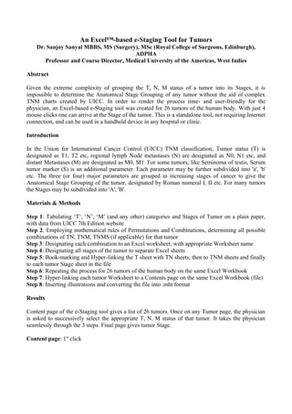
  • 1. An Excel™-based e-Staging Tool for Tumors Dr. Sanjoy Sanyal MBBS, MS (Surgery), MSc (Royal College of Surgeons, Edinburgh), ADPHA Professor and Course Director, Medical University of the Americas, West Indies Abstract Given the extreme complexity of grouping the T, N, M status of a tumor into its Stages, it is impossible to determine the Anatomical Stage Grouping of any tumor without the aid of complex TNM charts created by UICC. In order to render the process time- and user-friendly for the physician, an Excel-based e-Staging tool was created for 26 tumors of the human body. With just 4 mouse clicks one can arrive at the Stage of the tumor. This is a standalone tool, not requiring Internet connection, and can be used in a handheld device in any hospital or clinic. Introduction In the Union for International Cancer Control (UICC) TNM classification, Tumor status (T) is designated as T1, T2 etc, regional lymph Node metastases (N) are designated as N0, N1 etc, and distant Metastases (M) are designated as M0, M1. For some tumors, like Seminoma of testis, Serum tumor marker (S) is an additional parameter. Each parameter may be further subdivided into 'a', 'b' etc. The three (or four) major parameters are grouped in increasing stages of cancer to give the Anatomical Stage Grouping of the tumor, designated by Roman numeral I, II etc. For many tumors the Stages may be subdivided into 'A', 'B'. Materials & Methods Step 1: Tabulating ‘T’, ‘N’, ‘M’ (and any other) categories and Stages of Tumor on a plain paper, with data from UICC 7th Edition website Step 2: Employing mathematical rules of Permutations and Combinations, determining all possible combinations of TN, TNM, TNMS (if applicable) for that tumor Step 3: Designating each combination to an Excel worksheet, with appropriate Worksheet name Step 4: Designating all stages of the tumor to separate Excel sheets Step 5: Book-marking and Hyper-linking the T sheet with TN sheets, then to TNM sheets and finally to each tumor Stage sheet in the file Step 6: Repeating the process for 26 tumors of the human body on the same Excel Workbook Step 7: Hyper-linking each tumor Worksheet to a Contents page on the same Excel Workbook (file) Step 8: Inserting illustrations and converting the file into .mht format Results Content page of the e-Staging tool gives a list of 26 tumors. Once on any Tumor page, the physician is asked to successively select the appropriate T, N, M status of that tumor. It takes the physician seamlessly through the 3 steps. Final page gives tumor Stage. Content page: 1st click
  • 2. T page: 2nd click N page: 3rd click M page: 4th click
  • 3. Stage page: Final Discussion The permutations and combinations of T, N, M (and S) within a particular Anatomical Stage Grouping is extremely complex and varies considerably between different tumors. This complex classification renders it difficult for the oncologist to quickly determine the Stage of tumor without the aid of a TNM chart. This interactive e-Staging tool is standalone (in .mht format), simpler (requiring only 4 mouse clicks), quicker (just a few seconds) and easier than those from Stage CRAFT©, AJCC and Melanoma Center, which are all Web-based. It can be used by any physician with minimum computer skills. It is portable, can be used in any hand-held device anywhere in the hospital or clinic, and does not require Internet connection. It eliminates consulting complicated TNM charts, which are different for every tumor, and also the need to rely on memory, thus reducing errors and inconsistency between physicians. The tool can be incorporated in a hospital EMR with HL7. It is future-scalable, with options to add more tumor sites to the system. Acknowledgements Assistance of Medical University of the Americas in preparing the poster is gratefully acknowledged. Authorship and Conference Presentation This paper was authored by Dr Sanjoy Sanyal, Professor and Course Director of Neuroscience in Medical University of the Americas, Potworks, Charlestown, Nevis, St.Kitts-Nevis, West Indies. It was accepted at the international Stanford Medicine X Conference, and presented as a poster in
  • 4. Stanford University School of Medicine, LKSC Conference Center, 291 Campus Drive, Stanford, California, CA 94305-5101, USA, on 30 September, 2012. Poster Screenshot CANCER E-STAGING STANDALONE PROGRAM Process Flowchart for Implementation of Program
  • 7. Implementation Step 5 PATENT STATUS Provisional Patent Application accepted by United States Patent and Trademark Office (USPTO), giving it a 'Patent Pending' status.