Veterinary Dentistry for Technicians
•Descargar como PPTX, PDF•
11 recomendaciones•3,856 vistas

Veterinary Dentistry for Technicians -Dental cleaning and Oral Exam -Intraoral Radiogprahic Positioning -Oral Regional Nerve Blocks

Recomendados
Recomendados
Más contenido relacionado
La actualidad más candente
La actualidad más candente (20)
Destacado
Destacado (20)
Similar a Veterinary Dentistry for Technicians
Similar a Veterinary Dentistry for Technicians (20)
Último
Último (20)
Veterinary Dentistry for Technicians
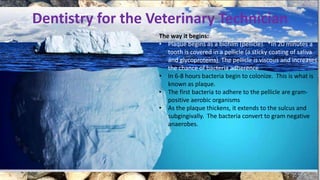
- 1. Dentistry for the Veterinary Technician The way it begins: • Plaque begins as a biofilm (pellicle). *In 20 minutes a tooth is covered in a pellicle (a sticky coating of saliva and glycoproteins). The pellicle is viscous and increases the chance of bacteria adherence. • In 6-8 hours bacteria begin to colonize. This is what is known as plaque. • The first bacteria to adhere to the pellicle are gram- positive aerobic organisms • As the plaque thickens, it extends to the sulcus and subgingivally. The bacteria convert to gram negative anaerobes.
- 2. Periodontal Disease = Septicemia • Blood flow (gingivitis) • Kidneys/Liver • Heart (Coronary Vessels)
- 6. Steps to a Dental Cleaning PPE **Protect yourself-----Protect your patient Exam Gloves Or face-shields instead of goggles Surgical Mask +/- Waterproof aprons? Safety goggles
- 7. Steps to a Dental Cleaning 1. Prolonged recovery 2. Bradycardia 3. Respiratory depression 4. Apnea 5. Ileus 6. Hypotension 7. Impaired clot function 8. Impaired immune function PPE Prevent Hypothermia
- 8. Steps to a Dental Cleaning Place pharyngeal pack
- 9. Steps to a Dental Cleaning Pre-rinse
- 11. Steps to a Dental Cleaning Remove bulky tartar Extraction or Tartar Removing Forceps
- 12. Steps to a Dental Cleaning Power Scale
- 14. Steps to a Dental Cleaning Hand Scale Jacquet (Sickle Scaler)
- 15. Hand Scalers
- 16. Steps to a Dental Cleaning Root Plane
- 17. Modified Pen Grasp with fulcrum Root Plane
- 18. Steps to a Dental Cleaning Modified pen grasp
- 19. Curettes Universal vs Area Specific
- 20. Curettes
- 21. Steps to a Dental Cleaning Rinse, polish, rinse
- 22. Steps to a Dental Cleaning Look for revealed tartar Rinse pumice
- 23. Steps to a Dental Cleaning Oral Exam/Radiograph Periodontal Probe Explorer
- 24. Oral Exam/Radiographs Probe and explore
- 25. Oral Exam/Radiographs Probe and explore Clinical Attachment
- 26. Some anatomy review Gingival Structures 409 (lower right first molar) Attached gingiva (Base of sulcus) Mucogingival junction (line) Gingival margin (Free Gingiva) Sulcus inside! Oral mucosa
- 28. Indications
- 29. Steps to a Dental Cleaning Chart findings
- 30. Charting
- 31. 5 Criteria for staging periodontal disease 1. Gingivitis and gingival index (GI) (grade 1-3) 2. Periodontal Probing Depth (P) in mm 3. Gingival recession (GR) in mm 4. Furcation exposure (FE) (Grade 1-3) 5. Tooth Mobility (M) (Grade 1-3) * Chart the stage of periodontal disease using the “worst tooth”. *Abnormal probing depth (pocket) + Gingival recession (from CEJ to gingival margin) = Total Attachment Loss Charting
- 33. Normal (PD 0): Clinically normal - no gingival inflammation or periodontitis clinically evident. Stage 1 (PD 1): Gingivitis only without attachment loss. The height and architecture of the alveolar margin are normal. Stage 2 (PD 2): Early periodontitis - less than 25% of attachment loss measured via probing or radiographs from CEJ to alveolar margin. Or stage 1 Furcation Exposure Stage 3 (PD 3): Moderate periodontitis - 25-50% of attachment loss measured via probing or radiographs from CEJ to alveolar margin or stage 2 Furcation Exposure. Stage 4 (PD 4): Advanced periodontitis - more than 50% of attachment loss measured via probing or radiographs from the CEJ to alveolar margin Or Stage 3 Furcation Exposure
- 34. Grade vs Stage Stage indicates a progressive condition Grade may be either progressive or reversible AVDC.org/nomenclature
- 35. 4 Clinical Signs of Periodontal Disease Depends on hosts’ response to the bacteria 1. Gingivitis 2. Calculus 3. Horizontal bone loss 4. Vertical bone loss
- 36. 4 Clinical Signs of Periodontal Disease Horizontal bone loss
- 37. 4 Clinical Signs of Periodontal Disease Vertical bone loss
- 38. Step 10. DVM Assessment/ Treatment Plan Calculate/Administer Nerve block(s) •Radiographs/Treatment plan •DVM views •Talk to client (via phone)? •Verbal estimate? •Plan/draw up Nerve block
- 39. 11. Periodontal Treatment INCLUDE: • Closed-Currettage -(debride pocket) • Open –surgical (flap) -root planing and currettage • Perioceutic - (Antibiotic pocket treatment) • Systemic antibiotics (BEFORE) cleaning - Clindamycin - “Pulse Therapy” • Extraction • Crown Reduction • Guided Tissue Regeneration -(Bone stimulant/Bone substitute) - Osteoallograft, Consil ®
- 40. 12. Fluoride Treatment •Desensitizes tooth •Helps minimize plaque adherence •Bacteriostatic •Its application is controversial because get fluoride from other sources
- 41. Questions?
- 44. Periodontal and Endodontic Structures
- 45. CEJ
- 46. Types Of Dentin: Primary Dentin Forms before tooth eruption Secondary Dentin The natural process of mastication stimulates production of more layers of dentin Tertiarty (Reparitive) Dentin Stimulates rapid formation as a result of pathology or injury
- 48. Maxilla
- 49. mandible
- 50. Regional Nerve Block Bupivicaine 0.5% Lidocaine 2% Onset 10-20 min 1-2 min Duration 4-8 hours ½ hour -1 hour
- 51. Regional Nerve Block Calculation for Nerve Block 1 mg/kg each drug Mix together 0.1mL/site –cats/sm dogs 0.3-0.5mL /site- med/large dogs ÷ how many nerve blocks (ie 4) •Don’t go over toxic dose of 1mg/kg each •ASPIRATE! •Monitor rhythm and blood pressure
- 52. Behavior response to pain Modulation Transduction Perception Transmission Nociception “The incision”
- 54. Regional Anesthesia Materials- 1mL or 3mL Syringe 25 x 5/8” needle unless large skeletal structure Warning- A less invasive approach= right outside the foramen vs inside Aspiration-3x (1/3 rotation and repeat) to check for blood Inject slowly. Apply digital pressure for 60 sec. Monitor patient.
- 55. Rostral Mandibular Nerve Block Middle Mental Foramen •Bone, teeth and soft tissue rostral to the mandibular pm/canine in cats •Dogs: Palpate foramen Landmark- labial frenulum & ventral to the mesial root of pm2 •Cats: Small foramen- palpate Landmark-Caudal to apex of canine
- 56. Mandibular Nerve Block (Inferior Alveolar Nerve) Mandibular Foramen •Bone, teeth and soft tissue of the ENTIRE mandible •Extraoral or Intraoral •Landmarks- ventral notch of mandible, lateral canthus of eye •Palpation of mandibular foramen- intraorally (Lingual surface 2/3 way from molar to angular process )
- 57. Mandibular Nerve Block (Inferior Alveolar Nerve) Intraoral Extraoral
- 58. Rostral Maxillary Nerve Block Infraorbital Foramen •Bone, teeth and soft tissue of the maxilla rostral to PM3 •Landmarks- Palpate juga of pm4- opening just rostral •Needle parallel to palate
- 59. Caudal Maxillary Nerve Block Infraorbital Nerve •Affects bone, teeth and soft tissue of the ENTIRE maxilla •Landmarks- Dogs: Max 2nd molar Cats: Divot caudal to max molar •Needle parallel to m root
- 61. Intraoral Radiography 3 Steps to remember 1. Patient positioning 2. Film placement within the patient’s mouth 3. Positioning the beam head
- 63. Positioning the beam head Parallel Technique Bisecting Angle (Vertical Angle)
- 64. Positioning the beam head Centering
- 65. Film or Sensor Placement
- 66. Bisecting Angle
- 67. Positioning5 areas of the mouth 1- Mandibular PM and M 2- Mandibular incisors/ canines 3- Maxillary incisors 4- Maxillary canines 5- Maxillary PM and M
- 68. Improper Beam Angle Beam Tooth FORSHORTENING •If the beam is pointing too close to the film or sensor •We have a short shadow when the sun is at noon fILm
- 69. Improper Beam Angle BeamTooth ELONGATION •If the beam is pointing too close to the tooth root •We have a long shadow when the sun is going down fILm
- 70. Improper Beam Angle HORIZONTAL ANGLE
- 72. Maxillary Canines Position as Max incisors with a 20° lateral (Horizontal) tilt
- 76. Why we love cats
- 77. Decreased Angle •Instead of Beam head perpendicular to BA •Angle is decreased by 20 ° •This purposefully elongates roots past Zygomatic Arch Special view to Avoid the Zygomatic Arch
- 78. Near Parallel Special view to Avoid the Zygomatic Arch
- 79. Simplified Method Relies on approximation instead of measurements Based on three basic angles: 45° Caudal maxillary teeth 60 ° Rostral teeth (incisors) 20 ° Horizontal tilt for Maxillary canines
- 81. Tooth Resorption
- 82. High Vitamin D Low Specific Gravity Dogs
- 83. Present in 65% of all cats TR1
- 84. Present in 65% of all cats
- 85. Present in 65% of all cats
- 86. TR4b Root>crown
- 87. Type II
- 88. TR4b Crown >root
- 89. TR5 aka “nubbin”
- 90. TR 5
- 97. TR4A- crown and root equally affected Type II