Dual Energy CT Detects Hepatocellular Carcinoma
•
0 recomendaciones•931 vistas

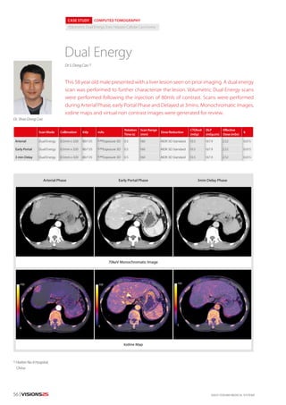
CASE: 58 year old male presented with a liver lesion seen on prior imaging. A dual energy scan was performed to further characterize the lesion. Volumetric Dual Energy scans were performed following the injection of 80mls of contrast. Scans were performed during Arterial Phase, early Portal Phase and Delayed at 3mins. Monochromatic images, iodine maps and virtual non contrast images were generated for review.

Recomendados
Más contenido relacionado
La actualidad más candente
La actualidad más candente (20)
Destacado
Destacado (9)
Similar a Dual Energy CT Detects Hepatocellular Carcinoma
Similar a Dual Energy CT Detects Hepatocellular Carcinoma (20)
Más de Canon Medical Systems Europe
Más de Canon Medical Systems Europe (20)
Último
Último (20)
Dual Energy CT Detects Hepatocellular Carcinoma
- 1. ©2015 TOSHIBA MEDICAL SYSTEMS56 | VISIONS25 1) Harbin No 4 Hospital, China CASE STUDY COMPUTED TOMOGRAPHY Volumetric Dual Energy, liver, Hepato-Callular Carcinoma Dual Energy ScanMode Collimation kVp mAs Rotation Time(s) ScanRange (mm) DoseReduction CTDIvol (mGy) DLP (mGy.cm) Effective Dose(mSv) k Arterial Dual Energy 0.5mm x 320 80/135 SUREExposure 3D 0.5 160 AIDR 3D Standard 10.5 167.9 2.52 0.015 Early Portal Dual Energy 0.5mm x 320 80/135 SUREExposure 3D 0.5 160 AIDR 3D Standard 10.5 167.9 2.52 0.015 3 min Delay Dual Energy 0.5mm x 320 80/135 SUREExposure 3D 0.5 160 AIDR 3D Standard 10.5 167.9 2.52 0.015 This 58 year old male presented with a liver lesion seen on prior imaging. A dual energy scan was performed to further characterize the lesion. Volumetric Dual Energy scans were performed following the injection of 80mls of contrast. Scans were performed during Arterial Phase, early Portal Phase and Delayed at 3mins. Monochromatic images, iodine maps and virtual non contrast images were generated for review. Dr S. Dong Cao 1) Dr. Shao Dong Coa Arterial Phase Early Portal Phase 70keV Monochromatic Image Iodine Map 3min Delay Phase
- 2. VISIONS25 | 57CTEU140097 SUMMARY The lesion demonstrates characteristics consistent with a Hepato-Cellular Carcinoma (HCC). These lesions have intense arterial enhancement, fast washout and then become homogeneous with normal liver. Raw data based dual energy provides high quality monochromatic images at 70keV which correspond to a routine 120kVp image allowing adjacent structures to be reviewed as usual. The iodine maps provide easy visualization and quantification of the lesion. This case demonstrates that volumetric dual energy can be implemented into dedicated liver imaging pro- tocols with good image quality in multiple phases at a reasonable radiation dose. Additional information is pro- vided by the iodine maps. Virtual Non Contrast images generated in the early venous phase demonstrate lesion homogeneity What is dual energy? CT imaging is based on the principle that various anatomical structures within the body attenuate X-rays differently. Unfortunately, structures with similar Hounsfield units (i.e. CT numbers) remain very difficult to differentiate from each other. However, structures that produce similar Hounsfield units at one beam energy may respond differentlyatadifferentbeamenergy.Dualenergyscanningcanincreasetheamount of information available from CT imaging. Iodine maps can be generated by analyz- ing images acquired at both high kVp and low kVp, providing functional information about tissue. As said Dual-energy helical scanning alternates between high and low kV with each gantryrotation.AlsothemAisautomaticallyadjustedforthetwodifferentenergiesto ensure a matched signal-to- noise ratio which increases the accuracy of dual energy analysis. Also the tube exposure can be manually turned OFF in the upper 180 degree ofthegantryrotationthatwouldexposetheventralsideofthepatientandpotentially more radiation sensitive areas such as breast tissue in females. Watch videos on the latest technology and educational lectures on our YouTube Channel: www.youtube.com/ToshibaMedicalEurope