High-Resolution Three-Dimensional Weight-Bearing Imaging of Lower Extremity Using Dedicated Cone Beam Computed Tomography (CBCT)

This paper addresses the benefits of a prototype (INVESTIGATIONAL – NOT FOR COMMERCIAL SALE) cone beam computed tomography system (hereafter referred to as the “CBCT system”) dedicated to extremity imaging. The CBCT system was co-developed by scientists at Carestream Health and John Hopkins University. The CBCT system has demonstrated spatial and contrast resolution beyond the limits of conventional multi-detector CT (MDCT) at a reduced radiation exposure1. The CBCT system was designed to image both upper and lower extremities, with the lower extremities also capable of being imaged in a weight-bearing configuration. This unique capability can unveil and better characterize certain pathologies in the knee and ankle joints such as meniscal extrusion, altered tibiofemoral joint space morphology, flatfoot deformity, and distal tibiofibular syndesmosis insufficiency. Learn more about Carestream's portfolio of products at http://www.carestream.com/medical

Recomendados
Recomendados
Más contenido relacionado
La actualidad más candente
La actualidad más candente (20)
Similar a High-Resolution Three-Dimensional Weight-Bearing Imaging of Lower Extremity Using Dedicated Cone Beam Computed Tomography (CBCT)
Similar a High-Resolution Three-Dimensional Weight-Bearing Imaging of Lower Extremity Using Dedicated Cone Beam Computed Tomography (CBCT) (20)
Más de Carestream
Más de Carestream (20)
Último
Último (20)
High-Resolution Three-Dimensional Weight-Bearing Imaging of Lower Extremity Using Dedicated Cone Beam Computed Tomography (CBCT)
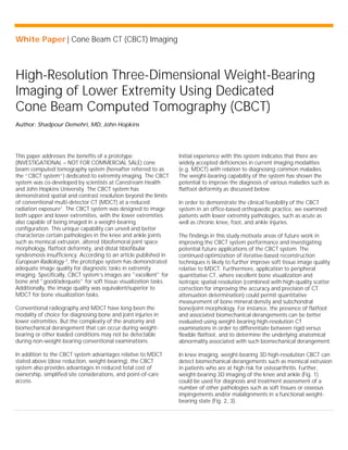
- 1. White Paper | Cone Beam CT (CBCT) Imaging High-Resolution Three-Dimensional Weight-Bearing Imaging of Lower Extremity Using Dedicated Cone Beam Computed Tomography (CBCT) Author: Shadpour Demehri, MD, John Hopkins This paper addresses the benefits of a prototype (INVESTIGATIONAL – NOT FOR COMMERCIAL SALE) cone beam computed tomography system (hereafter referred to as the “CBCT system”) dedicated to extremity imaging. The CBCT system was co-developed by scientists at Carestream Health and John Hopkins University. The CBCT system has demonstrated spatial and contrast resolution beyond the limits of conventional multi-detector CT (MDCT) at a reduced radiation exposure1 . The CBCT system was designed to image both upper and lower extremities, with the lower extremities also capable of being imaged in a weight-bearing configuration. This unique capability can unveil and better characterize certain pathologies in the knee and ankle joints such as meniscal extrusion, altered tibiofemoral joint space morphology, flatfoot deformity, and distal tibiofibular syndesmosis insufficiency. According to an article published in European Radiology 2 , the prototype system has demonstrated adequate image quality for diagnostic tasks in extremity imaging. Specifically, CBCT system’s images are "excellent" for bone and "good/adequate" for soft tissue visualization tasks. Additionally, the image quality was equivalent/superior to MDCT for bone visualization tasks. Conventional radiography and MDCT have long been the modality of choice for diagnosing bone and joint injuries in lower extremities. But the complexity of the anatomy and biomechanical derangement that can occur during weight- bearing or other loaded conditions may not be detectable during non-weight-bearing conventional examinations. In addition to the CBCT system advantages relative to MDCT stated above (dose reduction, weight-bearing), the CBCT system also provides advantages in reduced total cost of ownership, simplified site considerations, and point-of-care access. Initial experience with this system indicates that there are widely accepted deficiencies in current imaging modalities (e.g. MDCT) with relation to diagnosing common maladies. The weight-bearing capability of the system has shown the potential to improve the diagnosis of various maladies such as flatfoot deformity as discussed below. In order to demonstrate the clinical feasibility of the CBCT system in an office-based orthopaedic practice, we examined patients with lower extremity pathologies, such as acute as well as chronic knee, foot, and ankle injuries. The findings in this study motivate areas of future work in improving the CBCT system performance and investigating potential future applications of the CBCT system. The continued optimization of iterative-based reconstruction techniques is likely to further improve soft tissue image quality relative to MDCT. Furthermore, application to peripheral quantitative CT, where excellent bone visualization and isotropic spatial resolution (combined with high-quality scatter correction for improving the accuracy and precision of CT attenuation determination) could permit quantitative measurement of bone mineral density and subchondral bone/joint morphology. For instance, the presence of flatfoot and associated biomechanical derangements can be better evaluated using weight-bearing high-resolution CT examinations in order to differentiate between rigid versus flexible flatfoot, and to determine the underlying anatomical abnormality associated with such biomechanical derangement. In knee imaging, weight-bearing 3D high-resolution CBCT can detect biomechanical derangements such as meniscal extrusion in patients who are at high risk for osteoarthritis. Further, weight-bearing 3D imaging of the knee and ankle (Fig. 1) could be used for diagnosis and treatment assessment of a number of other pathologies such as soft tissues or osseous impingements and/or malalignments in a functional weight- bearing state (Fig. 2, 3).
- 2. White Paper | Cone Beam CT (CBCT) Imaging 2 Non-weight-bearing Weight-bearing Fig. 1: The MRI image (right) shows the presence of a fibrous band at the calcaneonavicular interface. CBCT 3D volumetric images (left) show the subtle flattening of the arch on the weight-bearing image associated with flatfoot. Fig. 2: High-resolution 3D CBCT of the ankle demonstrates no osseous coalition at the Calcaneo-navicular interface. Fig. 3: Weight-bearing and non-weight-bearing images show meniscal extrusion (small yellow lines) and biomechanical derangement in the weight-bearing image of this patient with osteoarthritis.
- 3. White Paper | Cone Beam CT (CBCT) Imaging carestream.com ©Carestream Health, Inc., 2015. CARESTREAM is a trademark of Carestream Health. CAT 2000140 09/15 References: 1. Carrino JA, Al Muhit A, Zbijewski W, Thawait GK, Stayman JW, Packard N, Senn R, Yang D, Foos DH, Yorkston J, Siewerdsen JH. Dedicated cone-beam CT system for extremity imaging. Radiology. 2014 Mar;270(3):816-24. 2. Demehri S, Muhit A, Zbijewski W, Stayman JW, Yorkston J, Packard N, Senn R, Yang D, Foos D, Thawait GK, Fayad LM, Chhabra A, Carrino JA, Siewerdsen JH. Assessment of image quality in soft tissue and bone visualization tasks for a dedicated extremity cone-beam CT system. Eur Radiol. 2015 Jun;25(6):1742-51. Dr. Demehri received his MD at Tehran University of Medical Sciences and completed Radiology residency and fellowship training in Musculoskeletal Imaging and intervention at Brigham and Women's Hospital. He joined the Department of Radiology at Johns Hopkins in 2012. His research interests are novel CT imaging modalities, 3D post-processing techniques and their application in Musculoskeletal imaging. He is currently principle investigator of the clinical trial assessing the feasibility of dedicated CBCT examinations for diagnosis and treatment of various pathologies involving the peripheral joints.