SlideShare una empresa de Scribd logo
1 de 35
Mature Oocyte


                Vitellogenic Phase
Lobulated Nuclear
Membrane            Germinal
                    Vesicle
      Nucleoli
Animal Pole


              Vegetal Pole
Yolk Platelet

  Yolk Nuclei
  Follicle Cells

Theca Interna
Animal
                  Hemisphere


                  Jelly Coat


                  Vegetal Pole

Before Cleavage
Cleavage
Furrow


    Blastomeres
Blastomeres


              Cleavage
              Furrow
Micromeres


3rd Cleavage
Furrow
(Equatorial)

 Developing
 Blastocoel


Macromeres
Fertilization
Membrane

Micromeres


Developing
Blastocoel


Macromeres
Animal
                 Hemisphere


                  Blastocoel filled
                  with albuminous
                  fluid


                  Marginal Zone,
                  Germ Ring,
                  Marginal Belt
                 Nucleus (darkly
                 staining body)
                 Vegetal
                 Hemisphere
Early Blastula
Nervous Layer
                 Animal
                 Hemisphere

                Epidermal Layer


                 Blastocoel filled
                 with albuminous
                 fluid


                Vegetal
                Hemisphere
Late Blastula
Dorsal lip of the
                 Blastopore
                 Blastopore
                 Ventral lip of the
                 Blastopore


                 Disintegrating
                 Blastocoel


Early Gastrula
A – Anterior End (Point of
            11   Sperm Entry)
                 P – Posterior End (Yolk
                 Plug)
                 1 - Ectoderm
                 2 - Endoderm
           10    3 - Mesoderm
                 4 - Disintegrating
                 Blastrocoel
                 5 - Archenteron /
                 Gastrocoel
                 6 - Dorsal Lip of the
                 Blastopore
                 7 - Blastopore
                 8 - Vental Lip of the
                 blastopore
                 9 - Endodermal Mass
                 10 - Completion Bridge
                 11 - Chordamesoderm


Gastrula
Neural Plate
Neural Groove
         Neural Fold
         Neural Crest
         Notochord

         Somatic
Midgut   Mesoderm
         Splanchnic
         Mesoderm
         Gut Endoderm

         Ectoderm
         Endoderm
         Lateral Plate
         Mesoderm
Neural Crest
                       Nerve Cord
                       Neural Canal
                       Notochord
                       Somite mesoderm
                       Subnotochordal Notch
              Midgut   Somatopleure
                       Splanchnopleure
                       Splanchnic
                       Mesoderm
                       Endoderm


Neural Tube
Encircled - Notochord
                             1               1 – Lateral Ventricles
      10                                     enclosed by the Cerebral
                 2                           Hemispheres
                                     1
                                             2 – Olfactory Membrane
           11    3                           3 – Olfactory Tube
                     4                       4 – Parachordal cartilage
                                             5 – Pharynx
                                             6 – Mandibular Cartilages
                                         2   7 – Oral Sucker
                             4
                                             8 – Lip
                                         3
                                             9 – Tooth Germ
                     5                       10 – Epidermis
                                             11 - Melanophores
                                 6
       7
            8
                 9
                         8
Level of Telencephalon
Jacobson’s Organ
1 – Diencephalon (cavity: III
                                                             ventricle or diocoel; roof
                                       1                     Anterior Choroid Plexus)
                                                             2 – Infundibulum
                                                             3 – Hypophysis
   10                                                        4 – Pharynx
                              2                              5 – Lens
                                                             6 – Retina
                          3
                                                 6       7   7 – Cornea
                                           8         5       8 – Optic Nerve
                      4
                                                             9 – Thyroid Bodies
                     12                                      10 – Lymph Sinus
                 9                                           11 – Ceratohyal Cartilage
                                           13                12 – Basibranchial Cartilage
                                  11                         13 – Mandibular Muscles
                                                             14 – Hyoid Muscle
                                  14
                                            10


Level of Diencephalon
2
         4                 5    4        1 – Myelencephalon
                                         2 – Rhombocoel or IV
         3             1        3        ventricle
              6             6            3 – Saccule
     7                              7    4 – Anterior Semicircular
                                         Canal
              8                          5 – Endolymphatic Duct
                       9                 6 – Ganglion VIII
                                         7 – Anterior Cardinal Vein
                                         8 – Pharynx
         10                              9 – Velar Plate
                   11               10
                                         10 – Gill rakers
 15                                 15   11 – Atrium
                                         12 – Ventricle
                           13            13 – Pericardial Cavity
14                                  14
                  12                     14 – Opercular Chamber
                                         15 – Internal Gills
Aortic Arch
Sinus Venosus
Duct of Cuvier


Ventricle
1 – Esophagus
                            2 – Lung Bud
                            3 – Nephrostome
                            4 – Glomus
                            5 – Liver
                            6 – Peritoneal Cavity
                        4
                            7 - Spiracle
2
        1           3

    6           2


                        7
            5
Reviewer
Reviewer

Más contenido relacionado

La actualidad más candente

Zoology- Phylum Porifera
Zoology- Phylum PoriferaZoology- Phylum Porifera
Zoology- Phylum PoriferaMc Nash Tanjili
 
Phylum chordata - zoology
Phylum chordata -  zoologyPhylum chordata -  zoology
Phylum chordata - zoologySijo A
 
Comparative Anatomy - Digestive System
Comparative Anatomy - Digestive SystemComparative Anatomy - Digestive System
Comparative Anatomy - Digestive SystemEmsi Onairpic
 
Animal Morphology : Cells, Tissues, Organs and Organ Aystems of Animals
Animal Morphology : Cells, Tissues, Organs and Organ Aystems of AnimalsAnimal Morphology : Cells, Tissues, Organs and Organ Aystems of Animals
Animal Morphology : Cells, Tissues, Organs and Organ Aystems of AnimalsTrixie Piloton
 
Canal system in porifera-different type of canal system
Canal system in porifera-different type of canal systemCanal system in porifera-different type of canal system
Canal system in porifera-different type of canal systemSoniaBajaj10
 
cleavage and gastrulation in amphibians
cleavage and gastrulation in amphibianscleavage and gastrulation in amphibians
cleavage and gastrulation in amphibiansAmina Ayub
 
Phylum Ctenophora , Comb Jellies pptx
Phylum Ctenophora , Comb Jellies pptxPhylum Ctenophora , Comb Jellies pptx
Phylum Ctenophora , Comb Jellies pptxDr. Muhammad Moosa
 
IB Biology Core 2.5: Cell Division
IB Biology Core 2.5: Cell DivisionIB Biology Core 2.5: Cell Division
IB Biology Core 2.5: Cell DivisionJason de Nys
 
Comparative Anatomy - Reproductive System
Comparative Anatomy - Reproductive SystemComparative Anatomy - Reproductive System
Comparative Anatomy - Reproductive SystemEmsi Onairpic
 

La actualidad más candente (20)

Frog dissecting
Frog dissectingFrog dissecting
Frog dissecting
 
Zoology- Phylum Porifera
Zoology- Phylum PoriferaZoology- Phylum Porifera
Zoology- Phylum Porifera
 
Scoliodon
ScoliodonScoliodon
Scoliodon
 
Phylum chordata - zoology
Phylum chordata -  zoologyPhylum chordata -  zoology
Phylum chordata - zoology
 
Comparative Anatomy - Digestive System
Comparative Anatomy - Digestive SystemComparative Anatomy - Digestive System
Comparative Anatomy - Digestive System
 
Animal Morphology : Cells, Tissues, Organs and Organ Aystems of Animals
Animal Morphology : Cells, Tissues, Organs and Organ Aystems of AnimalsAnimal Morphology : Cells, Tissues, Organs and Organ Aystems of Animals
Animal Morphology : Cells, Tissues, Organs and Organ Aystems of Animals
 
Lesson 11 animal tissues
Lesson 11 animal tissuesLesson 11 animal tissues
Lesson 11 animal tissues
 
Porifera phylum
Porifera phylumPorifera phylum
Porifera phylum
 
Canal system in porifera-different type of canal system
Canal system in porifera-different type of canal systemCanal system in porifera-different type of canal system
Canal system in porifera-different type of canal system
 
cleavage and gastrulation in amphibians
cleavage and gastrulation in amphibianscleavage and gastrulation in amphibians
cleavage and gastrulation in amphibians
 
Cleavage 1
Cleavage 1Cleavage 1
Cleavage 1
 
Amount and distribution of yolk and types of egg
Amount and distribution of yolk and types of egg Amount and distribution of yolk and types of egg
Amount and distribution of yolk and types of egg
 
Phylum Ctenophora , Comb Jellies pptx
Phylum Ctenophora , Comb Jellies pptxPhylum Ctenophora , Comb Jellies pptx
Phylum Ctenophora , Comb Jellies pptx
 
IB Biology Core 2.5: Cell Division
IB Biology Core 2.5: Cell DivisionIB Biology Core 2.5: Cell Division
IB Biology Core 2.5: Cell Division
 
Amphioxus (Branchiostoma)
Amphioxus (Branchiostoma)Amphioxus (Branchiostoma)
Amphioxus (Branchiostoma)
 
Chordata
ChordataChordata
Chordata
 
phylum porifera
phylum poriferaphylum porifera
phylum porifera
 
Comparative Anatomy - Reproductive System
Comparative Anatomy - Reproductive SystemComparative Anatomy - Reproductive System
Comparative Anatomy - Reproductive System
 
Phylum Porifera - Sycon
Phylum Porifera - SyconPhylum Porifera - Sycon
Phylum Porifera - Sycon
 
Phylum Porifera
Phylum  PoriferaPhylum  Porifera
Phylum Porifera
 

Similar a Reviewer

Virtual disection artifact
Virtual disection artifactVirtual disection artifact
Virtual disection artifactzs4033bn
 
Muscles of facial expression
Muscles of facial expressionMuscles of facial expression
Muscles of facial expressionDr. SHEETAL KAPSE
 
Roentgenology of skull
Roentgenology of skullRoentgenology of skull
Roentgenology of skullakshay_gursale
 
ชีววิทยา1
ชีววิทยา1ชีววิทยา1
ชีววิทยา1Darika Kanhala
 
Orofacial development
Orofacial developmentOrofacial development
Orofacial developmentRano Ya Naso
 
Mead & Fordyce. Therian skull. A lexicon with emphasis on the odontocetes.
Mead & Fordyce. Therian skull. A lexicon with emphasis on the odontocetes.Mead & Fordyce. Therian skull. A lexicon with emphasis on the odontocetes.
Mead & Fordyce. Therian skull. A lexicon with emphasis on the odontocetes.Ismael Mijan
 
Facial anatomy
Facial anatomyFacial anatomy
Facial anatomywebzforu
 
Non keratinocytes&specialized mucosa
Non keratinocytes&specialized mucosaNon keratinocytes&specialized mucosa
Non keratinocytes&specialized mucosaAhmed Abulata
 
Neural Induction in human brain: principles and processes
Neural Induction in human brain: principles and processesNeural Induction in human brain: principles and processes
Neural Induction in human brain: principles and processesNitish kumar
 
microscopic structure of ovary with diagram
microscopic structure of ovary with diagrammicroscopic structure of ovary with diagram
microscopic structure of ovary with diagramMuthuRoshni1
 
3.1_Third_Week_of_Development[1].pptx
3.1_Third_Week_of_Development[1].pptx3.1_Third_Week_of_Development[1].pptx
3.1_Third_Week_of_Development[1].pptxCALEBKIRUI1
 
Skeletal Identification
Skeletal IdentificationSkeletal Identification
Skeletal IdentificationMyika957
 

Similar a Reviewer (20)

Reviewer 2
Reviewer 2Reviewer 2
Reviewer 2
 
cerebrum sections for revision
cerebrum sections for revisioncerebrum sections for revision
cerebrum sections for revision
 
Virtual disection artifact
Virtual disection artifactVirtual disection artifact
Virtual disection artifact
 
Muscles of facial expression
Muscles of facial expressionMuscles of facial expression
Muscles of facial expression
 
Roentgenology of skull
Roentgenology of skullRoentgenology of skull
Roentgenology of skull
 
ชีววิทยา1
ชีววิทยา1ชีววิทยา1
ชีววิทยา1
 
Orofacial development
Orofacial developmentOrofacial development
Orofacial development
 
Mead & Fordyce. Therian skull. A lexicon with emphasis on the odontocetes.
Mead & Fordyce. Therian skull. A lexicon with emphasis on the odontocetes.Mead & Fordyce. Therian skull. A lexicon with emphasis on the odontocetes.
Mead & Fordyce. Therian skull. A lexicon with emphasis on the odontocetes.
 
Facial anatomy
Facial anatomyFacial anatomy
Facial anatomy
 
The neural crest
The neural crestThe neural crest
The neural crest
 
Non keratinocytes&specialized mucosa
Non keratinocytes&specialized mucosaNon keratinocytes&specialized mucosa
Non keratinocytes&specialized mucosa
 
Embryology minimals
Embryology minimalsEmbryology minimals
Embryology minimals
 
OLFACTORY NERVE
OLFACTORY NERVEOLFACTORY NERVE
OLFACTORY NERVE
 
Neural Induction in human brain: principles and processes
Neural Induction in human brain: principles and processesNeural Induction in human brain: principles and processes
Neural Induction in human brain: principles and processes
 
Embryology Overview
Embryology OverviewEmbryology Overview
Embryology Overview
 
Mollusks
MollusksMollusks
Mollusks
 
microscopic structure of ovary with diagram
microscopic structure of ovary with diagrammicroscopic structure of ovary with diagram
microscopic structure of ovary with diagram
 
Face mfe
Face mfeFace mfe
Face mfe
 
3.1_Third_Week_of_Development[1].pptx
3.1_Third_Week_of_Development[1].pptx3.1_Third_Week_of_Development[1].pptx
3.1_Third_Week_of_Development[1].pptx
 
Skeletal Identification
Skeletal IdentificationSkeletal Identification
Skeletal Identification
 

Más de Jay Edneil Olivar (17)

Microarray
Microarray Microarray
Microarray
 
A118G SNP
A118G SNPA118G SNP
A118G SNP
 
on GMOs
on GMOson GMOs
on GMOs
 
Bacon and Kuhn
Bacon and KuhnBacon and Kuhn
Bacon and Kuhn
 
150 2nd journal report
150 2nd journal report150 2nd journal report
150 2nd journal report
 
De extinction
De extinctionDe extinction
De extinction
 
Ordovician
OrdovicianOrdovician
Ordovician
 
150 journal reprt
150  journal reprt150  journal reprt
150 journal reprt
 
Carrot callus formation
Carrot callus formationCarrot callus formation
Carrot callus formation
 
Proprioception and spatial orientation
Proprioception and spatial orientationProprioception and spatial orientation
Proprioception and spatial orientation
 
120 journal reporting
120 journal reporting120 journal reporting
120 journal reporting
 
Hemoglobin and hematocrit determination
Hemoglobin and hematocrit determinationHemoglobin and hematocrit determination
Hemoglobin and hematocrit determination
 
Patterns of biodiveristy
Patterns of biodiveristyPatterns of biodiveristy
Patterns of biodiveristy
 
Journal reporting 161
Journal reporting 161Journal reporting 161
Journal reporting 161
 
122 journal reporting
122 journal reporting122 journal reporting
122 journal reporting
 
In vitro intestinal absorption
In vitro intestinal absorptionIn vitro intestinal absorption
In vitro intestinal absorption
 
Reviewer 2
Reviewer 2Reviewer 2
Reviewer 2
 

Reviewer

  • 1.
  • 2.
  • 3.
  • 4.
  • 5.
  • 6.
  • 7.
  • 8.
  • 9. Mature Oocyte Vitellogenic Phase
  • 10. Lobulated Nuclear Membrane Germinal Vesicle Nucleoli
  • 11. Animal Pole Vegetal Pole
  • 12. Yolk Platelet Yolk Nuclei Follicle Cells Theca Interna
  • 13. Animal Hemisphere Jelly Coat Vegetal Pole Before Cleavage
  • 14. Cleavage Furrow Blastomeres
  • 15. Blastomeres Cleavage Furrow
  • 18. Animal Hemisphere Blastocoel filled with albuminous fluid Marginal Zone, Germ Ring, Marginal Belt Nucleus (darkly staining body) Vegetal Hemisphere Early Blastula
  • 19. Nervous Layer Animal Hemisphere Epidermal Layer Blastocoel filled with albuminous fluid Vegetal Hemisphere Late Blastula
  • 20. Dorsal lip of the Blastopore Blastopore Ventral lip of the Blastopore Disintegrating Blastocoel Early Gastrula
  • 21. A – Anterior End (Point of 11 Sperm Entry) P – Posterior End (Yolk Plug) 1 - Ectoderm 2 - Endoderm 10 3 - Mesoderm 4 - Disintegrating Blastrocoel 5 - Archenteron / Gastrocoel 6 - Dorsal Lip of the Blastopore 7 - Blastopore 8 - Vental Lip of the blastopore 9 - Endodermal Mass 10 - Completion Bridge 11 - Chordamesoderm Gastrula
  • 23. Neural Groove Neural Fold Neural Crest Notochord Somatic Midgut Mesoderm Splanchnic Mesoderm Gut Endoderm Ectoderm Endoderm Lateral Plate Mesoderm
  • 24. Neural Crest Nerve Cord Neural Canal Notochord Somite mesoderm Subnotochordal Notch Midgut Somatopleure Splanchnopleure Splanchnic Mesoderm Endoderm Neural Tube
  • 25.
  • 26. Encircled - Notochord 1 1 – Lateral Ventricles 10 enclosed by the Cerebral 2 Hemispheres 1 2 – Olfactory Membrane 11 3 3 – Olfactory Tube 4 4 – Parachordal cartilage 5 – Pharynx 6 – Mandibular Cartilages 2 7 – Oral Sucker 4 8 – Lip 3 9 – Tooth Germ 5 10 – Epidermis 11 - Melanophores 6 7 8 9 8 Level of Telencephalon
  • 28. 1 – Diencephalon (cavity: III ventricle or diocoel; roof 1 Anterior Choroid Plexus) 2 – Infundibulum 3 – Hypophysis 10 4 – Pharynx 2 5 – Lens 6 – Retina 3 6 7 7 – Cornea 8 5 8 – Optic Nerve 4 9 – Thyroid Bodies 12 10 – Lymph Sinus 9 11 – Ceratohyal Cartilage 13 12 – Basibranchial Cartilage 11 13 – Mandibular Muscles 14 – Hyoid Muscle 14 10 Level of Diencephalon
  • 29.
  • 30. 2 4 5 4 1 – Myelencephalon 2 – Rhombocoel or IV 3 1 3 ventricle 6 6 3 – Saccule 7 7 4 – Anterior Semicircular Canal 8 5 – Endolymphatic Duct 9 6 – Ganglion VIII 7 – Anterior Cardinal Vein 8 – Pharynx 10 9 – Velar Plate 11 10 10 – Gill rakers 15 15 11 – Atrium 12 – Ventricle 13 13 – Pericardial Cavity 14 14 12 14 – Opercular Chamber 15 – Internal Gills
  • 32. Sinus Venosus Duct of Cuvier Ventricle
  • 33. 1 – Esophagus 2 – Lung Bud 3 – Nephrostome 4 – Glomus 5 – Liver 6 – Peritoneal Cavity 4 7 - Spiracle 2 1 3 6 2 7 5

Notas del editor

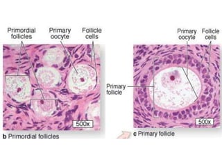
  1. cortex, a region filled with a highly cellular connective tissue stroma and many ovarian follicles, which in the adult ovary vary greatly in size The most internal part of the ovary is the medulla, which contains loose connective tissue and blood vessels entering the organprimordial follicles—consist of a primary oocyte enveloped by a single layer of the flattened follicular cellsunilaminar primary follicle  stratified follicular epithelium, the granulosa, in which the cells communicate through gap junctions. Follicle cells are now termed granulosa cells and the follicle is a multilayered primary follicle (cuboidal cells)
  2. Multilaminar Primary FolliceZonapellucida:four glycoproteins secreted by the oocyte;bind proteins on the surfaces of sperm and induce acrosomal activation. Filopodia of follicular cells and microvilli of the oocyte penetrate the zonapellucida, allowing communication between these cells via gap junctions.
  3. Each ovary is covered by a simple cuboidal epithelium, the germinal epithelium, continuous with the mesothelium and overlying a layer of dense connective tissue capsule, the tunica albugineagranulosa layer as the cells secrete follicular fluid  accumulates, the spaces gradually coalesce antrum Follicular Fluid : hyaluronate, growth factors, plasminogen, fibrinogen, the anticoagulant heparansulfate proteoglycan, and high concentrations of steroids (progesterone, androstenedione, and estrogens) with binding proteins.Membranagranulosa (columnar)
  4. Developing Secondary FollicleTunica Interna : Steroid Secreting
  5. Follicular WallsGranulosa Cells – Stratified ColumnarBM – separates TI and GTI: vacuolated and lightly stained because of their cytoplasmic lipid droplets, a characteristic of steroid-producing cells (androstenedione which is transformed in the granulosa cells as estradiol)TE: fibroblasts and smooth muscle cells continuous with stroma
  6. Corpus luteum after ovulation (super folded)Corpus Albicans:  scar of dense connective tissue called corpus albicans  in the absence of pregnancy (less convoluted)
  7. After ovulation:Granulosa cells  granulosa lutein cellsaromatase conversion of androstenedione into estradiolTheca interna cells  Theca lutein cells stain more darkly, with cytoplasmic ultrastructural features of steroid-synthesizing cells stimulated by LH
  8. Atretic follicle: oocyte is detached from granulosa cellsAtresia involves apoptosis and detachment of the granulosa cells, autolysis of the oocyte and collapse of the zonapellucida. Early in this process, macrophages invade the degenerating follicle and phagocytose the debris. Occurs anytime
  9. Mesolecithal specifically Telolecithal egg Animal Pole: Superficial Melanin, Cytoplasm; NucleusVegetal Pole: Yolk
  10. Vitellogenic Stage
  11. Mature OocyteAchievement of Polarity and Radial Symmetry
  12. Frog OvarySource of Yolk: Digested FoodYolk Nuclei from germinal vesicle
  13. Unfertilized egg
  14. Second Cleavage furrow relatively shallow compared to first
  15. Yolk is resistant to cleavage forces
  16. CoeloblastulaEpidermal layer (highly pigmented)  Skin Epithelium or Lining of nervous systemNervous Layer  Neuroblasts of the Nervous SystemMarginal Zone: active conversion of yolk to cytoplasm; involved in formation of the lips of the blastoporeStill seen is the fertilization membrane (not labeled)
  17. CoeloblastulaEpidermal layer (highly pigmented)  Skin Epithelium or Lining of nervous systemNervous Layer  Neuroblasts of the Nervous SystemMarginal Zone: active conversion of yolk to cytoplasm; involved in formation of the lips of the blastoporeStill seen is the fertilization membrane (not labeled)
  18. Process: InvolutionVentral limit of the gray crescent  region of formation of the dorsal lip of the blastopore
  19. Ring of involuted marginal zone (lip) cells are collectively known as the yolk plugInvoluted dorsal lip cells endodermMore ventral and lateral to the chordamesoderm is the mesodermChordamesoderm  notochord
  20. notochord was derived from cells indistinguishable from the mesoderm at the region of the dorsal lipSomites become: 1. Dermatome  dermis and appendage musculature 2. Sclerotome vertebral skeleton 3. Myotome skeletal musclesSomatic mesoderm in conjuction with the ectoderm  skin with its blood and connective tissuesSplanchnic mesoderm in conjuction with gut endoderm  lining epithelium, muscles, and blood vessels of the entire mid- and hindgut.
  21. Lateral Ventricles: Right (first); Left (second)
  22. Detail of Olfactory Placode