SlideShare una empresa de Scribd logo
1 de 166
1. How does the heart generate electricity and make
itself contract?
2. How does our body control the rate and strength of
those contractions?
3. How can we, as paramedics, affect this process using
medications?
Paramedic Cardiology
Electrophysiology
Part 0 – Cardiac Electrophysiology - 0 Fundamentals
• In this lecture we'll be going over some basic biology to get you ready for
cardiac electrophysiology.
• https://youtu.be/gCbHyK6y4Ms
Part 1 – Cardiovascular Electrophysiology 1 - Movement through the membrane
• In this lecture we're going to describe how different ions are able to move in
and out of the cell.
• https://youtu.be/IW3J2Y2ty6w
Part 2 – Cardiovascular Electrophysiology 2 - Electrical Flow In The Heart
• In this lecture we're going to talk about how a hunk of meat in your chest can
generate electricity.
• https://youtu.be/5A_lEW1suAM
Paramedic Cardiology
Electrophysiology
Part 3 - Cardiovascular Electrophysiology 3 - Action Potential of the Myocytes
• In this lecture, we're going to go over the pattern of how ions move in and out of the
cell in a regular, repeating pattern - called the Action Potential.
• https://youtu.be/KV3AaXWrNeY
Part 4 - Cardiovascular Electrophysiology 4 - Action Potential of the Pacemaker Cells
• In this lecture we're going to describe how 'pacemaker' cells can spontaneously
depolarize without anything stimulating them externally.
• https://youtu.be/M39wMZFd22o
Part 5 - Cardiovascular Electrophysiology 5 - Anatomy and Physiology of Myocytes
• In this lecture we describe how cardiac cells physically contract.
• https://youtu.be/qqnL0Fy3e2g
Paramedic Cardiology
Electrophysiology
Part 6 - Cardiovascular Electrophysiology 6 - Excitation Contraction Coupling
• In this video we're going to go over how depolarisation initiates contraction of
the heart.
• https://youtu.be/UBINkoW6JzM
Part 7 - Cardiovascular Electrophysiology 7 - ANS Influence on the Heart
• In this lecture we cover how our body changes the rate and strength of our
heart, going from external stimuli to the actual ionic changes that take place
in the myocardial cells. If I yell 'boo!', your heart speeds up. Why? How?
• https://youtu.be/JmAaibgOrLw
Part 8 - 8 Paramedic Pharmacology – Antidysrhythmics
• In this final lecture, we review cardiac antidysrhythmics with a fairly
introductory discussion of how they are classified and how they work.
• https://youtu.be/oY0XVEkfKr4
Paramedic Cardiology
Electrophysiology
Electrophysiology
Part 0
Fundamentals
Marc Colbeck
Paramedic Cardiology
© Marc Colbeck and Paramedicine.com, used by permission of the author. See:
https://creativecommons.org/licenses/by-nc-sa/4.0/
Maintaining this note in the file is sufficient attribution of authorship as per the author.
Topics
0. Fundamentals
1. Movement Through the Membrane
2. Electrical Flow in the Heart
3. The Action Potential – Myocytes
4. The Action Potential – Pacemaker cells
5. Anatomy and Physiology of Myocytes
6. Excitation-Contraction Coupling
7. ANS Influence on the Heart
8. Paramedic Pharmacology - Antidysrhythmics
Priming Questions
1. What is an ‘atom’
2. What is an ‘element’?
3. What is an ‘ion’?
4. How about anions and cations, what are those?
5. What is a cell?
6. Where did cells come from?
7. What are the walls of a cell made out of?
8. What are the major elements that humans are made out of?
9. Why is phosphorous so important to humans?
10. What are the ions that are important in cardiac electrophysiology?
You Should Already Know
Atoms
• Basic building block
– Protons+ and neutrons in nucleus
– Electrons- in orbit
Element
• Substance made entirely of one
type of atom (e.g. H2)
Ions
• An atom or element with a lack or
surplus of either protons or
electrons
– Anions: negative charge
– Cations: positive charge
The Elements of Life
99% CHON (by weight)
http://bookofresearch.com/images/earth-transparent.png
<1% P, as well as some S, Na, Mg, Cl, K, Ca and Fe
10
The Elements of Life
http://1.bp.blogspot.com/-mrMWO9w3UvE/TdE67QdLgOI/AAAAAAAAAEY/Bpu6nJpEBgk/s1600/periodic_table_of_elements1.jpg
composition (by weight)
11
The Elements of Life - Phosphorus
• Highly reactive, usually found as ‘red phosphorus’
• Used in explosives (napalm), poisons and nerve agents
• ‘Organophosphates’ are used as fertilizers, but can also (along with the
more potent fluorophosphates) be used as neurotoxins.
• 13th Element discovered (in 1669)
• Distilled from urine (100L ≈ 5mg) (later discovered in bones)
• Now mined from rocks
Essential to life:
• Part of DNA, RNA and ATP (‘phosphorylation) as well as phospholipids
http://0.tqn.com/d/chemistry/1/0/Q/4/1/phosphorus_allotropes.jpg
12
The Elements of Life
• Phosphorous
– binds to 4 oxygen
– hydrophilic
• Hydrocarbon tails
– hydrophobic
http://bioweb.wku.edu/courses/biol115/wyatt/biochem/lipid/P-lipid.gif
13
Cell Membranes
http://www.freethought-forum.com/images/anatomy2/phospholipid_bilayer.jpg
14
Cell Membranes
Liposomes
• “Inside/Outside”
https://upload.wikimedia.org/wikipedia/commons/0/01/Liposome_scheme-en.svg
15
Cell Contents
What’s inside liposomes?
http://www.fossilmuseum.net/Paleobiology/Paleobiology_images/seawater.jpg
16
Cell Contents
http://1.bp.blogspot.com/-mrMWO9w3UvE/TdE67QdLgOI/AAAAAAAAAEY/Bpu6nJpEBgk/s1600/periodic_table_of_elements1.jpg
What’s in sea water?
17
Cell Contents
What’s inside single celled organisms? Same stuff ...
• Cations
– Na+
– K+
– Mg+2
– Ca+2
• Anions
– Cl-
– HCO3
- (Bicarbonate)
– SO4
-2 (Sulphate)
http://www.salmonellablog.com/uploads/image/bacteria.jpg
18
Cell Contents
Single celled life became multi-cellular life, surrounding
each cell with fluid similar to the sea.
http://www.pnas.org/content/104/suppl.1/8613/F1.large.jpg
19
Cell Contents
Multi-cellular life came on land
http://2.bp.blogspot.com/_2ocgSdkueHs/Rh1viEauffI/AAAAAAAAAVk/viHbhiBj2OY/s400/cartoon-drfunmrevolution.gif
20
Cell Contents
Cytosol
Na+
K +
Ca 2+
Cl -
Extracelluar Fluid
Na+
K +
Ca 2+
Cl -
21
Cell Contents – passing through the membrane
• Small, non charged
particles/molecules can move
through the cell membrane easily,
(e.g. O2 and CO2, lipids, hormones,
anesthetic agents).
• Most ions and large molecules (like
proteins) or glucose (which is
hydrophilic) can’t get through.
P
P
P
P
P
P
P
P
P
P
22
Cations
23
Cations
0.6 nm* 0.8 nm* 1.2 nm*
*aqueous values (bonded with H2O)24
Electrophysiology
Part 1
Movement Through the
Membrane
Marc Colbeck
Paramedic Cardiology
© Marc Colbeck and Paramedicine.com, used by permission of the author. See:
https://creativecommons.org/licenses/by-nc-sa/4.0/
Maintaining this note in the file is sufficient attribution of authorship as per the author.
Topics
0. Fundamentals
1. Movement Through the Membrane
2. Electrical Flow in the Heart
3. The Action Potential – Myocytes
4. The Action Potential – Pacemaker cells
5. Anatomy and Physiology of Myocytes
6. Excitation-Contraction Coupling
7. ANS Influence on the Heart
8. Paramedic Pharmacology - Antidysrhythmics
Priming Questions
1. Describe where Na, K and Ca are normally found in the polarised cell.
2. Describe what a voltage gated channel is and what role it plays in depolarisation
3. Describe what a g-coupled protein receptor is and describe how it effects changes
in the cell.
4. Describe what a ligand gated channel is and give one example of a ligand.
5. What are the two primary active transport pumps and describe the ions they
exchange and what drives the movement of those ions.
6. Referencing the ion exchangers, describe how cellular hypoxia leads to irritable
dysrhythmias.
7. What are the two ion exchangers and describe the ions they exchange and what
drives the movement of those ions.
Movement Through the Membrane
How does stuff
get in and
out??
electrochemical
K+electrochemical
electrochemical
Ca2+
Na+
30
Movement Through the Membrane
31
Movement Through the Membrane
Voltage Gated
Channels
(VGCs)
“Gating” is the process of
opening the channel.
A “voltage gated”
channel is a channel that
opens due to electrical
(voltage) stimuli.
Time Dependent
• Open when ‘gated’
• Stay open ≈ ‘fast/slow’
• Must close to reset
(which is also voltage
dependent)
I = “intensite de courant”
INa
K
Na
Ca
32
Movement Through the Membrane
Receptor Gated Channels
VGCs
hormone or
neurotransmitter
(“ligand”)
receptor
RGC
K
Na
Ca
33
Examples:
• Insulin
• Hormones
• Steroids
• Prostaglandins
Movement Through the Membrane
RGCs
VGCs
G proteins signal
the inside of the cell
to do other stuff
(secondary
messenger systems)
“G coupled
protein receptor”
GCPR
RGC
K
Na
Ca
34
Examples:
• Neurotransmitters
• Adrenaline
• Narcotics
• 1/3 of drugs!
Movement Through the Membrane
VGCs
Primary Active
Transport
Pumps
3 Na+ out
2 K+ in
ATP
ADP
Na/K
• 1/3 of all ATP use in the body
• 1000 pumps per square micrometre
GCPR
RGCs
RGC
K
Na
Ca
35
Movement Through the Membrane
VGCs
Primary Active
Transport
Pumps
3 Na+ out
2 K+ in
ATP
ADP
Na/K
Ca2+ out
ATP
ADP
Ca/ATPase
GCPR
RGCs
RGC
K
Na
Ca
36
Movement Through the Membrane
VGCs
Ion Exchangers
Rely on the
concentration gradient
created by the Na/K
pump
Na/Ca*
3 Na+ in
1 Ca2+ out
1 Na+ in
1 H+ out
Na/Proton
GCPR
RGCs
RGC
K
Na
Ca
*The Na/Ca exchanger is inhibited by acidosis, causing an intracellular hypercalcemia,
increasing irritability in the cell. This is how hypoxia leads to irritable dysrhythmias.
37
Really Important Concept to Understand!
Movement Through the Membrane
RGCs
VGCs
Na/Ca
Na/Proton
3 Na+ out
2 K+ in
ATP
ADP
Na/K
Ca2+ out
ATP
ADP
Ca/ATPase
Really Important Concept to Understand!
3 Na+ in
1 Ca2+ out
1 Na+ in
1 H+ out
GCPR
RGC
K
Na
Ca
Electrophysiology
Part 2
Electrical Flow
in the Heart
Marc Colbeck
Paramedic Cardiology
© Marc Colbeck and Paramedicine.com, used by permission of the author. See:
https://creativecommons.org/licenses/by-nc-sa/4.0/
Maintaining this note in the file is sufficient attribution of authorship as per the author.
Topics
0. Fundamentals
1. Movement Through the Membrane
2. Electrical Flow in the Heart
3. The Action Potential – Myocytes
4. The Action Potential – Pacemaker cells
5. Anatomy and Physiology of Myocytes
6. Excitation-Contraction Coupling
7. ANS Influence on the Heart (!)
8. Paramedic Pharmacology - Antidysrhythmics
Priming Questions
1. What is electricity?
2. How does the heart create an electrical current?
3. Specifically what are the roles of ions and gap junctions.
cell
Sodium
(Na+)
Potassium
(K+)
+ -
There’s more sodium (+) outside,
and more potassium (-) inside
Electrical Flow in the Heart
44
There are also
negatively charged
proteins trapped inside
the cell.
To make electricity …
cell
+
-
Na+
K+
Electricity
“The collection of physical
effects related to the force
and motion of electrically
charged particles, typically
electrons, through or across
matter and space.”
http://www.thefreedictionary.com/electricity
Electrical Flow in the Heart
45
Electrical Flow in the Heart
The exchange takes place in a wave across the cell
46
Electrical Flow in the Heart
Gap Junctions allow Na to flow between cells
47
Electrical Flow in the Heart
depolarization
conduction
→
at overshoot
conduction
→
at overshoot
conduction
→
at overshoot
depolarization
48
Electrical flow
Electrical Flow in the Heart
- - - - - - - - -+ -+++++++++
49
Electrical Flow in the Heart
Heart cells
- - -
----------- --
--
---
-
--------
----------
-
------------
----------
----------
--
--------
-
-
---------- ---------
++
++
+
+
+++
++
+++++
+++++
++
+
++++++++
+
++++++
+
+
+
+
+++++++
+
+
++
++++++
+++
+
+++++
+++
+
+
+++++
+
+
++
+
++++
+
++
++++
+
+
+
++
+
+++
Electrophysiology
Part 3
The Action Potential:
Myocytes
Marc Colbeck
Paramedic Cardiology
© Marc Colbeck and Paramedicine.com, used by permission of the author. See:
https://creativecommons.org/licenses/by-nc-sa/4.0/
Maintaining this note in the file is sufficient attribution of authorship as per the author.
Topics
0. Fundamentals
1. Movement Through the Membrane
2. Electrical Flow in the Heart
3. The Action Potential – Myocytes
4. The Action Potential – Pacemaker cells
5. Anatomy and Physiology of Myocytes
6. Excitation-Contraction Coupling
7. ANS Influence on the Heart
8. Paramedic Pharmacology - Antidysrhythmics
Priming Questions
1. What is meant by ‘resting membrane potential’?
2. Label this diagram appropriately, using the given terms.
1. Polarised
2. Hyperpolarised
3. Depolarisation
4. Repolarisation
3. Also label the phases using the numbers 0-4.
4. Describe in details the movement of ions in phase 0-4 of
the depolarisation.
5. What is a ‘potassium rectifier channel’?
The Action Potential - Myocytes
56
0
RMP: Resting Membrane Potential
The charge of the inside of the cell compared to the outside.
Voltage(mV)
- 90
+ 20
Polarised
Equal internal and external charges
Hyperpolarised
The Action Potential - Myocytes
VGCs
K
Na
Ca
57
Neuron
Myocyte
- 90
+ 20
The Action Potential - Myocytes
Polarised
Depolarisation
Hyperpolarised
Plateau
Voltage(mV)
- 90
+ 20
Polarised
58
The Action Potential - Myocytes
59
Voltage(mV)
- 90
+ 20
Voltage(mV)
- 90
+ 20
PHASE 0
• rapid influx of Na through some Na channels
• Once an area of the cell wall reaches about 10-20mV
less negative than the resting membrane potential
(RMP) all the local voltage-gated Na channels open.
• Sodium ions rapidly diffuse inward through the Na
channels, and depolarisation occurs.
• Sodium channels open at about -70 mV, close at
about +20 mV
• They won’t open again until the RMP falls under
about -65 mV again (they have to ‘reset’)
Extracellular
Intracellular
Phase 0
Na+
membrane
Voltage(mV)
- 90
+ 20
PHASE 1
• Voltage-gated Na channels close
• Na influx stops
• “Fast” K channels open
• A very brief K efflux begins (causing a
brief decrease in the RMP)
Phase 0 Phase 1
Na+
K+
Extracellular
Intracellular
membrane
Voltage(mV)
- 90
+ 20 PHASE 2
• Ca influx begins at -60 to
-50 mV through Ca channels
• The efflux of K is slowed
(small arrow) so an
equilibrium is maintained
• There’s no sodium
movement
Phase 0 Phase 1 Phase 2
Na+
K+
Ca++
K+
Extracellular
Intracellular
membrane
Voltage(mV)
- 90
+ 20
PHASE 3
• Ca Channels close
• An efflux of K from K continues
• The Na and Ca channels reset
Phase 0 Phase 1 Phase 3Phase 2
Na+
K+
Ca++
Intracellular
membrane
Extracellular
K+K+
Voltage(mV)
- 90
+ 20
Phase 4
• The cell returns to -80/-90 mV
• The K rectifier channel keeps the transmembrane
potential stable at -90 mV
• The ATP pump restores K into the cell and Na out
of the cell
• Ca/ATP pump restores Ca to outside the cell
• Na/Ca exchanger restores Na and Ca
• Na/H exchanger removes H from cells
Phase 0 Phase 1 Phase 3Phase 2
Na+
K+
Ca++
Phase 4
Intracellular
membrane
Extracellular
K+K+ K+
Voltage(mV)
- 90
+ 20
Really Important Concept to Understand!
Phase 0 Phase 1 Phase 3Phase 2
Na+
K+ K+
Ca++
K+ K+
Phase 4
Intracellular
membrane
Extracellular
The Action Potential - Myocytes
https://www.youtube.com/watch?v=tLt3ZTeVzEY
https://www.khanacademy.org/science/health-and-medicine/circulatory-system
Electrophysiology
Part 4
The Action Potential:
Pacemaker Cells
Marc Colbeck
Paramedic Cardiology
© Marc Colbeck and Paramedicine.com, used by permission of the author. See:
https://creativecommons.org/licenses/by-nc-sa/4.0/
Maintaining this note in the file is sufficient attribution of authorship as per the author.
Topics
0. Fundamentals
1. Movement Through the Membrane
2. Electrical Flow in the Heart
3. The Action Potential – Myocytes
4. The Action Potential – Pacemaker cells
5. Anatomy and Physiology of Myocytes
6. Excitation-Contraction Coupling
7. ANS Influence on the Heart
8. Paramedic Pharmacology - Antidysrhythmics
Priming Questions
1. How can SA and AV node cells depolarize without being externally
stimulated?
2. What are ‘funny channels’?
3. What ions flow through the funny channels?
4. Why does a pacemaker cell only have phase 4, 0, and 3?
5. Compare the action potential of a pacemaker myocyte versus and
ventricular myocyte.
The Action Potential – Pacemaker Cells
Different tissue in the heart
depolarizes in different ways.
The SA and AV nodes are
different than the rest of the
heart.
They are the pacemakers or
‘auto-rhythmic’ cells.
73
The Action Potential – Pacemaker Cells
RMP
• Spontaneous phase 4 rise
(pacemaker potential)
• Due to “Funny Channels” (IF) and
Ca(T) channels
• IF = nonspecific cation channels
permeable to both Na+ (influx)
and K+ (efflux):
• At –ve membrane potential Na
influx > K efflux → gradual
depolarization
• AP in SA and AV nodal cells is similar,
but pacemaker potential in SA cells
reaches threshold more rapidly
Pacemaker
Potential
Action Potential
Na+
influx
Ca2+ influx
-40
Resting membrane potential
•Spontaneous phase 4 rise (pacemaker
potential)
•Due to “Funny Channels” (IF) and Ca(T)
channels
• IF = nonspecific cation channels
permeable to both Na+ (influx) and
K+ (efflux):
• At –ve membrane potential Na + Ca
influx > K efflux → gradual
depolarization
• AP in SA and AV nodal cells is similar – but
pacemaker potential in SA cells reaches
threshold more rapidly
4
0 3
Na/Ca
Na/Proton
3 Na+ out
2 K+ in
ATP
ADP
Na/K
Ca2+ out
ATP
ADP
Ca/ATPase
Really Important Concept to Understand!
If
Funny Channels
(pacemaker cells only)
3 Na+ in
1 Ca2+ out
1 Na+ out
1 H+ in
GCPR
RGC
K
Na
Ca
No fast Na channels in
pacemaker cells!
Na+
K+
The Action Potential – Pacemaker Cells
75
Na
PHASE 4
• If type channels gradually close as
membrane potential becomes more
+ve
• Membrane potential threshold @ -
40mV, results in spontaneous AP
PHASE 0
• Depolarization phase caused by
opening of L-type Ca2+ channels
(rather than Na+ in contractile cells)
• Ca2+ channels slower than Na+
• No fast Na channels = no Phase 1
• Rapid deactivation of Ca channels =
no Phase 2
The Action Potential – Pacemaker Cells
4
0 3
4
- 40
(IF)
Ca(T)
http://rezidentiat.3x.ro/eng/tulbritmeng.files/image001.gif
Pacemaker
Potential
Action Potential
K+ effluxCa2+ influx
- 40mV
PHASE 3
• Repolarisation occurs when Ca2+
channels close and slow K+ channels
open leading to K+ efflux
• Return to –ve potential activates
pacemaker mechanism
Polarized State
• Closure of “slow” K+ channels
(opened during repolarisation of
previous AP)
• Cyclic/rhythmic excitation of the cells
= “autorhythmicity”
The Action Potential – Pacemaker Cells
Cell has to
hyperpolarize in
order for F channels
to open again.
Cellular hypoxia
obstructs this and so
leads to bradycardia.
The Action Potential
‘Normal’ Myocytes Pacemaker Myocytes
Membrane Potential Stable at – 90 mV Unstable pacemaker potential;
usually starts at – 60 mV
Events leading to threshold
potential
Depolarization enters via gap
junctions
Net Na entry through If channels,
reinforced by CaT entry
Rise phase of action potential Na entry Ca entry
Repolarization phase Extended plateau caused by Ca
entry; rapid phase casued by K efflux
Rapid; caused by K efflux
Hyperpolarization None; resting potential is – 90 mV,
the equilibrium potential for K
Normally none; when repolarization
hits -60 mV the If channels open
again.
Duration of action potential Extended: 200+ msec Variable: generally 150+ msec
Refractory period Long because resetting of Na
channel gates delayed until end of
action potential
None
78
Putting it All Together
Comparison of Action Potential’s across the heart
Top
Bottom
Marieb Fig 18-14
Electrophysiology
Part 5
Anatomy & Physiology
of Myocytes
Marc Colbeck
Paramedic Cardiology
© Marc Colbeck and Paramedicine.com, used by permission of the author. See:
https://creativecommons.org/licenses/by-nc-sa/4.0/
Maintaining this note in the file is sufficient attribution of authorship as per the author.
Topics
0. Fundamentals
1. Movement Through the Membrane
2. Electrical Flow in the Heart
3. The Action Potential – Myocytes
4. The Action Potential – Pacemaker cells
5. Anatomy and Physiology of Myocytes
6. Excitation-Contraction Coupling
7. ANS Influence on the Heart
8. Paramedic Pharmacology - Antidysrhythmics
Priming Questions
1. What percent of myocytes are contractile and what percent are
pacemaker cells?
2. What are the two structures of the intercalated disks? Describe
their function?
3. What is meant by the term ‘functional syncytium’?
4. What are the two main filaments involved in muscular contraction
(which are attached to the M and Z structures)
5. What is tropomyosin? What is its role in contraction? Describe this
by explaining the roles of three elements of the troponin complex,
Ca and ATP.
Myocytes
Myocytes
Myocytes
• ≈99% of cardiomyocytes are contractile
• ≈1% specialised autorhythmic (pacemaker) cells
Myocytes
Myocytes
Myocytes
Z disks: Z stands for “zwischen” which means “between” in German
M line: M stands for “Mittle” which means “middle” in German
A bands: A stand for “anisotropic”
I bands: I stands for “isotropic”,
both A & I have to do with how the structures interact with polarized light
H zone: H stands for “helle” which means “bright” in German
Myocytes
Myocytes
Myocytes
Myocytes
Troponin C
• has a strong affinity for Calcium, when bound it moves the tropomyosin
strand off the actin so that the actin can interact with the myosin
Troponin I
• Inactivates the actin/myosing binding
• Defibrillation and (brief) CPR don’t elevate TnI levels
• TnI is more specific to cardiac muscle, so is theoretically better to test for
Tropinin T
• Ties the TnC and TnI together
• TnT is analyzed in the ER to determine the presence of A.M.I.
• Current research into ‘high sensitivity TnT’ lab tests are being conducted
Myocytes
Free Ca ions binding to
the actin TnC sites (thus
revealing the tropomyosin)
plus ATP to myosin S1 heads
produces the power stroke.
Myocytes
No ATP→ cell unable to
dissociate actin and
myosin filaments
Relaxation of the power
stroke (by decoupling the
actin and myosin) also
requires ATP-supplied
energy.
This is relevant
to APO – any
idea how?
Myocytes
An animated movie (very short) of the power stroke
http://www.sci.sdsu.edu/movies/actin_myosin.html
Electrophysiology
Part 6
Excitation-Contraction
Coupling
Marc Colbeck
Paramedic Cardiology
© Marc Colbeck and Paramedicine.com, used by permission of the author. See:
https://creativecommons.org/licenses/by-nc-sa/4.0/
Maintaining this note in the file is sufficient attribution of authorship as per the author.
Topics
0. Fundamentals
1. Movement Through the Membrane
2. Electrical Flow in the Heart
3. The Action Potential – Myocytes
4. The Action Potential – Pacemaker cells
5. Anatomy and Physiology of Myocytes
6. Excitation-Contraction Coupling
7. ANS Influence on the Heart
8. Paramedic Pharmacology - Antidysrhythmics
Priming Questions
1. What are the ten stages of excitation contraction
coupling?
2. Which ion is the ‘driver’ of contraction?
3. Label the diagram on the following slide
Excitation-Contraction Coupling
How do we go from depoloarizing conductive tissue
(for example, purkinje fibres)
to getting the actin and myosin
in a myocyte to contract ???
This process is called “excitation-contraction coupling”.
Excitation-Contraction Coupling
The 3 ways a myocyte is usually depolarized
1. Stimulation by a
pacemaker cell (in a node).
2. From an adjacent myocyte,
injecting sodium through
the gap junction.
3. From an adjacent Purkinje
fibre injecting sodium
through the gap junction.
Excitation-Contraction Coupling
How does getting the membrane of a myocyte to
depolarize cause the actin and the myosin on the inside
to contract?
Excitation-Contraction Coupling
1. An action potential
enters from adjacent
cell, depolarizing the
plasma membrane.
This wave of
depolarization
continues deep into
the myocyte via the T-
tubules.
Excitation-Contraction Coupling
2. Voltage gated L-
type Ca channels
open, and Ca enters
the myocyte through
the plasma membrane
Verapamil (isoptin) partially blocks this entry -
acting as a negative inotrope.
Excitation-Contraction Coupling
3. Ca binds to a
receptor in the
sarcoplasmic
reticulum. This induces
Ca release from the
cisternae of the SR.
This is called Calcium
Induced Calcium
Release (CICR).
“Nice-to-know” (not tested):
These receptors are called “Ryanodine receptors”
because we found (experimentally) that the drug
Ryanodine will bind to them.
“Nice-to-know” (not tested):
These receptors are called “Dihydropiridine
receptors” because we found (experimentally) that
the drug Dihydropiridine will bind to them.
Excitation-Contraction Coupling
4. The local release of
Ca causes Ca ‘sparks’
(short lived, highly
local releases of Ca
throughout the cell)
Excitation-Contraction Coupling
5. Summated Ca
sparks create a Ca
signal which travels to
the actin and myosin.
Excitation-Contraction Coupling
6. Ca binds to
Troponin C (TnC) to
move the tropomyosin
sheath off the actin,
with power from ATP,
the myosin heads
initiate the ‘power
stroke’ and contraction
occurs.
Excitation-Contraction Coupling
7. Relaxation of the
actin/myosin complex
occurs when Troponin
I (TnI) initiates Ca
release from the TnC,
and the myosin heads
reset to their starting
position.
Excitation-Contraction Coupling
8. Ca is pumped back
into the SR for
storage.
Excitation-Contraction Coupling
9. Calcium in the
cytosol is moved out of
the cell by the Na/Ca
exchanger.
Excitation-Contraction Coupling
10. The Na brought
into the cell by the
Na/CA exchanger is
removed by Na/K-
ATPase pump.
Excitation-Contraction Coupling
Ca2+ entry in the
cell is the driver
of contraction
Electrophysiology
Part 7
ANS Influence
on The Heart
Marc Colbeck
Paramedic Cardiology
© Marc Colbeck and Paramedicine.com, used by permission of the author. See:
https://creativecommons.org/licenses/by-nc-sa/4.0/
Maintaining this note in the file is sufficient attribution of authorship as per the author.
Topics
0. Fundamentals
1. Movement Through the Membrane
2. Electrical Flow in the Heart
3. The Action Potential – Myocytes
4. The Action Potential – Pacemaker cells
5. Anatomy and Physiology of Myocytes
6. Excitation-Contraction Coupling
7. ANS Influence on the Heart
8. Paramedic Pharmacology - Antidysrhythmics
Priming Questions
1. Sketch the layout of the nervous system using the following terms:
1. Central and peripheral
2. Afferent and Efferent
3. Somatic and Autonomic
4. Sympathetic and Parasympathetic
2. Define the following terms. State the role of the SNS and the PSNS on each.
1. Inotropy
2. Chronotropy
3. Dromotropy
4. Irritability
3. Differentiate between the autonomic innervation of the atria and the ventricles.
4. List 5 important categories of factors that determine the autonomic tone in the body.
5. Sketch and label the transmission of signals from the brain to the heart through the nervous
system. Include the receptors found on the myocytes for both SNS and PSNS signals.
6. Section Review – label the following diagram
ANS Influence
Peripheral Nervous System
Central Nervous
System
Afferent
Nervous System
Efferent
Nervous System
Autonomic Nervous System
Somatic
Nervous
System
Sympathetic Nervous
System
Parasympathetic
Nervous System
The SNS and PSNS balance each other
S ensory is …
A fferent
M otor is …
E fferent
(SAME)
ANS Influence
Most involuntary organs and glands have both sympathetic
and parasympathetic innervation. The SNS and PSNS act
antagonistically to maintain or quickly restore homeostasis.
The sympathetic nervous system excites the receptors (‘fight
or flight’ response).
The parasympathetic nervous system relaxes the receptors
(‘rest and digest’ response).
ANS Influence
If you’re a bit vague on the ANS there’s a lecture on it here:
https://www.youtube.com/watch?v=7lVt0ABcnM4
ANS Influence
Sympathetic Stimulation
(thoraco-lumbar)
SNS innervates both atria and ventricles
↑ heart rate (↑ chronotropy)
↑ conduction velocity (↑ dromotropy)
↑ strength of contraction (↑ inotropy)
↑ irritability
ANS Influence
Parasympathetic Stimulation
(cranio-saccral)
PSNS innervates only the atria
↓ heart rate (↓ chronotropy)
↓ conduction velocity (↓ dromotropy)
↓ strength of contraction (↓ inotropy)
↓ irritability
The sympathetic branch influences both the atria
(i.e. SA Node, the Intraatraial and internodal
pathways, and the AV junction) and the
ventricles.
The parasympathetic branch influences only the
atria.
parasympathetic
sympathetic
ANS Influence
TheAutonomicNervousSystem
This is important!
The SNS can speed up the whole heart, but the PSNS can only slow
down the atria.
If the ventricles are going too fast, it’s no use trying to activate the
PSNS to try and slow them down. The PSNS has no effect on the
ventricles.
ANS Influence
Contractile cells of the:
1. atrial myocardium
2. ventricular myocardium
AV Node cells:
3. velocity of conduction
4. length of refractoriness
Automaticity cells of the:
5. SA node
6. AV node
7. atria
8. ventricular conduction
system (not velocity, just
automaticity)
SNS
Stimulates
PSNS
Inhibits
Really Important Concept to Understand!
ANS Influence
What controls the autonomic balance?
1. Nervous Regulation
• cortex and hypothalamus
2. Circulatory Regulation
• venous and arterial
3. Respiratory Regulation
• inspiration and expiration
4. Other Somatic Regulation
• muscles, pain, temp., etc.
5. Chemical Regulation
• O2, CO2 , H+
• Adrenaline, Noradrenaline, Thyroxin & Histamine
• Poisons and Drugs
ANS Influence
What controls the autonomic balance?
Thoughts
Feelings
Hypothalamus
Blood pressure
Respiration
Movement
Pain
Temperature
pH
Hormones
Chemicals
Brain
MedullaOblongata
Cardiac
Acceleratory
Centre
Cardiac
Inhibitory
Centre
Really Important Concept to Understand!
ANS Influence
How does the brain send messages to the heart?
MedullaOblongata
CIC&CAC
Preganglionic neuron
R. & L. Vagus Nerve
Sympathetic
Chain Ganglia
Cardiac
Plexus
Postganglionic nerve
HEART
ANS Influence
SNS Ganglia Chain
Cardiac Plexus
ANS Influence
How does the ANS affect the heart?
http://www.thecolor.com/images/Magician.gif
“...and then the
rabbit makes the
heart speed up ...”
ANS Influence
Let’s connect this ...
With this ...
HOW IT
DOES IT
WHAT IT
DOES
ANS InfluenceMedullaOblongata
CIC&CAC
Preganglionic neuron
R. & L. Vagus Nerve
Sympathetic
Chain Ganglia
Cardiac
Ganglia
Postganglionic nerve
NAd
Ach
M2
Ach
Really Important Concept to Understand!
β1NAd
M2Ach
Na/K
ATP
ADP
Ca2+
ATP
ADP
Ca/Na
Na/H
GCPR
RGC
K
Ca
Clinical Effects of SNS Activation:
1. ↑ Chronotropy: The pumps and
exchangers work more quickly, allowing
the heart to ‘reset’ more quickly,
allowing an increased HR, and an
increased resistance to acidosis.
2. ↑ Dromotropy: Ion channels work
more quickly, increasing the speed of
depolorization.
3. ↑ Inotropy: The increased amount of
calcium entering the cell causes an
increased release of calcium from the
sarcoplasmic reticulum, causing more
actin and myosin to bridge, increasing
the strength of contraction.
β1NAd
Na
ANS Influence
①
②
③
Really Important Concept to Understand!
ATP
ADP
ATP
ADP
GCPR
RGC
K
Ca
K+
IKAch
M2Ach
Na/K
Ca2+
Ca/Na
Na/H
2-5x more M2 in the atria than in the ventricles
Clinical Effects of PSNS activation:
1. ↓ Chronotropy: The pumps and
exchangers work more slowly, causing
the heart to ‘reset’ more slowly, causing
a decreased HR, and an decreased
resistance to acidosis.
2. ↓ Dromotropy: Ion channels work more
slowly, decreasing the speed of
depolorization.
3. ↓ Inotropy: The decreased amount of
calcium entering the cell causes a
decreased release of calcium from the
sarcoplasmic reticulum, causing less actin
and myosin to bridge, decreasing the
strength of contraction.
Na
ANS Influence
①
②
③
Really Important Concept to Understand!
Under ischemia or cardiac stress
ATP (Adenosine Triphosphate)
is degraded down to just
Adenosine (Ado)
by losing all 3 of its Pi.
Adenosine
(Ado)
P P P
Adenosine
(Ado)
ANS Influence
Ado inhibits NAd release from the SNS
postganglionic nerve terminal.
Ado inhibits the β1 secondary messenger
systems and so acts as a sympatholytic.
Ado ↑K efflux and ↓Ca influx,
hyperpolarizing (mostly) the AVN,
slowing and/or temporarily inactivating it
(up to 30s!).
It’s kind of like an internal,
cardioprotective “emergency brake”. In
effect, it works like the PSNS without
having to invoke the PSNS.
Possibly the nociceptive (pain) pathway
for angina.
ATP
ADP
ATP
ADP
GCPR
RGC
K
If
Na+
K+
K+
IKAch
Na/K
Ca2+
Ca/Na
Na/H
Na
β1
Ca
NAd
ANS Influence
Really Important Concept to Understand!
Quick Review
• The SNS speeds things up by affecting various
structures in the plasma membrane.
• The PSNS slows things down by affecting various
structures in the plasma membrane.
• Adenosine kind of does a lot of what the PSNS does,
without having to stimulate the PSNS.
Really Important Concept to Understand!
ANS Influence
ATP
ADP
ATP
ADP
GCPR
RGC
K
Na/K
Ca2+
Ca/Na
Na/H
Na
β1
Ca
NAd
Ach M2
MedullaOblongata
CIC&CAC
Preganglionic
neuron
R. & L.
Vagus Nerve
Sympathetic
Chain Ganglia
Cardiac
Plexus
Postganglionic
nerve
Postganglionic
nerve
ANS Influence - Overview
M2
Really Important Concept to Understand!
ATP
ADP
ATP
ADP
GCPR
RGC
K
If
Na+
K+
Na/K
Ca2+
Ca/Na
Na/H
Na
β1
Ca
NAd
Ach M2
MedullaOblongata
CIC&CAC
Preganglionic
neuron
R. & L.
Vagus Nerve
Sympathetic
Chain Ganglia
Cardiac
Plexus
Postganglionic
nerve
Postganglionic
nerve
ANS Influence - Overview
M2
Summary of the ANS Influence in One Diagram
Electrophysiology
Part 8
Antidysrhythmic
Pharmacology
Marc Colbeck
Paramedic Cardiology
© Marc Colbeck and Paramedicine.com, used by permission of the author. See:
https://creativecommons.org/licenses/by-nc-sa/4.0/
Maintaining this note in the file is sufficient attribution of authorship as per the author.
Topics
0. Fundamentals
1. Movement Through the Membrane
2. Electrical Flow in the Heart
3. The Action Potential – Myocytes
4. The Action Potential – Pacemaker cells
5. Anatomy and Physiology of Myocytes
6. Excitation-Contraction Coupling
7. ANS Influence on the Heart
8. Paramedic Pharmacology - Antidysrhythmics
Priming Questions
1. Explain the Singh/Vaughan classifications of drugs acting on the heart and indicate where on the graph they act.
a. Class 1:
b. Class 2:
c. Class 3:
d. Class 4
e. Class 5:
2. Explain the actions of the following antidysrhythmic medications
• Adenosine
• Amiodarone
• Beta Blockers
• Calcium Channel Blockers
• Digoxin
• Magnesium Sulphate
• Xylocaine/Lidocaine
3. Why might ‘The Sicilian Gambit’ be a better method to describe ventricular antidysrhythmics?
Antidysrhythmics
• Singh / Vaughan Williams classification
• “Sicilian Gambit” classification
Antidysrhythmics
Singh / Vaughan Williams classification
Introduced in 1970 by doctoral candidate at Oxford
University, Dr. Bramah Singh (a cardiologist)
(his supervisor was EM Vaughan Williams)
Antidysrhythmics – Singh/Vaughan Williams
Na/K pump
Na/Ca exchanger
Na influx through
gap junctions
Fast K
channel
efflux
T and L channel Ca influx
Voltage(mV)
- 90
+ 10
Ca influx stops
K efflux through “slow K” channels
Class II
Beta Blockers
(↓ SNS tone)
Class I (a,b,c)
Fast Na channel
blockers
Class III
Block K efflux
Class IV
Block Ca
Channels
Class V
Does other
stuff
Class I agents: Sodium Channel Blockers
are divided into three groups based upon their effect on the
length of the action potential.
• Ia lengthens the action potential (right shift) Procainamide
• Ib shortens the action potential (left shift) Xylocaine
• Ic does not significantly affect the action potential (no shift) Propafenone
Antidysrhythmics – Class I
http://en.wikipedia.org/wiki/Antiarrhythmic_agent
Antidysrhythmics – Class I
http://en.wikipedia.org/wiki/Antiarrhythmic_agent
Class Ia Disopyramide, Quinidine, Procainamide Double Quarter Pounder
Class Ib Lidocaine, Mexiletine, Tocainide Lettuce, Mayo*, Tomato
Class Ic Moricizine, Flecainide, Propafenone More* Fries Please.
*note there are two "M"s in the mnemonic, but morcizine and more can clarify
which is which)
How to remember the Class 1 drugs.
Class II agents are conventional beta blockers. They act
by blocking the effects of catecholamines at the β1-
adrenergic receptors, thereby decreasing sympathetic
activity on the heart. They decrease conduction through
the AV node.
Antidysrhythmics – Class II
http://en.wikipedia.org/wiki/Antiarrhythmic_agent
Cardioselective
Acebutolol
Atenolol
Esmolol
Metoprolol
Non -Cardioselective
Aprenolol
Labetalol
Propranolol
Sotalol
Class III agents predominantly block the potassium
channels, thereby prolonging repolarization. Since these
agents do not affect the sodium channel, conduction
velocity is not decreased.
Antidysrhythmics – Class III
http://en.wikipedia.org/wiki/Antiarrhythmic_agent
Amiodarone
Class IV agents are slow calcium channel blockers.
They decrease conduction through the AV node, and
shorten phase two (the plateau) of the cardiac action
potential. They thus reduce the contractility of the heart.
However, in contrast to beta blockers, they allow the
body to retain adrenergic control of heart rate and
contractility.
Antidysrhythmics – Class IV
http://en.wikipedia.org/wiki/Antiarrhythmic_agent
Diltiazem
Verapamil
Class V agents
Since the development of the original Vaughan-Williams
classification system, additional agents have been used that don't fit
cleanly into categories I through IV. Some sources use the term
"Class V". However, they are more frequently identified by their
precise mechanism.
Agents include:
• Digoxin, which decreases conduction of electrical impulses
through the AV node (by inhibiting the Na/K pump) and
increases vagal activity via its central action on the central
nervous system.
• Adenosine, which helps block signals through the AV node
• Magnesium sulfate, which helps block ventricular
dysrhythmias
Antidysrhythmics – Class V
http://en.wikipedia.org/wiki/Antiarrhythmic_agent
Antidysrhythmics – The Sicilian Gambit
Classification by site of action
https://academic.oup.com/eurheartj/article-abstract/12/10/1112/440813
Antidysrhythmics – The Sicilian Gambit
Classification by site of action
Drugs are classified by the…
• voltage gated channels,
• receptor gated channels,
• pumps
• or exchangers
… that they affect, and how they affect them.
Adenosine - V
Amiodarone - III
Diltiazem - IV
Labetalol - II
Magnesium Sulphate - V
Metoprolol - II
Nifedipine - IV
(Oxygen)
(Phenytoin - I)
Verapamil - IV
Australasian Paramedic Antidysrhythmics
Topics
0. Fundamentals
1. Movement Through the Membrane
2. Electrical Flow in the Heart
3. The Action Potential – Myocytes
4. The Action Potential – Pacemaker cells
5. Anatomy and Physiology of Myocytes
6. Excitation-Contraction Coupling
7. ANS Influence on the Heart
8. Paramedic Pharmacology - Antidysrhythmics
Paramedic Electrophysiology. Colbeck

Más contenido relacionado

Similar a Paramedic Electrophysiology. Colbeck

Sources of bioelectric potentials
Sources of bioelectric potentialsSources of bioelectric potentials
Sources of bioelectric potentialsGhansyam Rathod
 
MEDICAL ELECTRONICS_EC8073_Unit 1 session 2
MEDICAL ELECTRONICS_EC8073_Unit 1 session 2MEDICAL ELECTRONICS_EC8073_Unit 1 session 2
MEDICAL ELECTRONICS_EC8073_Unit 1 session 2SIVALAKSHMIPANNEERSE
 
Biosynthetic reactions of amino acids and Gel Electrophoresis
Biosynthetic reactions of amino acids and Gel Electrophoresis Biosynthetic reactions of amino acids and Gel Electrophoresis
Biosynthetic reactions of amino acids and Gel Electrophoresis Amany Elsayed
 
Lecture 4 (transport of substances through plasmallema)
Lecture 4 (transport of substances through plasmallema)Lecture 4 (transport of substances through plasmallema)
Lecture 4 (transport of substances through plasmallema)Ayub Abdi
 
6-Membrane Potential.pptx
6-Membrane Potential.pptx6-Membrane Potential.pptx
6-Membrane Potential.pptxTemam1
 
Membranes And Transport
Membranes And TransportMembranes And Transport
Membranes And Transportraj kumar
 
Membrane Transport&Membrane Potential
Membrane Transport&Membrane PotentialMembrane Transport&Membrane Potential
Membrane Transport&Membrane Potentialraj kumar
 
ECG graduation project
ECG graduation projectECG graduation project
ECG graduation projecteslam aboeliel
 
Cardiac muscle (The Guyton and Hall Physiology)
Cardiac muscle (The Guyton and Hall Physiology)Cardiac muscle (The Guyton and Hall Physiology)
Cardiac muscle (The Guyton and Hall Physiology)Maryam Fida
 
Cell and its organelles MK MBBS SPIHER.pptx
Cell and its organelles MK MBBS SPIHER.pptxCell and its organelles MK MBBS SPIHER.pptx
Cell and its organelles MK MBBS SPIHER.pptxManinder Kour
 
Blood properties, components and composition
Blood properties, components and compositionBlood properties, components and composition
Blood properties, components and compositionVamsi kumar
 
Session no. 3.2. energy transformation cellular respiration
Session no. 3.2. energy transformation  cellular respirationSession no. 3.2. energy transformation  cellular respiration
Session no. 3.2. energy transformation cellular respirationanonymous143
 
Biomedical Engineering
Biomedical EngineeringBiomedical Engineering
Biomedical Engineeringsubkal
 
Excitable tissue Physiology
Excitable tissue PhysiologyExcitable tissue Physiology
Excitable tissue Physiologyaliagr
 
Capillary electrophoresis
Capillary electrophoresisCapillary electrophoresis
Capillary electrophoresisPratik Parikh
 
Biocausality, the cell and the Schroedinger Machine concept
Biocausality, the cell and the Schroedinger Machine conceptBiocausality, the cell and the Schroedinger Machine concept
Biocausality, the cell and the Schroedinger Machine conceptAlexander Powell
 

Similar a Paramedic Electrophysiology. Colbeck (20)

Sources of bioelectric potentials
Sources of bioelectric potentialsSources of bioelectric potentials
Sources of bioelectric potentials
 
MEDICAL ELECTRONICS_EC8073_Unit 1 session 2
MEDICAL ELECTRONICS_EC8073_Unit 1 session 2MEDICAL ELECTRONICS_EC8073_Unit 1 session 2
MEDICAL ELECTRONICS_EC8073_Unit 1 session 2
 
Biosynthetic reactions of amino acids and Gel Electrophoresis
Biosynthetic reactions of amino acids and Gel Electrophoresis Biosynthetic reactions of amino acids and Gel Electrophoresis
Biosynthetic reactions of amino acids and Gel Electrophoresis
 
Cellular aspects
Cellular aspectsCellular aspects
Cellular aspects
 
Nuclear imaging
Nuclear imagingNuclear imaging
Nuclear imaging
 
Lecture 4 (transport of substances through plasmallema)
Lecture 4 (transport of substances through plasmallema)Lecture 4 (transport of substances through plasmallema)
Lecture 4 (transport of substances through plasmallema)
 
6-Membrane Potential.pptx
6-Membrane Potential.pptx6-Membrane Potential.pptx
6-Membrane Potential.pptx
 
Membranes And Transport
Membranes And TransportMembranes And Transport
Membranes And Transport
 
Membrane Transport&Membrane Potential
Membrane Transport&Membrane PotentialMembrane Transport&Membrane Potential
Membrane Transport&Membrane Potential
 
ECG graduation project
ECG graduation projectECG graduation project
ECG graduation project
 
Cardiac muscle (The Guyton and Hall Physiology)
Cardiac muscle (The Guyton and Hall Physiology)Cardiac muscle (The Guyton and Hall Physiology)
Cardiac muscle (The Guyton and Hall Physiology)
 
Cell and its organelles MK MBBS SPIHER.pptx
Cell and its organelles MK MBBS SPIHER.pptxCell and its organelles MK MBBS SPIHER.pptx
Cell and its organelles MK MBBS SPIHER.pptx
 
Blood properties, components and composition
Blood properties, components and compositionBlood properties, components and composition
Blood properties, components and composition
 
Tissues notes
Tissues notesTissues notes
Tissues notes
 
Session no. 3.2. energy transformation cellular respiration
Session no. 3.2. energy transformation  cellular respirationSession no. 3.2. energy transformation  cellular respiration
Session no. 3.2. energy transformation cellular respiration
 
Biomedical Engineering
Biomedical EngineeringBiomedical Engineering
Biomedical Engineering
 
Basics of electro physiology of heart
Basics of electro physiology of heartBasics of electro physiology of heart
Basics of electro physiology of heart
 
Excitable tissue Physiology
Excitable tissue PhysiologyExcitable tissue Physiology
Excitable tissue Physiology
 
Capillary electrophoresis
Capillary electrophoresisCapillary electrophoresis
Capillary electrophoresis
 
Biocausality, the cell and the Schroedinger Machine concept
Biocausality, the cell and the Schroedinger Machine conceptBiocausality, the cell and the Schroedinger Machine concept
Biocausality, the cell and the Schroedinger Machine concept
 

Último

Call Girls Rishikesh Just Call 9667172968 Top Class Call Girl Service Available
Call Girls Rishikesh Just Call 9667172968 Top Class Call Girl Service AvailableCall Girls Rishikesh Just Call 9667172968 Top Class Call Girl Service Available
Call Girls Rishikesh Just Call 9667172968 Top Class Call Girl Service Availableperfect solution
 
Jogeshwari ! Call Girls Service Mumbai - 450+ Call Girl Cash Payment 90042684...
Jogeshwari ! Call Girls Service Mumbai - 450+ Call Girl Cash Payment 90042684...Jogeshwari ! Call Girls Service Mumbai - 450+ Call Girl Cash Payment 90042684...
Jogeshwari ! Call Girls Service Mumbai - 450+ Call Girl Cash Payment 90042684...Anamika Rawat
 
Russian Call Girls Service Jaipur {8445551418} ❤️PALLAVI VIP Jaipur Call Gir...
Russian Call Girls Service  Jaipur {8445551418} ❤️PALLAVI VIP Jaipur Call Gir...Russian Call Girls Service  Jaipur {8445551418} ❤️PALLAVI VIP Jaipur Call Gir...
Russian Call Girls Service Jaipur {8445551418} ❤️PALLAVI VIP Jaipur Call Gir...parulsinha
 
9630942363 Genuine Call Girls In Ahmedabad Gujarat Call Girls Service
9630942363 Genuine Call Girls In Ahmedabad Gujarat Call Girls Service9630942363 Genuine Call Girls In Ahmedabad Gujarat Call Girls Service
9630942363 Genuine Call Girls In Ahmedabad Gujarat Call Girls ServiceGENUINE ESCORT AGENCY
 
Mumbai ] (Call Girls) in Mumbai 10k @ I'm VIP Independent Escorts Girls 98333...
Mumbai ] (Call Girls) in Mumbai 10k @ I'm VIP Independent Escorts Girls 98333...Mumbai ] (Call Girls) in Mumbai 10k @ I'm VIP Independent Escorts Girls 98333...
Mumbai ] (Call Girls) in Mumbai 10k @ I'm VIP Independent Escorts Girls 98333...Ishani Gupta
 
Coimbatore Call Girls in Thudiyalur : 7427069034 High Profile Model Escorts |...
Coimbatore Call Girls in Thudiyalur : 7427069034 High Profile Model Escorts |...Coimbatore Call Girls in Thudiyalur : 7427069034 High Profile Model Escorts |...
Coimbatore Call Girls in Thudiyalur : 7427069034 High Profile Model Escorts |...chennailover
 
Premium Call Girls In Jaipur {8445551418} ❤️VVIP SEEMA Call Girl in Jaipur Ra...
Premium Call Girls In Jaipur {8445551418} ❤️VVIP SEEMA Call Girl in Jaipur Ra...Premium Call Girls In Jaipur {8445551418} ❤️VVIP SEEMA Call Girl in Jaipur Ra...
Premium Call Girls In Jaipur {8445551418} ❤️VVIP SEEMA Call Girl in Jaipur Ra...parulsinha
 
Call Girls Service Jaipur {8445551418} ❤️VVIP BHAWNA Call Girl in Jaipur Raja...
Call Girls Service Jaipur {8445551418} ❤️VVIP BHAWNA Call Girl in Jaipur Raja...Call Girls Service Jaipur {8445551418} ❤️VVIP BHAWNA Call Girl in Jaipur Raja...
Call Girls Service Jaipur {8445551418} ❤️VVIP BHAWNA Call Girl in Jaipur Raja...parulsinha
 
Call Girls Service Jaipur {9521753030} ❤️VVIP RIDDHI Call Girl in Jaipur Raja...
Call Girls Service Jaipur {9521753030} ❤️VVIP RIDDHI Call Girl in Jaipur Raja...Call Girls Service Jaipur {9521753030} ❤️VVIP RIDDHI Call Girl in Jaipur Raja...
Call Girls Service Jaipur {9521753030} ❤️VVIP RIDDHI Call Girl in Jaipur Raja...Sheetaleventcompany
 
Call Girls Vasai Virar Just Call 9630942363 Top Class Call Girl Service Avail...
Call Girls Vasai Virar Just Call 9630942363 Top Class Call Girl Service Avail...Call Girls Vasai Virar Just Call 9630942363 Top Class Call Girl Service Avail...
Call Girls Vasai Virar Just Call 9630942363 Top Class Call Girl Service Avail...GENUINE ESCORT AGENCY
 
8980367676 Call Girls In Ahmedabad Escort Service Available 24×7 In Ahmedabad
8980367676 Call Girls In Ahmedabad Escort Service Available 24×7 In Ahmedabad8980367676 Call Girls In Ahmedabad Escort Service Available 24×7 In Ahmedabad
8980367676 Call Girls In Ahmedabad Escort Service Available 24×7 In AhmedabadGENUINE ESCORT AGENCY
 
(Low Rate RASHMI ) Rate Of Call Girls Jaipur ❣ 8445551418 ❣ Elite Models & Ce...
(Low Rate RASHMI ) Rate Of Call Girls Jaipur ❣ 8445551418 ❣ Elite Models & Ce...(Low Rate RASHMI ) Rate Of Call Girls Jaipur ❣ 8445551418 ❣ Elite Models & Ce...
(Low Rate RASHMI ) Rate Of Call Girls Jaipur ❣ 8445551418 ❣ Elite Models & Ce...parulsinha
 
Call Girls Hosur Just Call 9630942363 Top Class Call Girl Service Available
Call Girls Hosur Just Call 9630942363 Top Class Call Girl Service AvailableCall Girls Hosur Just Call 9630942363 Top Class Call Girl Service Available
Call Girls Hosur Just Call 9630942363 Top Class Call Girl Service AvailableGENUINE ESCORT AGENCY
 
Best Rate (Patna ) Call Girls Patna ⟟ 8617370543 ⟟ High Class Call Girl In 5 ...
Best Rate (Patna ) Call Girls Patna ⟟ 8617370543 ⟟ High Class Call Girl In 5 ...Best Rate (Patna ) Call Girls Patna ⟟ 8617370543 ⟟ High Class Call Girl In 5 ...
Best Rate (Patna ) Call Girls Patna ⟟ 8617370543 ⟟ High Class Call Girl In 5 ...Dipal Arora
 
Low Rate Call Girls Bangalore {7304373326} ❤️VVIP NISHA Call Girls in Bangalo...
Low Rate Call Girls Bangalore {7304373326} ❤️VVIP NISHA Call Girls in Bangalo...Low Rate Call Girls Bangalore {7304373326} ❤️VVIP NISHA Call Girls in Bangalo...
Low Rate Call Girls Bangalore {7304373326} ❤️VVIP NISHA Call Girls in Bangalo...Sheetaleventcompany
 
The Most Attractive Hyderabad Call Girls Kothapet 𖠋 9332606886 𖠋 Will You Mis...
The Most Attractive Hyderabad Call Girls Kothapet 𖠋 9332606886 𖠋 Will You Mis...The Most Attractive Hyderabad Call Girls Kothapet 𖠋 9332606886 𖠋 Will You Mis...
The Most Attractive Hyderabad Call Girls Kothapet 𖠋 9332606886 𖠋 Will You Mis...chandars293
 
Trichy Call Girls Book Now 9630942363 Top Class Trichy Escort Service Available
Trichy Call Girls Book Now 9630942363 Top Class Trichy Escort Service AvailableTrichy Call Girls Book Now 9630942363 Top Class Trichy Escort Service Available
Trichy Call Girls Book Now 9630942363 Top Class Trichy Escort Service AvailableGENUINE ESCORT AGENCY
 
Independent Call Girls In Jaipur { 8445551418 } ✔ ANIKA MEHTA ✔ Get High Prof...
Independent Call Girls In Jaipur { 8445551418 } ✔ ANIKA MEHTA ✔ Get High Prof...Independent Call Girls In Jaipur { 8445551418 } ✔ ANIKA MEHTA ✔ Get High Prof...
Independent Call Girls In Jaipur { 8445551418 } ✔ ANIKA MEHTA ✔ Get High Prof...parulsinha
 
Call Girls Madurai Just Call 9630942363 Top Class Call Girl Service Available
Call Girls Madurai Just Call 9630942363 Top Class Call Girl Service AvailableCall Girls Madurai Just Call 9630942363 Top Class Call Girl Service Available
Call Girls Madurai Just Call 9630942363 Top Class Call Girl Service AvailableGENUINE ESCORT AGENCY
 

Último (20)

Call Girls Rishikesh Just Call 9667172968 Top Class Call Girl Service Available
Call Girls Rishikesh Just Call 9667172968 Top Class Call Girl Service AvailableCall Girls Rishikesh Just Call 9667172968 Top Class Call Girl Service Available
Call Girls Rishikesh Just Call 9667172968 Top Class Call Girl Service Available
 
Jogeshwari ! Call Girls Service Mumbai - 450+ Call Girl Cash Payment 90042684...
Jogeshwari ! Call Girls Service Mumbai - 450+ Call Girl Cash Payment 90042684...Jogeshwari ! Call Girls Service Mumbai - 450+ Call Girl Cash Payment 90042684...
Jogeshwari ! Call Girls Service Mumbai - 450+ Call Girl Cash Payment 90042684...
 
Russian Call Girls Service Jaipur {8445551418} ❤️PALLAVI VIP Jaipur Call Gir...
Russian Call Girls Service  Jaipur {8445551418} ❤️PALLAVI VIP Jaipur Call Gir...Russian Call Girls Service  Jaipur {8445551418} ❤️PALLAVI VIP Jaipur Call Gir...
Russian Call Girls Service Jaipur {8445551418} ❤️PALLAVI VIP Jaipur Call Gir...
 
9630942363 Genuine Call Girls In Ahmedabad Gujarat Call Girls Service
9630942363 Genuine Call Girls In Ahmedabad Gujarat Call Girls Service9630942363 Genuine Call Girls In Ahmedabad Gujarat Call Girls Service
9630942363 Genuine Call Girls In Ahmedabad Gujarat Call Girls Service
 
Mumbai ] (Call Girls) in Mumbai 10k @ I'm VIP Independent Escorts Girls 98333...
Mumbai ] (Call Girls) in Mumbai 10k @ I'm VIP Independent Escorts Girls 98333...Mumbai ] (Call Girls) in Mumbai 10k @ I'm VIP Independent Escorts Girls 98333...
Mumbai ] (Call Girls) in Mumbai 10k @ I'm VIP Independent Escorts Girls 98333...
 
Coimbatore Call Girls in Thudiyalur : 7427069034 High Profile Model Escorts |...
Coimbatore Call Girls in Thudiyalur : 7427069034 High Profile Model Escorts |...Coimbatore Call Girls in Thudiyalur : 7427069034 High Profile Model Escorts |...
Coimbatore Call Girls in Thudiyalur : 7427069034 High Profile Model Escorts |...
 
Premium Call Girls In Jaipur {8445551418} ❤️VVIP SEEMA Call Girl in Jaipur Ra...
Premium Call Girls In Jaipur {8445551418} ❤️VVIP SEEMA Call Girl in Jaipur Ra...Premium Call Girls In Jaipur {8445551418} ❤️VVIP SEEMA Call Girl in Jaipur Ra...
Premium Call Girls In Jaipur {8445551418} ❤️VVIP SEEMA Call Girl in Jaipur Ra...
 
Call Girls Service Jaipur {8445551418} ❤️VVIP BHAWNA Call Girl in Jaipur Raja...
Call Girls Service Jaipur {8445551418} ❤️VVIP BHAWNA Call Girl in Jaipur Raja...Call Girls Service Jaipur {8445551418} ❤️VVIP BHAWNA Call Girl in Jaipur Raja...
Call Girls Service Jaipur {8445551418} ❤️VVIP BHAWNA Call Girl in Jaipur Raja...
 
Call Girls Service Jaipur {9521753030} ❤️VVIP RIDDHI Call Girl in Jaipur Raja...
Call Girls Service Jaipur {9521753030} ❤️VVIP RIDDHI Call Girl in Jaipur Raja...Call Girls Service Jaipur {9521753030} ❤️VVIP RIDDHI Call Girl in Jaipur Raja...
Call Girls Service Jaipur {9521753030} ❤️VVIP RIDDHI Call Girl in Jaipur Raja...
 
Call Girls Vasai Virar Just Call 9630942363 Top Class Call Girl Service Avail...
Call Girls Vasai Virar Just Call 9630942363 Top Class Call Girl Service Avail...Call Girls Vasai Virar Just Call 9630942363 Top Class Call Girl Service Avail...
Call Girls Vasai Virar Just Call 9630942363 Top Class Call Girl Service Avail...
 
8980367676 Call Girls In Ahmedabad Escort Service Available 24×7 In Ahmedabad
8980367676 Call Girls In Ahmedabad Escort Service Available 24×7 In Ahmedabad8980367676 Call Girls In Ahmedabad Escort Service Available 24×7 In Ahmedabad
8980367676 Call Girls In Ahmedabad Escort Service Available 24×7 In Ahmedabad
 
(Low Rate RASHMI ) Rate Of Call Girls Jaipur ❣ 8445551418 ❣ Elite Models & Ce...
(Low Rate RASHMI ) Rate Of Call Girls Jaipur ❣ 8445551418 ❣ Elite Models & Ce...(Low Rate RASHMI ) Rate Of Call Girls Jaipur ❣ 8445551418 ❣ Elite Models & Ce...
(Low Rate RASHMI ) Rate Of Call Girls Jaipur ❣ 8445551418 ❣ Elite Models & Ce...
 
Call Girls Hosur Just Call 9630942363 Top Class Call Girl Service Available
Call Girls Hosur Just Call 9630942363 Top Class Call Girl Service AvailableCall Girls Hosur Just Call 9630942363 Top Class Call Girl Service Available
Call Girls Hosur Just Call 9630942363 Top Class Call Girl Service Available
 
Call Girls in Gagan Vihar (delhi) call me [🔝 9953056974 🔝] escort service 24X7
Call Girls in Gagan Vihar (delhi) call me [🔝  9953056974 🔝] escort service 24X7Call Girls in Gagan Vihar (delhi) call me [🔝  9953056974 🔝] escort service 24X7
Call Girls in Gagan Vihar (delhi) call me [🔝 9953056974 🔝] escort service 24X7
 
Best Rate (Patna ) Call Girls Patna ⟟ 8617370543 ⟟ High Class Call Girl In 5 ...
Best Rate (Patna ) Call Girls Patna ⟟ 8617370543 ⟟ High Class Call Girl In 5 ...Best Rate (Patna ) Call Girls Patna ⟟ 8617370543 ⟟ High Class Call Girl In 5 ...
Best Rate (Patna ) Call Girls Patna ⟟ 8617370543 ⟟ High Class Call Girl In 5 ...
 
Low Rate Call Girls Bangalore {7304373326} ❤️VVIP NISHA Call Girls in Bangalo...
Low Rate Call Girls Bangalore {7304373326} ❤️VVIP NISHA Call Girls in Bangalo...Low Rate Call Girls Bangalore {7304373326} ❤️VVIP NISHA Call Girls in Bangalo...
Low Rate Call Girls Bangalore {7304373326} ❤️VVIP NISHA Call Girls in Bangalo...
 
The Most Attractive Hyderabad Call Girls Kothapet 𖠋 9332606886 𖠋 Will You Mis...
The Most Attractive Hyderabad Call Girls Kothapet 𖠋 9332606886 𖠋 Will You Mis...The Most Attractive Hyderabad Call Girls Kothapet 𖠋 9332606886 𖠋 Will You Mis...
The Most Attractive Hyderabad Call Girls Kothapet 𖠋 9332606886 𖠋 Will You Mis...
 
Trichy Call Girls Book Now 9630942363 Top Class Trichy Escort Service Available
Trichy Call Girls Book Now 9630942363 Top Class Trichy Escort Service AvailableTrichy Call Girls Book Now 9630942363 Top Class Trichy Escort Service Available
Trichy Call Girls Book Now 9630942363 Top Class Trichy Escort Service Available
 
Independent Call Girls In Jaipur { 8445551418 } ✔ ANIKA MEHTA ✔ Get High Prof...
Independent Call Girls In Jaipur { 8445551418 } ✔ ANIKA MEHTA ✔ Get High Prof...Independent Call Girls In Jaipur { 8445551418 } ✔ ANIKA MEHTA ✔ Get High Prof...
Independent Call Girls In Jaipur { 8445551418 } ✔ ANIKA MEHTA ✔ Get High Prof...
 
Call Girls Madurai Just Call 9630942363 Top Class Call Girl Service Available
Call Girls Madurai Just Call 9630942363 Top Class Call Girl Service AvailableCall Girls Madurai Just Call 9630942363 Top Class Call Girl Service Available
Call Girls Madurai Just Call 9630942363 Top Class Call Girl Service Available
 

Paramedic Electrophysiology. Colbeck

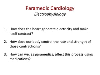
  • 1. 1. How does the heart generate electricity and make itself contract? 2. How does our body control the rate and strength of those contractions? 3. How can we, as paramedics, affect this process using medications? Paramedic Cardiology Electrophysiology
  • 2. Part 0 – Cardiac Electrophysiology - 0 Fundamentals • In this lecture we'll be going over some basic biology to get you ready for cardiac electrophysiology. • https://youtu.be/gCbHyK6y4Ms Part 1 – Cardiovascular Electrophysiology 1 - Movement through the membrane • In this lecture we're going to describe how different ions are able to move in and out of the cell. • https://youtu.be/IW3J2Y2ty6w Part 2 – Cardiovascular Electrophysiology 2 - Electrical Flow In The Heart • In this lecture we're going to talk about how a hunk of meat in your chest can generate electricity. • https://youtu.be/5A_lEW1suAM Paramedic Cardiology Electrophysiology
  • 3. Part 3 - Cardiovascular Electrophysiology 3 - Action Potential of the Myocytes • In this lecture, we're going to go over the pattern of how ions move in and out of the cell in a regular, repeating pattern - called the Action Potential. • https://youtu.be/KV3AaXWrNeY Part 4 - Cardiovascular Electrophysiology 4 - Action Potential of the Pacemaker Cells • In this lecture we're going to describe how 'pacemaker' cells can spontaneously depolarize without anything stimulating them externally. • https://youtu.be/M39wMZFd22o Part 5 - Cardiovascular Electrophysiology 5 - Anatomy and Physiology of Myocytes • In this lecture we describe how cardiac cells physically contract. • https://youtu.be/qqnL0Fy3e2g Paramedic Cardiology Electrophysiology
  • 4. Part 6 - Cardiovascular Electrophysiology 6 - Excitation Contraction Coupling • In this video we're going to go over how depolarisation initiates contraction of the heart. • https://youtu.be/UBINkoW6JzM Part 7 - Cardiovascular Electrophysiology 7 - ANS Influence on the Heart • In this lecture we cover how our body changes the rate and strength of our heart, going from external stimuli to the actual ionic changes that take place in the myocardial cells. If I yell 'boo!', your heart speeds up. Why? How? • https://youtu.be/JmAaibgOrLw Part 8 - 8 Paramedic Pharmacology – Antidysrhythmics • In this final lecture, we review cardiac antidysrhythmics with a fairly introductory discussion of how they are classified and how they work. • https://youtu.be/oY0XVEkfKr4 Paramedic Cardiology Electrophysiology
  • 6. © Marc Colbeck and Paramedicine.com, used by permission of the author. See: https://creativecommons.org/licenses/by-nc-sa/4.0/ Maintaining this note in the file is sufficient attribution of authorship as per the author.
  • 7. Topics 0. Fundamentals 1. Movement Through the Membrane 2. Electrical Flow in the Heart 3. The Action Potential – Myocytes 4. The Action Potential – Pacemaker cells 5. Anatomy and Physiology of Myocytes 6. Excitation-Contraction Coupling 7. ANS Influence on the Heart 8. Paramedic Pharmacology - Antidysrhythmics
  • 8. Priming Questions 1. What is an ‘atom’ 2. What is an ‘element’? 3. What is an ‘ion’? 4. How about anions and cations, what are those? 5. What is a cell? 6. Where did cells come from? 7. What are the walls of a cell made out of? 8. What are the major elements that humans are made out of? 9. Why is phosphorous so important to humans? 10. What are the ions that are important in cardiac electrophysiology?
  • 9. You Should Already Know Atoms • Basic building block – Protons+ and neutrons in nucleus – Electrons- in orbit Element • Substance made entirely of one type of atom (e.g. H2) Ions • An atom or element with a lack or surplus of either protons or electrons – Anions: negative charge – Cations: positive charge
  • 10. The Elements of Life 99% CHON (by weight) http://bookofresearch.com/images/earth-transparent.png <1% P, as well as some S, Na, Mg, Cl, K, Ca and Fe 10
  • 11. The Elements of Life http://1.bp.blogspot.com/-mrMWO9w3UvE/TdE67QdLgOI/AAAAAAAAAEY/Bpu6nJpEBgk/s1600/periodic_table_of_elements1.jpg composition (by weight) 11
  • 12. The Elements of Life - Phosphorus • Highly reactive, usually found as ‘red phosphorus’ • Used in explosives (napalm), poisons and nerve agents • ‘Organophosphates’ are used as fertilizers, but can also (along with the more potent fluorophosphates) be used as neurotoxins. • 13th Element discovered (in 1669) • Distilled from urine (100L ≈ 5mg) (later discovered in bones) • Now mined from rocks Essential to life: • Part of DNA, RNA and ATP (‘phosphorylation) as well as phospholipids http://0.tqn.com/d/chemistry/1/0/Q/4/1/phosphorus_allotropes.jpg 12
  • 13. The Elements of Life • Phosphorous – binds to 4 oxygen – hydrophilic • Hydrocarbon tails – hydrophobic http://bioweb.wku.edu/courses/biol115/wyatt/biochem/lipid/P-lipid.gif 13
  • 16. Cell Contents What’s inside liposomes? http://www.fossilmuseum.net/Paleobiology/Paleobiology_images/seawater.jpg 16
  • 18. Cell Contents What’s inside single celled organisms? Same stuff ... • Cations – Na+ – K+ – Mg+2 – Ca+2 • Anions – Cl- – HCO3 - (Bicarbonate) – SO4 -2 (Sulphate) http://www.salmonellablog.com/uploads/image/bacteria.jpg 18
  • 19. Cell Contents Single celled life became multi-cellular life, surrounding each cell with fluid similar to the sea. http://www.pnas.org/content/104/suppl.1/8613/F1.large.jpg 19
  • 20. Cell Contents Multi-cellular life came on land http://2.bp.blogspot.com/_2ocgSdkueHs/Rh1viEauffI/AAAAAAAAAVk/viHbhiBj2OY/s400/cartoon-drfunmrevolution.gif 20
  • 21. Cell Contents Cytosol Na+ K + Ca 2+ Cl - Extracelluar Fluid Na+ K + Ca 2+ Cl - 21
  • 22. Cell Contents – passing through the membrane • Small, non charged particles/molecules can move through the cell membrane easily, (e.g. O2 and CO2, lipids, hormones, anesthetic agents). • Most ions and large molecules (like proteins) or glucose (which is hydrophilic) can’t get through. P P P P P P P P P P 22
  • 24. Cations 0.6 nm* 0.8 nm* 1.2 nm* *aqueous values (bonded with H2O)24
  • 25.
  • 26. Electrophysiology Part 1 Movement Through the Membrane Marc Colbeck Paramedic Cardiology
  • 27. © Marc Colbeck and Paramedicine.com, used by permission of the author. See: https://creativecommons.org/licenses/by-nc-sa/4.0/ Maintaining this note in the file is sufficient attribution of authorship as per the author.
  • 28. Topics 0. Fundamentals 1. Movement Through the Membrane 2. Electrical Flow in the Heart 3. The Action Potential – Myocytes 4. The Action Potential – Pacemaker cells 5. Anatomy and Physiology of Myocytes 6. Excitation-Contraction Coupling 7. ANS Influence on the Heart 8. Paramedic Pharmacology - Antidysrhythmics
  • 29. Priming Questions 1. Describe where Na, K and Ca are normally found in the polarised cell. 2. Describe what a voltage gated channel is and what role it plays in depolarisation 3. Describe what a g-coupled protein receptor is and describe how it effects changes in the cell. 4. Describe what a ligand gated channel is and give one example of a ligand. 5. What are the two primary active transport pumps and describe the ions they exchange and what drives the movement of those ions. 6. Referencing the ion exchangers, describe how cellular hypoxia leads to irritable dysrhythmias. 7. What are the two ion exchangers and describe the ions they exchange and what drives the movement of those ions.
  • 30. Movement Through the Membrane How does stuff get in and out?? electrochemical K+electrochemical electrochemical Ca2+ Na+ 30
  • 31. Movement Through the Membrane 31
  • 32. Movement Through the Membrane Voltage Gated Channels (VGCs) “Gating” is the process of opening the channel. A “voltage gated” channel is a channel that opens due to electrical (voltage) stimuli. Time Dependent • Open when ‘gated’ • Stay open ≈ ‘fast/slow’ • Must close to reset (which is also voltage dependent) I = “intensite de courant” INa K Na Ca 32
  • 33. Movement Through the Membrane Receptor Gated Channels VGCs hormone or neurotransmitter (“ligand”) receptor RGC K Na Ca 33 Examples: • Insulin • Hormones • Steroids • Prostaglandins
  • 34. Movement Through the Membrane RGCs VGCs G proteins signal the inside of the cell to do other stuff (secondary messenger systems) “G coupled protein receptor” GCPR RGC K Na Ca 34 Examples: • Neurotransmitters • Adrenaline • Narcotics • 1/3 of drugs!
  • 35. Movement Through the Membrane VGCs Primary Active Transport Pumps 3 Na+ out 2 K+ in ATP ADP Na/K • 1/3 of all ATP use in the body • 1000 pumps per square micrometre GCPR RGCs RGC K Na Ca 35
  • 36. Movement Through the Membrane VGCs Primary Active Transport Pumps 3 Na+ out 2 K+ in ATP ADP Na/K Ca2+ out ATP ADP Ca/ATPase GCPR RGCs RGC K Na Ca 36
  • 37. Movement Through the Membrane VGCs Ion Exchangers Rely on the concentration gradient created by the Na/K pump Na/Ca* 3 Na+ in 1 Ca2+ out 1 Na+ in 1 H+ out Na/Proton GCPR RGCs RGC K Na Ca *The Na/Ca exchanger is inhibited by acidosis, causing an intracellular hypercalcemia, increasing irritability in the cell. This is how hypoxia leads to irritable dysrhythmias. 37
  • 38. Really Important Concept to Understand! Movement Through the Membrane RGCs VGCs Na/Ca Na/Proton 3 Na+ out 2 K+ in ATP ADP Na/K Ca2+ out ATP ADP Ca/ATPase Really Important Concept to Understand! 3 Na+ in 1 Ca2+ out 1 Na+ in 1 H+ out GCPR RGC K Na Ca
  • 39.
  • 40. Electrophysiology Part 2 Electrical Flow in the Heart Marc Colbeck Paramedic Cardiology
  • 41. © Marc Colbeck and Paramedicine.com, used by permission of the author. See: https://creativecommons.org/licenses/by-nc-sa/4.0/ Maintaining this note in the file is sufficient attribution of authorship as per the author.
  • 42. Topics 0. Fundamentals 1. Movement Through the Membrane 2. Electrical Flow in the Heart 3. The Action Potential – Myocytes 4. The Action Potential – Pacemaker cells 5. Anatomy and Physiology of Myocytes 6. Excitation-Contraction Coupling 7. ANS Influence on the Heart (!) 8. Paramedic Pharmacology - Antidysrhythmics
  • 43. Priming Questions 1. What is electricity? 2. How does the heart create an electrical current? 3. Specifically what are the roles of ions and gap junctions.
  • 44. cell Sodium (Na+) Potassium (K+) + - There’s more sodium (+) outside, and more potassium (-) inside Electrical Flow in the Heart 44 There are also negatively charged proteins trapped inside the cell.
  • 45. To make electricity … cell + - Na+ K+ Electricity “The collection of physical effects related to the force and motion of electrically charged particles, typically electrons, through or across matter and space.” http://www.thefreedictionary.com/electricity Electrical Flow in the Heart 45
  • 46. Electrical Flow in the Heart The exchange takes place in a wave across the cell 46
  • 47. Electrical Flow in the Heart Gap Junctions allow Na to flow between cells 47
  • 48. Electrical Flow in the Heart depolarization conduction → at overshoot conduction → at overshoot conduction → at overshoot depolarization 48
  • 49. Electrical flow Electrical Flow in the Heart - - - - - - - - -+ -+++++++++ 49
  • 50. Electrical Flow in the Heart Heart cells - - - ----------- -- -- --- - -------- ---------- - ------------ ---------- ---------- -- -------- - - ---------- --------- ++ ++ + + +++ ++ +++++ +++++ ++ + ++++++++ + ++++++ + + + + +++++++ + + ++ ++++++ +++ + +++++ +++ + + +++++ + + ++ + ++++ + ++ ++++ + + + ++ + +++
  • 51.
  • 52. Electrophysiology Part 3 The Action Potential: Myocytes Marc Colbeck Paramedic Cardiology
  • 53. © Marc Colbeck and Paramedicine.com, used by permission of the author. See: https://creativecommons.org/licenses/by-nc-sa/4.0/ Maintaining this note in the file is sufficient attribution of authorship as per the author.
  • 54. Topics 0. Fundamentals 1. Movement Through the Membrane 2. Electrical Flow in the Heart 3. The Action Potential – Myocytes 4. The Action Potential – Pacemaker cells 5. Anatomy and Physiology of Myocytes 6. Excitation-Contraction Coupling 7. ANS Influence on the Heart 8. Paramedic Pharmacology - Antidysrhythmics
  • 55. Priming Questions 1. What is meant by ‘resting membrane potential’? 2. Label this diagram appropriately, using the given terms. 1. Polarised 2. Hyperpolarised 3. Depolarisation 4. Repolarisation 3. Also label the phases using the numbers 0-4. 4. Describe in details the movement of ions in phase 0-4 of the depolarisation. 5. What is a ‘potassium rectifier channel’?
  • 56. The Action Potential - Myocytes 56 0 RMP: Resting Membrane Potential The charge of the inside of the cell compared to the outside. Voltage(mV) - 90 + 20 Polarised Equal internal and external charges Hyperpolarised
  • 57. The Action Potential - Myocytes VGCs K Na Ca 57 Neuron Myocyte - 90 + 20
  • 58. The Action Potential - Myocytes Polarised Depolarisation Hyperpolarised Plateau Voltage(mV) - 90 + 20 Polarised 58
  • 59. The Action Potential - Myocytes 59 Voltage(mV) - 90 + 20
  • 60. Voltage(mV) - 90 + 20 PHASE 0 • rapid influx of Na through some Na channels • Once an area of the cell wall reaches about 10-20mV less negative than the resting membrane potential (RMP) all the local voltage-gated Na channels open. • Sodium ions rapidly diffuse inward through the Na channels, and depolarisation occurs. • Sodium channels open at about -70 mV, close at about +20 mV • They won’t open again until the RMP falls under about -65 mV again (they have to ‘reset’) Extracellular Intracellular Phase 0 Na+ membrane
  • 61. Voltage(mV) - 90 + 20 PHASE 1 • Voltage-gated Na channels close • Na influx stops • “Fast” K channels open • A very brief K efflux begins (causing a brief decrease in the RMP) Phase 0 Phase 1 Na+ K+ Extracellular Intracellular membrane
  • 62. Voltage(mV) - 90 + 20 PHASE 2 • Ca influx begins at -60 to -50 mV through Ca channels • The efflux of K is slowed (small arrow) so an equilibrium is maintained • There’s no sodium movement Phase 0 Phase 1 Phase 2 Na+ K+ Ca++ K+ Extracellular Intracellular membrane
  • 63. Voltage(mV) - 90 + 20 PHASE 3 • Ca Channels close • An efflux of K from K continues • The Na and Ca channels reset Phase 0 Phase 1 Phase 3Phase 2 Na+ K+ Ca++ Intracellular membrane Extracellular K+K+
  • 64. Voltage(mV) - 90 + 20 Phase 4 • The cell returns to -80/-90 mV • The K rectifier channel keeps the transmembrane potential stable at -90 mV • The ATP pump restores K into the cell and Na out of the cell • Ca/ATP pump restores Ca to outside the cell • Na/Ca exchanger restores Na and Ca • Na/H exchanger removes H from cells Phase 0 Phase 1 Phase 3Phase 2 Na+ K+ Ca++ Phase 4 Intracellular membrane Extracellular K+K+ K+
  • 65. Voltage(mV) - 90 + 20 Really Important Concept to Understand! Phase 0 Phase 1 Phase 3Phase 2 Na+ K+ K+ Ca++ K+ K+ Phase 4 Intracellular membrane Extracellular
  • 66. The Action Potential - Myocytes https://www.youtube.com/watch?v=tLt3ZTeVzEY
  • 68.
  • 69. Electrophysiology Part 4 The Action Potential: Pacemaker Cells Marc Colbeck Paramedic Cardiology
  • 70. © Marc Colbeck and Paramedicine.com, used by permission of the author. See: https://creativecommons.org/licenses/by-nc-sa/4.0/ Maintaining this note in the file is sufficient attribution of authorship as per the author.
  • 71. Topics 0. Fundamentals 1. Movement Through the Membrane 2. Electrical Flow in the Heart 3. The Action Potential – Myocytes 4. The Action Potential – Pacemaker cells 5. Anatomy and Physiology of Myocytes 6. Excitation-Contraction Coupling 7. ANS Influence on the Heart 8. Paramedic Pharmacology - Antidysrhythmics
  • 72. Priming Questions 1. How can SA and AV node cells depolarize without being externally stimulated? 2. What are ‘funny channels’? 3. What ions flow through the funny channels? 4. Why does a pacemaker cell only have phase 4, 0, and 3? 5. Compare the action potential of a pacemaker myocyte versus and ventricular myocyte.
  • 73. The Action Potential – Pacemaker Cells Different tissue in the heart depolarizes in different ways. The SA and AV nodes are different than the rest of the heart. They are the pacemakers or ‘auto-rhythmic’ cells. 73
  • 74. The Action Potential – Pacemaker Cells RMP • Spontaneous phase 4 rise (pacemaker potential) • Due to “Funny Channels” (IF) and Ca(T) channels • IF = nonspecific cation channels permeable to both Na+ (influx) and K+ (efflux): • At –ve membrane potential Na influx > K efflux → gradual depolarization • AP in SA and AV nodal cells is similar, but pacemaker potential in SA cells reaches threshold more rapidly Pacemaker Potential Action Potential Na+ influx Ca2+ influx -40 Resting membrane potential •Spontaneous phase 4 rise (pacemaker potential) •Due to “Funny Channels” (IF) and Ca(T) channels • IF = nonspecific cation channels permeable to both Na+ (influx) and K+ (efflux): • At –ve membrane potential Na + Ca influx > K efflux → gradual depolarization • AP in SA and AV nodal cells is similar – but pacemaker potential in SA cells reaches threshold more rapidly 4 0 3
  • 75. Na/Ca Na/Proton 3 Na+ out 2 K+ in ATP ADP Na/K Ca2+ out ATP ADP Ca/ATPase Really Important Concept to Understand! If Funny Channels (pacemaker cells only) 3 Na+ in 1 Ca2+ out 1 Na+ out 1 H+ in GCPR RGC K Na Ca No fast Na channels in pacemaker cells! Na+ K+ The Action Potential – Pacemaker Cells 75 Na
  • 76. PHASE 4 • If type channels gradually close as membrane potential becomes more +ve • Membrane potential threshold @ - 40mV, results in spontaneous AP PHASE 0 • Depolarization phase caused by opening of L-type Ca2+ channels (rather than Na+ in contractile cells) • Ca2+ channels slower than Na+ • No fast Na channels = no Phase 1 • Rapid deactivation of Ca channels = no Phase 2 The Action Potential – Pacemaker Cells 4 0 3 4 - 40 (IF) Ca(T) http://rezidentiat.3x.ro/eng/tulbritmeng.files/image001.gif
  • 77. Pacemaker Potential Action Potential K+ effluxCa2+ influx - 40mV PHASE 3 • Repolarisation occurs when Ca2+ channels close and slow K+ channels open leading to K+ efflux • Return to –ve potential activates pacemaker mechanism Polarized State • Closure of “slow” K+ channels (opened during repolarisation of previous AP) • Cyclic/rhythmic excitation of the cells = “autorhythmicity” The Action Potential – Pacemaker Cells Cell has to hyperpolarize in order for F channels to open again. Cellular hypoxia obstructs this and so leads to bradycardia.
  • 78. The Action Potential ‘Normal’ Myocytes Pacemaker Myocytes Membrane Potential Stable at – 90 mV Unstable pacemaker potential; usually starts at – 60 mV Events leading to threshold potential Depolarization enters via gap junctions Net Na entry through If channels, reinforced by CaT entry Rise phase of action potential Na entry Ca entry Repolarization phase Extended plateau caused by Ca entry; rapid phase casued by K efflux Rapid; caused by K efflux Hyperpolarization None; resting potential is – 90 mV, the equilibrium potential for K Normally none; when repolarization hits -60 mV the If channels open again. Duration of action potential Extended: 200+ msec Variable: generally 150+ msec Refractory period Long because resetting of Na channel gates delayed until end of action potential None 78
  • 79. Putting it All Together Comparison of Action Potential’s across the heart Top Bottom Marieb Fig 18-14
  • 80.
  • 81. Electrophysiology Part 5 Anatomy & Physiology of Myocytes Marc Colbeck Paramedic Cardiology
  • 82. © Marc Colbeck and Paramedicine.com, used by permission of the author. See: https://creativecommons.org/licenses/by-nc-sa/4.0/ Maintaining this note in the file is sufficient attribution of authorship as per the author.
  • 83. Topics 0. Fundamentals 1. Movement Through the Membrane 2. Electrical Flow in the Heart 3. The Action Potential – Myocytes 4. The Action Potential – Pacemaker cells 5. Anatomy and Physiology of Myocytes 6. Excitation-Contraction Coupling 7. ANS Influence on the Heart 8. Paramedic Pharmacology - Antidysrhythmics
  • 84. Priming Questions 1. What percent of myocytes are contractile and what percent are pacemaker cells? 2. What are the two structures of the intercalated disks? Describe their function? 3. What is meant by the term ‘functional syncytium’? 4. What are the two main filaments involved in muscular contraction (which are attached to the M and Z structures) 5. What is tropomyosin? What is its role in contraction? Describe this by explaining the roles of three elements of the troponin complex, Ca and ATP.
  • 87. Myocytes • ≈99% of cardiomyocytes are contractile • ≈1% specialised autorhythmic (pacemaker) cells
  • 89.
  • 91. Myocytes Z disks: Z stands for “zwischen” which means “between” in German M line: M stands for “Mittle” which means “middle” in German A bands: A stand for “anisotropic” I bands: I stands for “isotropic”, both A & I have to do with how the structures interact with polarized light H zone: H stands for “helle” which means “bright” in German
  • 95. Myocytes Troponin C • has a strong affinity for Calcium, when bound it moves the tropomyosin strand off the actin so that the actin can interact with the myosin Troponin I • Inactivates the actin/myosing binding • Defibrillation and (brief) CPR don’t elevate TnI levels • TnI is more specific to cardiac muscle, so is theoretically better to test for Tropinin T • Ties the TnC and TnI together • TnT is analyzed in the ER to determine the presence of A.M.I. • Current research into ‘high sensitivity TnT’ lab tests are being conducted
  • 96. Myocytes Free Ca ions binding to the actin TnC sites (thus revealing the tropomyosin) plus ATP to myosin S1 heads produces the power stroke.
  • 97. Myocytes No ATP→ cell unable to dissociate actin and myosin filaments Relaxation of the power stroke (by decoupling the actin and myosin) also requires ATP-supplied energy. This is relevant to APO – any idea how?
  • 98. Myocytes An animated movie (very short) of the power stroke http://www.sci.sdsu.edu/movies/actin_myosin.html
  • 99.
  • 101. © Marc Colbeck and Paramedicine.com, used by permission of the author. See: https://creativecommons.org/licenses/by-nc-sa/4.0/ Maintaining this note in the file is sufficient attribution of authorship as per the author.
  • 102. Topics 0. Fundamentals 1. Movement Through the Membrane 2. Electrical Flow in the Heart 3. The Action Potential – Myocytes 4. The Action Potential – Pacemaker cells 5. Anatomy and Physiology of Myocytes 6. Excitation-Contraction Coupling 7. ANS Influence on the Heart 8. Paramedic Pharmacology - Antidysrhythmics
  • 103. Priming Questions 1. What are the ten stages of excitation contraction coupling? 2. Which ion is the ‘driver’ of contraction? 3. Label the diagram on the following slide
  • 104.
  • 105. Excitation-Contraction Coupling How do we go from depoloarizing conductive tissue (for example, purkinje fibres) to getting the actin and myosin in a myocyte to contract ??? This process is called “excitation-contraction coupling”.
  • 106. Excitation-Contraction Coupling The 3 ways a myocyte is usually depolarized 1. Stimulation by a pacemaker cell (in a node). 2. From an adjacent myocyte, injecting sodium through the gap junction. 3. From an adjacent Purkinje fibre injecting sodium through the gap junction.
  • 107. Excitation-Contraction Coupling How does getting the membrane of a myocyte to depolarize cause the actin and the myosin on the inside to contract?
  • 108. Excitation-Contraction Coupling 1. An action potential enters from adjacent cell, depolarizing the plasma membrane. This wave of depolarization continues deep into the myocyte via the T- tubules.
  • 109. Excitation-Contraction Coupling 2. Voltage gated L- type Ca channels open, and Ca enters the myocyte through the plasma membrane Verapamil (isoptin) partially blocks this entry - acting as a negative inotrope.
  • 110. Excitation-Contraction Coupling 3. Ca binds to a receptor in the sarcoplasmic reticulum. This induces Ca release from the cisternae of the SR. This is called Calcium Induced Calcium Release (CICR). “Nice-to-know” (not tested): These receptors are called “Ryanodine receptors” because we found (experimentally) that the drug Ryanodine will bind to them. “Nice-to-know” (not tested): These receptors are called “Dihydropiridine receptors” because we found (experimentally) that the drug Dihydropiridine will bind to them.
  • 111. Excitation-Contraction Coupling 4. The local release of Ca causes Ca ‘sparks’ (short lived, highly local releases of Ca throughout the cell)
  • 112. Excitation-Contraction Coupling 5. Summated Ca sparks create a Ca signal which travels to the actin and myosin.
  • 113. Excitation-Contraction Coupling 6. Ca binds to Troponin C (TnC) to move the tropomyosin sheath off the actin, with power from ATP, the myosin heads initiate the ‘power stroke’ and contraction occurs.
  • 114. Excitation-Contraction Coupling 7. Relaxation of the actin/myosin complex occurs when Troponin I (TnI) initiates Ca release from the TnC, and the myosin heads reset to their starting position.
  • 115. Excitation-Contraction Coupling 8. Ca is pumped back into the SR for storage.
  • 116. Excitation-Contraction Coupling 9. Calcium in the cytosol is moved out of the cell by the Na/Ca exchanger.
  • 117. Excitation-Contraction Coupling 10. The Na brought into the cell by the Na/CA exchanger is removed by Na/K- ATPase pump.
  • 118. Excitation-Contraction Coupling Ca2+ entry in the cell is the driver of contraction
  • 119.
  • 120. Electrophysiology Part 7 ANS Influence on The Heart Marc Colbeck Paramedic Cardiology
  • 121. © Marc Colbeck and Paramedicine.com, used by permission of the author. See: https://creativecommons.org/licenses/by-nc-sa/4.0/ Maintaining this note in the file is sufficient attribution of authorship as per the author.
  • 122. Topics 0. Fundamentals 1. Movement Through the Membrane 2. Electrical Flow in the Heart 3. The Action Potential – Myocytes 4. The Action Potential – Pacemaker cells 5. Anatomy and Physiology of Myocytes 6. Excitation-Contraction Coupling 7. ANS Influence on the Heart 8. Paramedic Pharmacology - Antidysrhythmics
  • 123. Priming Questions 1. Sketch the layout of the nervous system using the following terms: 1. Central and peripheral 2. Afferent and Efferent 3. Somatic and Autonomic 4. Sympathetic and Parasympathetic 2. Define the following terms. State the role of the SNS and the PSNS on each. 1. Inotropy 2. Chronotropy 3. Dromotropy 4. Irritability 3. Differentiate between the autonomic innervation of the atria and the ventricles. 4. List 5 important categories of factors that determine the autonomic tone in the body. 5. Sketch and label the transmission of signals from the brain to the heart through the nervous system. Include the receptors found on the myocytes for both SNS and PSNS signals. 6. Section Review – label the following diagram
  • 124.
  • 125. ANS Influence Peripheral Nervous System Central Nervous System Afferent Nervous System Efferent Nervous System Autonomic Nervous System Somatic Nervous System Sympathetic Nervous System Parasympathetic Nervous System The SNS and PSNS balance each other S ensory is … A fferent M otor is … E fferent (SAME)
  • 126. ANS Influence Most involuntary organs and glands have both sympathetic and parasympathetic innervation. The SNS and PSNS act antagonistically to maintain or quickly restore homeostasis. The sympathetic nervous system excites the receptors (‘fight or flight’ response). The parasympathetic nervous system relaxes the receptors (‘rest and digest’ response).
  • 127. ANS Influence If you’re a bit vague on the ANS there’s a lecture on it here: https://www.youtube.com/watch?v=7lVt0ABcnM4
  • 128. ANS Influence Sympathetic Stimulation (thoraco-lumbar) SNS innervates both atria and ventricles ↑ heart rate (↑ chronotropy) ↑ conduction velocity (↑ dromotropy) ↑ strength of contraction (↑ inotropy) ↑ irritability
  • 129. ANS Influence Parasympathetic Stimulation (cranio-saccral) PSNS innervates only the atria ↓ heart rate (↓ chronotropy) ↓ conduction velocity (↓ dromotropy) ↓ strength of contraction (↓ inotropy) ↓ irritability
  • 130. The sympathetic branch influences both the atria (i.e. SA Node, the Intraatraial and internodal pathways, and the AV junction) and the ventricles. The parasympathetic branch influences only the atria. parasympathetic sympathetic ANS Influence
  • 131. TheAutonomicNervousSystem This is important! The SNS can speed up the whole heart, but the PSNS can only slow down the atria. If the ventricles are going too fast, it’s no use trying to activate the PSNS to try and slow them down. The PSNS has no effect on the ventricles.
  • 132. ANS Influence Contractile cells of the: 1. atrial myocardium 2. ventricular myocardium AV Node cells: 3. velocity of conduction 4. length of refractoriness Automaticity cells of the: 5. SA node 6. AV node 7. atria 8. ventricular conduction system (not velocity, just automaticity) SNS Stimulates PSNS Inhibits Really Important Concept to Understand!
  • 133. ANS Influence What controls the autonomic balance? 1. Nervous Regulation • cortex and hypothalamus 2. Circulatory Regulation • venous and arterial 3. Respiratory Regulation • inspiration and expiration 4. Other Somatic Regulation • muscles, pain, temp., etc. 5. Chemical Regulation • O2, CO2 , H+ • Adrenaline, Noradrenaline, Thyroxin & Histamine • Poisons and Drugs
  • 134. ANS Influence What controls the autonomic balance? Thoughts Feelings Hypothalamus Blood pressure Respiration Movement Pain Temperature pH Hormones Chemicals Brain MedullaOblongata Cardiac Acceleratory Centre Cardiac Inhibitory Centre Really Important Concept to Understand!
  • 135. ANS Influence How does the brain send messages to the heart? MedullaOblongata CIC&CAC Preganglionic neuron R. & L. Vagus Nerve Sympathetic Chain Ganglia Cardiac Plexus Postganglionic nerve Postganglionic nerve HEART
  • 136. ANS Influence SNS Ganglia Chain Cardiac Plexus
  • 137. ANS Influence How does the ANS affect the heart? http://www.thecolor.com/images/Magician.gif “...and then the rabbit makes the heart speed up ...”
  • 138. ANS Influence Let’s connect this ... With this ... HOW IT DOES IT WHAT IT DOES
  • 139. ANS InfluenceMedullaOblongata CIC&CAC Preganglionic neuron R. & L. Vagus Nerve Sympathetic Chain Ganglia Cardiac Ganglia Postganglionic nerve Postganglionic nerve NAd Ach M2 Ach Really Important Concept to Understand! β1NAd M2Ach
  • 140. Na/K ATP ADP Ca2+ ATP ADP Ca/Na Na/H GCPR RGC K Ca Clinical Effects of SNS Activation: 1. ↑ Chronotropy: The pumps and exchangers work more quickly, allowing the heart to ‘reset’ more quickly, allowing an increased HR, and an increased resistance to acidosis. 2. ↑ Dromotropy: Ion channels work more quickly, increasing the speed of depolorization. 3. ↑ Inotropy: The increased amount of calcium entering the cell causes an increased release of calcium from the sarcoplasmic reticulum, causing more actin and myosin to bridge, increasing the strength of contraction. β1NAd Na ANS Influence ① ② ③ Really Important Concept to Understand!
  • 141. ATP ADP ATP ADP GCPR RGC K Ca K+ IKAch M2Ach Na/K Ca2+ Ca/Na Na/H 2-5x more M2 in the atria than in the ventricles Clinical Effects of PSNS activation: 1. ↓ Chronotropy: The pumps and exchangers work more slowly, causing the heart to ‘reset’ more slowly, causing a decreased HR, and an decreased resistance to acidosis. 2. ↓ Dromotropy: Ion channels work more slowly, decreasing the speed of depolorization. 3. ↓ Inotropy: The decreased amount of calcium entering the cell causes a decreased release of calcium from the sarcoplasmic reticulum, causing less actin and myosin to bridge, decreasing the strength of contraction. Na ANS Influence ① ② ③ Really Important Concept to Understand!
  • 142. Under ischemia or cardiac stress ATP (Adenosine Triphosphate) is degraded down to just Adenosine (Ado) by losing all 3 of its Pi. Adenosine (Ado) P P P Adenosine (Ado) ANS Influence
  • 143. Ado inhibits NAd release from the SNS postganglionic nerve terminal. Ado inhibits the β1 secondary messenger systems and so acts as a sympatholytic. Ado ↑K efflux and ↓Ca influx, hyperpolarizing (mostly) the AVN, slowing and/or temporarily inactivating it (up to 30s!). It’s kind of like an internal, cardioprotective “emergency brake”. In effect, it works like the PSNS without having to invoke the PSNS. Possibly the nociceptive (pain) pathway for angina. ATP ADP ATP ADP GCPR RGC K If Na+ K+ K+ IKAch Na/K Ca2+ Ca/Na Na/H Na β1 Ca NAd ANS Influence Really Important Concept to Understand!
  • 144. Quick Review • The SNS speeds things up by affecting various structures in the plasma membrane. • The PSNS slows things down by affecting various structures in the plasma membrane. • Adenosine kind of does a lot of what the PSNS does, without having to stimulate the PSNS. Really Important Concept to Understand! ANS Influence
  • 145. ATP ADP ATP ADP GCPR RGC K Na/K Ca2+ Ca/Na Na/H Na β1 Ca NAd Ach M2 MedullaOblongata CIC&CAC Preganglionic neuron R. & L. Vagus Nerve Sympathetic Chain Ganglia Cardiac Plexus Postganglionic nerve Postganglionic nerve ANS Influence - Overview M2 Really Important Concept to Understand!
  • 146. ATP ADP ATP ADP GCPR RGC K If Na+ K+ Na/K Ca2+ Ca/Na Na/H Na β1 Ca NAd Ach M2 MedullaOblongata CIC&CAC Preganglionic neuron R. & L. Vagus Nerve Sympathetic Chain Ganglia Cardiac Plexus Postganglionic nerve Postganglionic nerve ANS Influence - Overview M2 Summary of the ANS Influence in One Diagram
  • 147.
  • 149. © Marc Colbeck and Paramedicine.com, used by permission of the author. See: https://creativecommons.org/licenses/by-nc-sa/4.0/ Maintaining this note in the file is sufficient attribution of authorship as per the author.
  • 150. Topics 0. Fundamentals 1. Movement Through the Membrane 2. Electrical Flow in the Heart 3. The Action Potential – Myocytes 4. The Action Potential – Pacemaker cells 5. Anatomy and Physiology of Myocytes 6. Excitation-Contraction Coupling 7. ANS Influence on the Heart 8. Paramedic Pharmacology - Antidysrhythmics
  • 151. Priming Questions 1. Explain the Singh/Vaughan classifications of drugs acting on the heart and indicate where on the graph they act. a. Class 1: b. Class 2: c. Class 3: d. Class 4 e. Class 5: 2. Explain the actions of the following antidysrhythmic medications • Adenosine • Amiodarone • Beta Blockers • Calcium Channel Blockers • Digoxin • Magnesium Sulphate • Xylocaine/Lidocaine 3. Why might ‘The Sicilian Gambit’ be a better method to describe ventricular antidysrhythmics?
  • 152. Antidysrhythmics • Singh / Vaughan Williams classification • “Sicilian Gambit” classification
  • 153. Antidysrhythmics Singh / Vaughan Williams classification Introduced in 1970 by doctoral candidate at Oxford University, Dr. Bramah Singh (a cardiologist) (his supervisor was EM Vaughan Williams)
  • 154. Antidysrhythmics – Singh/Vaughan Williams Na/K pump Na/Ca exchanger Na influx through gap junctions Fast K channel efflux T and L channel Ca influx Voltage(mV) - 90 + 10 Ca influx stops K efflux through “slow K” channels Class II Beta Blockers (↓ SNS tone) Class I (a,b,c) Fast Na channel blockers Class III Block K efflux Class IV Block Ca Channels Class V Does other stuff
  • 155. Class I agents: Sodium Channel Blockers are divided into three groups based upon their effect on the length of the action potential. • Ia lengthens the action potential (right shift) Procainamide • Ib shortens the action potential (left shift) Xylocaine • Ic does not significantly affect the action potential (no shift) Propafenone Antidysrhythmics – Class I http://en.wikipedia.org/wiki/Antiarrhythmic_agent
  • 156. Antidysrhythmics – Class I http://en.wikipedia.org/wiki/Antiarrhythmic_agent Class Ia Disopyramide, Quinidine, Procainamide Double Quarter Pounder Class Ib Lidocaine, Mexiletine, Tocainide Lettuce, Mayo*, Tomato Class Ic Moricizine, Flecainide, Propafenone More* Fries Please. *note there are two "M"s in the mnemonic, but morcizine and more can clarify which is which) How to remember the Class 1 drugs.
  • 157. Class II agents are conventional beta blockers. They act by blocking the effects of catecholamines at the β1- adrenergic receptors, thereby decreasing sympathetic activity on the heart. They decrease conduction through the AV node. Antidysrhythmics – Class II http://en.wikipedia.org/wiki/Antiarrhythmic_agent Cardioselective Acebutolol Atenolol Esmolol Metoprolol Non -Cardioselective Aprenolol Labetalol Propranolol Sotalol
  • 158. Class III agents predominantly block the potassium channels, thereby prolonging repolarization. Since these agents do not affect the sodium channel, conduction velocity is not decreased. Antidysrhythmics – Class III http://en.wikipedia.org/wiki/Antiarrhythmic_agent Amiodarone
  • 159. Class IV agents are slow calcium channel blockers. They decrease conduction through the AV node, and shorten phase two (the plateau) of the cardiac action potential. They thus reduce the contractility of the heart. However, in contrast to beta blockers, they allow the body to retain adrenergic control of heart rate and contractility. Antidysrhythmics – Class IV http://en.wikipedia.org/wiki/Antiarrhythmic_agent Diltiazem Verapamil
  • 160. Class V agents Since the development of the original Vaughan-Williams classification system, additional agents have been used that don't fit cleanly into categories I through IV. Some sources use the term "Class V". However, they are more frequently identified by their precise mechanism. Agents include: • Digoxin, which decreases conduction of electrical impulses through the AV node (by inhibiting the Na/K pump) and increases vagal activity via its central action on the central nervous system. • Adenosine, which helps block signals through the AV node • Magnesium sulfate, which helps block ventricular dysrhythmias Antidysrhythmics – Class V http://en.wikipedia.org/wiki/Antiarrhythmic_agent
  • 161. Antidysrhythmics – The Sicilian Gambit Classification by site of action https://academic.oup.com/eurheartj/article-abstract/12/10/1112/440813
  • 162. Antidysrhythmics – The Sicilian Gambit Classification by site of action Drugs are classified by the… • voltage gated channels, • receptor gated channels, • pumps • or exchangers … that they affect, and how they affect them.
  • 163.
  • 164. Adenosine - V Amiodarone - III Diltiazem - IV Labetalol - II Magnesium Sulphate - V Metoprolol - II Nifedipine - IV (Oxygen) (Phenytoin - I) Verapamil - IV Australasian Paramedic Antidysrhythmics
  • 165. Topics 0. Fundamentals 1. Movement Through the Membrane 2. Electrical Flow in the Heart 3. The Action Potential – Myocytes 4. The Action Potential – Pacemaker cells 5. Anatomy and Physiology of Myocytes 6. Excitation-Contraction Coupling 7. ANS Influence on the Heart 8. Paramedic Pharmacology - Antidysrhythmics