2. Cerebrovascular
Atherosclerosis
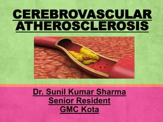
▪ Atherosclerosis – Athero=Fat
Sclerosis=Hardening
▪ Atherosclerosis - thickening & hardening of
arterial walls affecting primarily the intima of
large and medium-sized muscular arteries and
is characterized by the presence of fibro fatty
plaques or atheromas.
▪ Serious complications.
12. Cerebrovascular
Atherosclerosis
▪ Extracranial Vs Intracranial
▪ Symptomatic Vs Asymptomatic-Symptomatic carotid
disease is defined as focal neurologic symptoms that are
sudden in onset and referable to the appropriate carotid
artery distribution (I/L to significant carotid atherosclerotic
pathology), including one or more transient ischemic
attacks characterized by focal neurologic dysfunction or
transient monocular blindness, or one or more minor
(nondisabling) ischemic strokes
13. ▪ Atherothrombosis is multifactorial
▪ Comorbidities frequently overlap, and risk
factors are often additive
▪ The pathogenesis of stroke due to intracranial
arterial stenosis may be similar to stroke due to
extracranial arterial disease
14. Risk factor – Nonmodifiable
▪ Age(men >45, women>55)
▪ Gender - M>F(premenopausal)
▪ Race/ethnicity-
Intrcranial athero- Asians, blacks, Hispanics> whites
Extracranial- more in whites
▪ Family history
▪ Genetics- A/W Large artery ather. Dis.-HDAC9-1p13
(+CAD) and TSPAN2(-CAD)
17. Cerebrovascular
Atherosclerosis…
▪ Atherosclerosis primarily affects the larger
extracranial and intracranial vessels
eg.-
▪ Bifurcation of the common carotid artery
▪ Proximal internal carotid artery
▪ Carotid siphon
▪ MCA stem
▪ Origin of the vertebral arteries (V1)
▪ Intracranial segment of the vertebral arteries (V4)
▪ Basilar artery
18. ▪ ICAD - leading cause of ischemic stroke-8%-50%
of pts.
▪ Recent study from the US -
EICA atherosclerosis -11.5%
IICA atherosclerosis -1.1%
▪ Research from Korea -symptomatic
atherosclerosis of the EICA > IICA - 4:1
▪ A study from China -
symptomatic IICA atherosclerosis (4.1%)
symptomatic EICA atherosclerosis (3.8%).
Kposterior circulation disease. Stroke 2012;43:3313- 3318.
11. Flaherty ML, Kissela B, Khoury JC, et al. Carotid artery stenosis as a cause of stroke. Neuroepidemiology 2013;40:36-41
im JS, Nah HW, Park SM, et al. Risk factors and stroke mechanisms in atherosclerotic stroke: intracranial compared with extracranial and anterior compared
with
19. ▪ In an Indian study done by Dr. Trilochan
Srivastava- Out of 60 cases of stroke , 32
cases were positive for significant stenosis and
a total number of stenotic segments was 45.
▪ Out of 45 stenotic segments (single and
multiple), there were 24 (53.33%) intracranial
and 21 (46.67%) extracranial.
-CT Angiographic evaluation of pattern and distribution of stenosis and its association with risk factors among indian ischemic stroke patients Amit Shrivastava,
Trilochan Srivastava, Richa Saxena Pol J Radiol, 2016; 81: 357-362
20. Comparative Frequency of
ICAD in Patients With Stroke
▪ Chinese - 33–50
▪ Thai - 47
▪ Korean - 56
▪ South Asians - 54
▪ US Whites - 1
▪ US Blacks - 6
▪ US Hispanics - 11
-De Silva DA, Woon F-P, Lee M-P, Chen CPLH, Chang H-M, Wong M-C. South Asia patients with ischemic stroke. Intracranial large arteries are the
predominant site of disease. Stroke. 2007;38:2592–2594.
-Wong LKS. Global burden of intracranial atherosclerosis. Int J Stroke. 2006;1:158 –159
21. ▪ Early studies revealed that stroke patients with
IICA stenosis had very poor prognoses
▪ Mortality rate-was 7.8%-12.8% per year
▪ Rate of ipsilateral stroke - 7.6%-8.1% per year
▪ Cardiac disease –MC cause of death during
follow-up.
Klijn CJ, Kappelle LJ, Algra A, et al. Outcome in patients with symptomatic occlusion of the internal carotid artery or intracranial arterial lesions: a meta-analysis
of the role of baseline characteristics and type of antithrombotic treatment. Cerebrovasc Dis 2001;12:228-234.
22. ▪ Pt. with 50% to 99% stenosis of symptomatic
intracranial vessels -12% to 14% risk for a
recurrent stroke during a 2-year follow-up, in
spite of antiplatelet /anticoagulation therapy.
▪ The annual risk may > 20% in high-risk groups
▪ Silent MI in >50% of patients with ICAD
Gorelick PB,Wong KS, Bae HJ, et al. Large artery intracranial occlusive disease: a large worldwide burden but a relatively neglected frontier. Stroke
2008;39:2396-2399.
-. Sacco RL, Kargman DE, Gu Q, et al. Race-ethnicity and determinants of intracranial atherosclerotic cerebral infarction. TheNorthernManhattan Stroke Study.
Stroke 1995;26:14-20.
-Chimowitz MI, Lynn MJ, Derdeyn CP, et al. SAMMPRIS Trial Investigators. Stenting versus aggressive medical therapy in intracranial arterial stenosis. N Engl
J Med 2011;365:993-1003
25. Assessment of Carotid
Stenosis
Currently, three methods-
▪ NASCET-The North American Symptomatic
Carotid Endarterectomy Trial
▪ ECST-The European Carotid Surgery Trial
▪ CC-common carotid
26. NASCET
▪ Hemodynamically signi. carotid stenosis ≥60%
or a flow reduction distal to the lesion.
▪ Stenosis= (1-A/B) × 100%,
▪ A-diameter at the point of maximum stenosis
▪ B -diameter of the arterial segment distal to the
stenosis where the arterial walls first become
parallel
North American Symptomatic Carotid Endarterectomy Trial. Methods, patient characteristics, and progress. Stroke 1991; 22:711.
27. ▪ The European Carotid Surgery Trial (ECST)-
lumen diameter at the most stenotic portion
compared with the estimated probable original
diameter at the site of maximum stenosis.
▪ The common carotid (CC) method - lumen
diameter in the most stenotic portion
compared with the proxi. CCA.
-Rothwell PM, Gibson RJ, Slattery J, et al. Equivalence of measurements of carotid stenosis. A comparison of three methods on 1001 angiograms. European
Carotid Surgery Trialists' Collaborative Group. Stroke 1994; 25:2435.
-North American Symptomatic Carotid Endarterectomy Trial. Methods, patient characteristics, and progress. Stroke 1991; 22:711.
-MRC European Carotid Surgery Trial: interim results for symptomatic patients with severe (70-99%) or with mild (0-29%) carotid stenosis. European Carotid
Surgery Trialists' Collaborative Group. Lancet 1991; 337:1235.
-Wardlaw JM, Lewis SC, Humphrey P, et al. How does the degree of carotid stenosis affect the accuracy and interobserver variability of magnetic resonance
angiography? J Neurol Neurosurg Psychiatry 2001; 71:155.
28.
29. ▪ Equivalent measurements for the three
methods have been determined
▪ A 50% stenosis with the NASCET ≈ 65%
stenosis for both the ECST and CC methods
▪ A 70 % stenosis with the NASCET ≈ 82 percent
stenosis for both the ECST and CC methods
30. Assessment of thrombus burden-CBS
▪ CBS - for anterior circulation to quantify the extent of
ipsilateral intracranial thrombus,
▪ Allotting major arteries 10 points for the presence of
contrast opacification on CTA.
▪ Two points each were subtracted for absence of contrast
opacification in the complete cross-section of any part of
the proximal M1, distal M1 or supraclinoid ICA and 1 point
each for M2 branches, A1 segment and infraclinoid ICA
Puetz V, Dzialowski I, Hill MD, et al. Intracranial thrombus extent predicts clinical outcome, final infarct size and hemorrhagic transformation in ischemic stroke:
the clot burden score. Int J Stroke. 2008;3(4):230-236
31. CBS..
▪ Partial filling defects were rated as patent.
▪ A score of 10 - absence of a visible occlusion
▪ Score of 0 - occlusion of all major intracranial
anterior circulation arteries
32. Assessment of thrombus
burden-CBS
Ten-point clot burden score (CBS): one
or two points each (as indicated) are
subtracted for absent contrast
opacification on computed tomography
angiography (CTA) in the infraclinoid
internal carotid artery (ICA) (1),
supraclinoid ICA (2), proximal M1
segment (2), distal M1 segment (2),
M2branches (one each) and A1
segment (1).
The CBS applies only to the
symptomatic hemisphere.
33. CONVENTIONAL CEREBRAL
ANGIOGRAPHY
▪ Gold standard for imaging the carotid arteries.
▪ DSA has largely replaced conventional
angiography -less contrast/time & small cath.
▪ The quality of the angiogram depends upon
selective catheterization of the carotid artery with
at least two unimpeded views.
34. Cerebral angiography
Advantages —
▪ Permits an evaluation of the entire carotid artery
system
▪ Provides information about tandem atherosclerotic
disease, plaque morphology, and collateral
circulation which - may affect management
-Wolpert SM, Caplan LR. Current role of cerebral angiography in the diagnosis of cerebrovascular diseases. AJR Am J Roentgenol 1992; 159:191.
-Kappelle LJ, Eliasziw M, Fox AJ, et al. Importance of intracranial atherosclerotic disease in patients with symptomatic stenosis of the internal carotid artery. The
North American Symptomatic Carotid Endarterectomy Trail. Stroke 1999; 30:282
35. Disadvantages —
▪ Invasive nature,
▪ High cost/less availability
▪ Risk of morbidity and mortality.
▪ Risk of all neurologic complications -4 %
▪ Risk of serious neuro. complications or death -1%
▪ The risk of morbidity is increased with
cerebrovascular symptoms, advanced age,
diabetes, hypertension, elevated serum
creatinine, and peripheral vascular disease.
36. Disadvantages
▪ Limited number of projections, typically two or
three, depicting the carotid artery and
bifurcation-underestimation in asymmetrical
stenosis.
▪ Rotational angiography provides 16 to 32
projections and is better, but seldom used in
practice
Bendszus M, Koltzenburg M, Burger R, et al. Silent embolism in diagnostic cerebral angiography and neurointerventional procedures: a prospective study.
Lancet 1999; 354:1594
37. CAROTID DUPLEX
ULTRASOUND
▪ CDUS - detect focal increases in blood flow velocity - high
grade carotid stenosis .
▪ The peak systolic velocity is the most frequently used
measurement to assess the severity of the stenosis
▪ End-diastolic velocity and the carotid index (or peak internal
carotid artery velocity to common carotid artery velocity
ratio) provide additional information
38. ▪ A meta-analysis published in 2006 concluded that
CDUS compared with intra-arterial cerebral
angiography for the diagnosis of 70 to 99 percent
carotid stenosis
▪ Sensitivity of - 95%
▪ Specificity of - 95%
Wardlaw JM, Chappell FM, Best JJ, et al. Non-invasive imaging compared with intra-arterial angiography in the diagnosis of symptomatic carotid stenosis: a
meta-analysis. Lancet 2006; 367:1503.
39. Advantages —
▪ Noninvasive , safe, and relatively inexpensive
technique
▪ Carotid index (peak internal carotid artery velocity
÷ common carotid artery velocity) >4 provided the
highest accuracy (sensitivity 91 percent, specificity
87 percent)
40. ▪ Disadvantages — hairline residual lumens
can be missed on carotid duplex ultrasound
(CDUS) .
▪ In addition, several studies have found that
CDUS tends to overestimate the degree of
stenosis
Dawson DL, Zierler RE, Strandness DE Jr, et al. The role of duplex scanning and arteriography before carotid endarterectomy: a prospective study. J Vasc Surg
1993; 18:673.
41. TRANSCRANIAL DOPPLER
▪ TCD-As an adjunct to CDUS
▪ TCD examines the major intracerebral arteries through
the orbit and at the base of the brain.
▪ Improve the accuracy of CDUS in identifying surgical
carotid disease .
▪ Detection of middle cerebral artery microemboli that
arise from the heart or carotid artery .
▪ Visualized as high intensity signal transients (HITS)
Wilterdink JL, Furie KL, Benavides J, et al. Combined transcranial and carotid Duplex ultrasound optimizes screening for carotid artery stenosis. Can J Neurol
Sci 1993; 20:S205
Siebler M, Kleinschmidt A, Sitzer M, et al. Cerebral microembolism in symptomatic and asymptomatic high-grade internal carotid artery stenosis. Neurology
42. For the transorbital approach, the strongest
indicators of a residual lumen diameter <1.5 mm
are
▪ Reversed flow in the I/L ophthalmic artery
▪ >50 % PSV difference between the carotid siphons
(distal internal carotid arteries) in pts with unilateral
ICA origin stenosis.
▪ Specificity of 100 % for PSV >440 cm/sec, EDV
>155 cm/sec, or carotid index >10.
▪ Sensitivity- 31 percent and 26 percent
43. For the transtemporal approach in
patients with a unilateral stenosis
▪ >35 % difference in I/L MCA PSV relative to the C/L MCA
,or a >50 percent difference in C/L ACA PSV relative to
the I/L ACA were 100 percent specific for identifying a
residual lumen diameter of <1.5 mm.
▪ Sensitivities -32 % & 43 %.
▪ Regardless of C/ L stenosis, a >35 percent difference in
I/L MCA peak systolic velocity relative to the I/L PCA
▪ Specificity-100 %
▪ Sensitivity - 23 percent for detecting a <1.5 mm minimal
residual lumen diameter.
44. ADDITIONAL ULTRASOUND
MODALITIES
1-Contrast enhanced USG –
▪ IV inj. of a microbubble contrast agent.
▪ Useful for evaluating carotid plaque
neovascularization, a possible marker of plaque
instability
▪ May help distinguish complete carotid occlusion from
near occlusion in carotid arteries -technically
challenging by conventional CDUS.
-Partovi S, Loebe M, Aschwanden M, et al. Contrast-enhanced ultrasound for assessing carotid atherosclerotic plaque lesions. AJR Am J Roentgenol 2012;
198:W13.
-Staub D, Schinkel AF, Coll B, et al. Contrast-enhanced ultrasound imaging of the vasa vasorum: from early atherosclerosis to the identification of unstable
plaques. JACC Cardiovasc Imaging 2010; 3:761.
-Ten Kate GL, van den Oord SC, Sijbrands EJ, et al. Current status and future developments of contrastenhanced ultrasound of carotid atherosclerosis. J Vasc
Surg 2013; 57:539.
45. ▪ 2) 3D ultrasound-
▪ Improves visualization of vascular anatomy
▪ Advantages -quantitative monitoring of plaque
volume changes in all three directions [53].
▪ measurement of plaque volume change- a
more sensitive marker of plaque progression
Fenster A, Downey DB, Cardinal HN. Three-dimensional ultrasound imaging. Phys Med Biol 2001; 46:R67
Landry A, Spence JD, Fenster A. Measurement of carotid plaque volume by 3-dimensional ultrasound. Stroke 2004; 35:864.
46. 3) Compound ultrasound-
▪ compounding to average several images taken
from different perspectives [55].
▪ Advantages -improved visualization of plaque
texture and surface, as well as reduction of
artifacts
▪ Not widely utilized
Jespersen SK, Wilhjelm JE, Sillesen H. Multi-angle compound imaging. Ultrason Imaging 1998; 20:81
47. MRA
▪ MRA- most often employed for evaluating the
extracranial carotid arteries utilize either 2D/3D TOF
MRA or gadolinium-enhanced MRA (CEMRA).
▪ The use of a paramagnetic agent acting as a
vascular contrast allows for higher quality images
that are less prone to artifacts
▪ Accurate for high-grade carotid artery stenosis and
occlusion
48. MRA…
▪ Less accurate for detecting moderate stenosis
▪ Sensitivity - 91 to 99 %
▪ Specificities -88 to 99 %
▪ Compared with CDUS, MRA-less operator-
dependent and does produce an image of the
artery.
Debrey SM, Yu H, Lynch JK, et al. Diagnostic accuracy of magnetic resonance angiography for internal carotid artery disease: a systematic review and meta-
analysis. Stroke 2008; 39:2237.
49. MRA…
▪ More expensive and time-consuming than
CDUS
▪ Less readily available.
Contraindications –
▪ Critically ill pt.
▪ Unable to lie supine or has claustrophobia
▪ Pacemaker or ferromagnetic implants
50. CT ANGIOGRAPHY
▪ CTA- provides an anatomic depiction of the carotid
artery lumen, adjacent soft tissue and bony structures.
▪ 3D reconstruction - accurate measurements of residual
lumen diameter.
▪ CTA - particularly useful when CDUS is not reliable
(eg, in cases with severe kinking, severe calcification,
short neck, or high bifurcation) or when an overall view
of the vascular field is required
▪ Sensitivity -95%
▪ Specificity -95%
51. DIAGNOSIS OF COMPLETE
OCCLUSION
▪ No Sx. Rx has proven benefit for prevention of
subsequent stroke in complete carotid artery occlusion.
▪ Imp. to distinguish between completely occluded vessels
and those with some remaining flow
▪ In current practice the combi. of MRA and CDUS is
probably sufficient for pt. with carotid artery occlusion
▪ Complete occlusion in CDUS study and confirmed on
MRA- No further imaging is necessary
52. CHOICE OF IMAGING
TEST
▪ Conventional cerebral angiography - gold
standard for the evaluation of ECAD/ICAD
▪ However, angiography is associated with a
small but real risk of stroke, which makes it ill
suited for use as a screening test.
Rothwell PM. For severe carotid stenosis found on ultrasound, further arterial evaluation prior to carotid endarterectomy is unnecessary: the argument against.
Stroke 2003; 34:1817.
53. Patients are generally selected for angiography
using one of the noninvasive tests-
▪ Carotid duplex ultrasonography (CDUS)
▪ Time of flight magnetic resonance angiography
(TOF MRA)
▪ Contrast enhanced magnetic resonance
angiography (CEMRA)
▪ Computed tomography angiography (CTA)
54. ▪ CDUS, MRA, CEMRA, and CTA all have high
sensitivities and specificities for diagnosing 70 to
99 % ICA stenosis
▪ CEMRA may be marginally more accurate than
the other noninvasive methods
▪ The accuracy of the noninvasive tests for 50 to 69
% stenosis appears to be reduced compared with
79 to 99 percent stenosis.
Wardlaw JM, Chappell FM, Best JJ, et al. Non-invasive imaging compared with intra-arterial angiography in the diagnosis of symptomatic carotid stenosis: a
meta-analysis. Lancet 2006; 367:1503.
55. ▪ General approach -first perform CDUS.
▪ Stenosis <50 % - followed with serial annual exami.
▪ Stenosis ≥50%-evaluated with TCD and MRA.
▪ CTA –if MRA contraindicated & CDUS and MRA do
not agree.
56. ▪ Conventional angiography is rarely performed
Indications –
▪ Pt. who cannot tolerate an MRA
▪ Nonatherosclerotic disease -(eg, dissection,
vasculitis and aneurysm).
▪ Suspected disease affecting the proximal CCA or the
origins of the great vessels from the aortic arch
▪ Severe multi-vessel disease, such as combined
carotid and vertebral artery disease,
▪ Poor quality of noninvasive imaging
▪ Discordant results of noninvasive imaging
58. AHA/ASA Guideline-
Extracranial Carotid Disease
▪ For patients with a TIA or ischemic stroke within the past 6
months and ipsilateral severe (70%–99%) carotid artery
stenosis as documented by noninvasive imaging, carotid
endarterectomy (CEA) is recommended if the perioperative
morbidity and mortality risk is estimated to be <6% (Class
I; Level of Evidence A).
▪ For patients with recent TIA or ischemic stroke and
ipsilateral moderate (50%–69%) carotid stenosis as
documented by catheter-based imaging or noninvasive
imaging with corroboration (eg, MRA/CTA), CEA is
recommended depending on patient-specific factors, such
as age, sex, and comorbidities, if the perioperative
morbidity and mortality risk is estimated to be <6% (Class
I; Level of Evidence B).
59. AHA/ASA Guideline-
Extracranial Carotid Disease…
▪ When the degree of stenosis is <50%, CEA and CAS are
not recommended (Class III; Level of Evidence A).
▪ When revascularization is indicated for patients with TIA or
minor, nondisabling stroke, it is reasonable to perform the
procedure within 2 weeks of the index event rather than
delay surgery if there are no contraindications to early
revascularization (Class IIa; Level of Evidence B).
▪ CAS is indicated as an alternative to CEA for symptomatic
patients at average or low risk of complications associated
with endovascular intervention when the diameter of the
lumen of the internal carotid arteryis reduced by >70% by
noninvasive imaging or >50% by catheter- based imaging
or noninvasive imaging with corroboration and the
anticipated rate of periprocedural stroke or death is <6%
(Class IIa; Level of Evidence B). (Revised recomm.)
60. AHA/ASA Guideline-
Extracranial Carotid Disease…
▪ It is reasonable to consider patient age in choosing between
CAS and CEA. For older patients (ie, older than ≈70 years),
CEA may be associated with improved outcome compared
with CAS, particularly when arterial anatomy is unfavorable
for endovascular intervention. For younger patients, CAS is
equivalent to CEA in terms of risk for periprocedural
complications (ie, stroke, MI, or death) and long-term risk for
ipsilateral stroke (Class IIa; Level of Evidence B). (New
recommendation)
▪ Among patients with symptomatic severe stenosis (>70%) in
whom anatomic or medical conditions are present that
greatly increase the risk for surgery or when other specific
circumstances exist such as radiation- induced stenosis or
restenosis after CEA, CAS is reasonable (Class IIa; Level of
Evidence B). (Revised recommendation)
61. AHA/ASA Guideline-Extracranial
Carotid Disease…
▪ CAS and CEA in the above settings should be
performed by operators with established periprocedural
stroke and mortality rates of <6% for symptomatic
patients, similar to that observed in trials comparing
CEA to medical therapy and more recent observational
studies (Class I; Level of Evidence B). (Revised
recommendation)
▪ Routine, long-term follow-up imaging of the extracranial
carotid circulation with carotid duplex ultrasonography is
not recommended (Class III; Level of Evidence B). (New
recommendation)
62. AHA/ASA Guideline-Extracranial
Carotid Disease…
▪ For patients with a recent (within 6 months) TIA or ischemic
stroke ipsilateral to a stenosis or occlusion of the middle
cerebral or carotid artery, extracranial/intracranial (EC/IC)
bypass surgery is not recommended (Class III; Level of
Evidence A).
▪ Optimal medical therapy, which should include antiplatelet
therapy, statin therapy, and risk factor modification, is
recommended for all patients with carotid artery stenosis
and a TIA or stroke (Class I; Level of Evidence A).
63. Extracranial Vertebrobasilar
Disease
▪ Routine preventive therapy with emphasis on
antithrombotic therapy, lipid lowering, BP control, and
lifestyle optimization is recommended for all patients
with recently symptomatic extracranial vertebral artery
stenosis (Class I; Level of Evidence C).
▪ Endovascular stenting of patients with extracranial
vertebral stenosis may be considered when patients
are having symptoms despite optimal medical
treatment (Class IIb; Level of Evidence C).
64. Extracranial
Vertebrobasilar Disease…
▪ Open surgical procedures, including vertebral
endarterectomy and vertebral artery transposition,
may be considered when patients are having
symptoms despite optimal medical treatment
(Class IIb; Level of Evidence C).
65. Intracranial Atherosclerosis
▪ For patients with a stroke or TIA caused by 50% to
99% stenosis of a major intracranial artery, aspirin
325 mg/d is recommended in preference to warfarin
(Class I; Level of Evidence B). (Revised
recommendation)
▪ For patients with recent stroke or TIA (within 30
days) attributable to severe stenosis (70%–99%) of a
major intracranial artery, the addition of clopidogrel
75 mg/d to aspirin for 90 days might be reasonable
(Class Iib; Level of Evidence B). (New
recommendation)
66. ▪ For patients with a stroke or TIA attributable to 50%
to 99% stenosis of a major intracranial artery,
maintenance of SBP below 140 mm Hg and high-
intensity statin therapy are recommended (Class I;
Level of Evidence B). (Revised recommendation)
67. ▪ For patients with a stroke or TIA attributable to moderate
stenosis (50%–69%) of a major intracranial artery,
angioplasty or stenting is not recommended given the low
rate of stroke with medical management and the inherent
periprocedural risk of endovascular treatment (Class III;
Level of Evidence B). (New recommendation)
▪ For patients with stroke or TIA attributable to severe
stenosis (70%–99%) of a major intracranial artery,
stenting with the Wingspan stent system is not
recommended as an initial treatment, even for patients
who were taking an antithrombotic agent at the time of
the stroke or TIA (Class III; Level of Evidence B). (New
recommendation)
68. ▪ For patients with stroke or TIA attributable to severe
stenosis (70%–99%) of a major intracranial artery, the
usefulness of angioplasty alone or placement of stents
other than the Wingspan stent is unknown and is
considered investigational (Class IIb; Level of Evidence
C). (Revised recommendation)
▪ For patients with severe stenosis (70%–99%) of a major
intracranial artery and recurrent TIA or stroke after
institution of aspirin and clopidogrel therapy,
achievement of SBP <140 mm Hg, and high- intensity
statin therapy, the usefulness of angioplasty alone or
placement of a Wingspan stent or other stent is
unknown and is considered investigational (Class IIb;
Level of Evidence C). (New recommendation)
69. ▪ For patients with severe stenosis (70%–99%) of a major
intracranial artery and actively progressing symptoms
after institution of aspirin and clopidogrel therapy, the
usefulness of angioplasty alone or placement of a
Wingspan stent or other stents is unknown and is
considered investigational (Class IIb; Level of Evidence
C). (New recommendation
▪ For patients with stroke or TIA attributable to 50% to
99% stenosis of a major intracranial artery, EC/IC
bypass surgery is not recommended (Class III; Level of
Evidence B).
70. Conclusion
▪ Cerebovascular atherosclerosis are common cause of
stroke
▪ May be extra/intracranial
▪ ECAD More common in western popu. & ICAD in Asians
▪ May be symptomatic/asymptomatic
▪ DSA is gold standard
▪ Non invasive diagnostic measure are preferred
▪ Rx-Risk factor modification,Medical(IC) ,Sx-CEA and
CAS(EC)
72. References
▪ Bradley’s neurology in Clinical Practice;7th edi.
▪ Guidelines for the Prevention of Stroke in Patients With Stroke and Transient
Ischemic Attack A Guideline for Healthcare Professionals From the American
Heart Association/American Stroke AssociationStroke. ;Walter N. Kernan, et al
;MD,2014;45:2160-2236.
▪ Intracranial large artery atherosclerosis – UpToDate; As'ad Ehtisham, MD, MBBS
et al; last updated: May 25, 2017
▪ Evaluation of carotid artery stenosis – UpToDate; Karen L Furie, MD, MPH et al;
last updated: Dec 30, 2016
▪ Large Artery Atherosclerosis: Carotid Stenosis, Vertebral Artery Disease, and
Intracranial Atherosclerosis Seemant Chaturvedi, MD, et al; Continuum (Minneap
Minn) 2014;20(2):323–334.
▪ North American Symptomatic Carotid Endarterectomy Trial Collaborators, Barnett
HJM, Taylor DW, et al. Beneficial effect of carotid endarterectomy in symptomatic
patients with high-grade carotid stenosis. N Engl J Med 1991; 325:445.
▪ Harrison texbook of internal medicine;18th edi.