
Recomendados
Recomendados
Más contenido relacionado
La actualidad más candente
La actualidad más candente (18)
Destacado
Destacado (18)
Similar a Rozenchan_et_al-2009-International_Journal_of_Cancer
Similar a Rozenchan_et_al-2009-International_Journal_of_Cancer (20)
Rozenchan_et_al-2009-International_Journal_of_Cancer
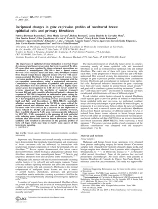
- 1. Reciprocal changes in gene expression profiles of cocultured breast epithelial cells and primary fibroblasts Patricia Bortman Rozenchan1 , Dirce Maria Carraro2 , Helena Brentani2 , Louise Danielle de Carvalho Mota2 , Elen Pereira Bastos1 , Elisa Napolitano e Ferreira2 , Cesar H. Torres2 , Maria Lucia Hirata Katayama1 , Rosimeire Aparecida Roela1 , Eduardo C. Lyra3 , Fernando Augusto Soares2 , Maria Aparecida Azevedo Koike Folgueira1 , Jo~ao Carlos Guedes Sampaio Goes3 and Maria Mitzi Brentani1* 1 Disciplina de Oncologia, Departamento de Radiologia, Faculdade de Medicina da Universidade de S~ao Paulo, Av. Dr. Arnaldo, 455, Sala 4112, S~ao Paulo, SP CEP 01246-903, Brazil 2 Centro de Ensino e Pesquisa, Hospital A.C. Camargo, R. Prof Antoˆnio Prudente, 211, Liberdade, CEP 01509-900, S~ao Paulo, SP, Brazil 3 Instituto Brasileiro de Controle do Caˆncer, Av. Alcaˆntara Machado 2576, S~ao Paulo, SP CEP 03102-002, Brazil The importance of epithelial-stroma interaction in normal breast development and tumor progression has been recognized. To iden- tify genes that were regulated by these reciprocal interactions, we cocultured a nonmalignant (MCF10A) and a breast cancer derived (MDA-MB231) basal cell lines, with fibroblasts isolated from breast benign-disease adjacent tissues (NAF) or with carci- noma-associated fibroblasts (CAF), in a transwell system. Gene expression profiles of each coculture pair were compared with the correspondent monocultures, using a customized microarray. Contrariwise to large alterations in epithelial cells genomic pro- files, fibroblasts were less affected. In MDA-MB231 highly repre- sented genes downregulated by CAF derived factors coded for proteins important for the specificity of vectorial transport between ER and golgi, possibly affecting cell polarity whereas the response of MCF10A comprised an induction of genes coding for stress responsive proteins, representing a prosurvival effect. While NAF downregulated genes encoding proteins associated to glyco- lipid and fatty acid biosynthesis in MDA-MB231, potentially affecting membrane biogenesis, in MCF10A, genes critical for growth control and adhesion were altered. NAFs responded to coculture with MDA-MB231 by a decrease in the expression of genes induced by TGFb1 and associated to motility. However, there was little change in NAFs gene expression profile influenced by MCF10A. CAFs responded to the presence of both epithelial cells inducing genes implicated in cell proliferation. Our data indicate that interactions between breast fibroblasts and basal epithelial cells resulted in alterations in the genomic profiles of both cell types which may help to clarify some aspects of this heterotypic signaling. ' 2009 UICC Key words: breast cancer; fibroblasts; microenvironment; coculture; microarray Important early literature and several recently reviewed studies have demonstrated that neoplastic transformation and progression of breast carcinoma cells are influenced by interactions with neighboring stromal components of which the principal cells are fibroblasts.1–6 Recent reports demonstrated that breast cancer derived fibroblasts exhibit biological characteristics distinct from fibroblasts obtained from nonmalignant breast.7–13 Genetic and epigenetic alterations were also described in the stromal cells.14–19 Malignant cells interact with the microenvironment by secreting soluble factors or by cell–cell and cell–matrix contacts. Tumor cells may release signaling molecules that affect the transcription of genes in nearby cells and conversely, fibroblasts in the stromal compartment of malignant breast lesions are also a source of growth factors, chemokines, proteases and provisional extracellular matrix molecules, creating a permissive field for the malignant cells and potentially affecting nontumorigenic epithelial cells.2,7,20-26 A number of reports have also demonstrated fibroblast effects on growth differentiation and invasion of normal/malignant breast epithelial cells.9,27–34 The importance of the functional state of the stromal cell compartment for the clinical behavior of tumors was recently reported.35 The microenvironment on which the tumor grows is complex consisting mainly of tumor epithelial cells and associated fibroblasts as well as nontransformed epithelial cells and normal fibroblasts. The exact role of these cell types, interacting with each other, in the progression of breast cancer has yet to be fully understood. One approach to study this interaction is to determine changes in gene expression profiles resulting from combination between fibroblasts and nonmalignant or malignant breast epithe- lial cells. Reciprocal inductive interactions on gene expression have been previously studied in animal breast cancer models36 and analyzed in coculture systems involving melanoma,37 pancre- atic38 and lung cancer cells39 and recently in mammary cell lines cocultivated with fibroblasts cell lines of different origins.40 To ask whether soluble factors released by stromal fibroblasts could regulate gene expression profile of normal or transformed breast epithelial cells and vice-versa, we performed coculture assays and analyzed changes in gene profile in both cell types, in response to 2-way interactions between them. In our experimental approach, we used a transwell system and cocultivated fibroblasts derived from breast carcinomas of different patients (CAFs) or isolated from breast tissues adjacent to breast benign tumors (NAFs) with either an spontaneously immortalized but noncancer- ous breast epithelial cell line (MCF10A) or an invasive immortal- ized breast carcinoma (MDA-MB231), both expressing a basal signature.41 The gene expression profile of each component of the different combinations was compared with the respective monocultures (Fig. 1). Material and methods Tissue samples Malignant and benign breast tissue specimens were obtained from patients undergoing surgery for breast disease. Carcinoma samples were obtained from 6 patients clinically staged as IIa, and benign samples were obtained from 6 patients with a diagnosis of fibroadenoma (n 5 4) or fibrocystic disease (n 5 2) (hereafter called normal fibroblasts). None of the patients had received preoperative chemotherapy. All tissue donors were patients at Instituto Brasileiro de Controle do Caˆncer, S~ao Paulo, Brazil, a reference center for cancer treatment. This study was approved by Additional Supporting Information may be found in the online version of this article. Grant sponsor: FAPESP; Grant numbers: 01/13515-1, 05/51593-5, 04/ 04607-8; Grant sponsor: CNPQ; Grant number: 47.7538/2003-7. *Correspondence to: Faculdade de Medicina da USP, Departamento de Radiologia, Disciplina de Oncologia, Av. Dr. Arnaldo, 455, 4 Andar, Sala 4112, S~ao Paulo, SP, Brazil. Fax: 55-11-3082.6580. E-mail: mbrentani@lim24.fm.usp.br Received 13 October 2008; Accepted after revision 2 June 2009 DOI 10.1002/ijc.24646 Published online 15 June 2009 in Wiley InterScience (www.interscience. wiley.com). Int. J. Cancer: 125, 2767–2777 (2009) ' 2009 UICC Publication of the International Union Against Cancer
- 2. the Institutional Committee and written informed consent was obtained by all participants. Invasive breast cancer was confirmed histopathologically. See Table I for patient characteristics. For validation studies, fresh-frozen human breast tumor samples were retrieved from the Tumor Tissue Biobank of the Medical and Research Center - Hospital A. C. Camargo, S~ao Paulo. Sections of 5-lm thick from the fresh-frozen tumor blocks were cut onto glass slides, stained with hematoxylin and eosin (HE), and reviewed by a pathologist. The HE-stained sections were used to evaluate and select appropriate tumor areas corresponding to each histolog- ical component, only the stromal component of all samples was collected. Four matched samples were evaluated, consisting of 4 IDC samples and 4 non-neoplastic samples, these samples were obtained from perilesional mammary specimens from patients obtained during resection of tumoral lesions. A pathologist sub- jected all slides representative of IDC to a careful histopathologi- cal analysis. Cell lines The immortalized human breast carcinoma MDA-MB231 and the spontaneously immortalized but nontumorigenic breast epithe- lial MCF10A cell line42 were purchased from American Type Culture Collection (ATCC, Manassas, VA) and maintained as indicated by the supplier. Briefly, MDA-MB231 cells were culti- vated with Leibovitz L-15 medium (Gibco, Invitrogen, Carlsbad, CA) supplemented with 10% FBS (Gibco, Invitrogen). MCF10A cells were cultivated in DMEM/Ham’s F-12 medium (Gibco, Invitrogen) supplemented with 100 ng/ml cholera toxin (Calbio- chem, La Jolla, CA), 0.01 mg/ml insulin (Sigma, Saint Louis, MO), 500 ng/ml hydrocortisone (Sigma), 20 ng/ml epidermal growth factor (Sigma) and 5% horse serum (Gibco, Invitrogen). The luminal mammary epithelial cells, HB4A, were generously donated by Drs. Michael J. O’Hare and Alan McKay, from Lud- wig Institute for Cancer Research, London, UK, and maintained with RPMI 1640 (Gibco, Invitrogen) supplemented with 10% FBS, 5 lg/ml insulin and 5 lg/ml hydrocortisone and was used as reference for cDNA microarray assays and validation assays. All cell lines were maintained at 37°C in a humidified atmosphere containing 5% CO2. Subconfluent cultures (80–90%) were used in the experiments. Primary cell culture Fibroblasts were obtained from normal adjacent tissue samples from patients with benign breast diseases (NAF) or with primary invasive breast cancer tumors (CAF). HE-stained, frozen histo- logical sections were prepared from each tissue sample to confirm benignity or malignancy. After adipose tissue removal, tissue was minced in PBS, washed twice in PBS and in culture medium, and then chopped into small 1–4 mm3 pieces under sterile conditions. A total of 15–30 fragments were transferred to a T25 culture flask and covered with DMEM, 20% FBS, 100 lg/ml ampicillin, 100 lg/ml streptomycin, 2.5 lg/ml Fungizone and maintained at 37°C in a humidified atmosphere containing 5% CO2. Outgrowth of cells was recorded after 10–20 days and medium was renewed once or twice a week thereafter. After sufficient outgrowth, the tissue fragments were removed and the cells were passaged by mild trypsinization. Characterization of fibroblasts Primary cells cultures of breast fibroblast were characterized by immunofluorescence. Briefly, cells in early passages (passage 3) were platted in circular slides (U 5 13 mm, Glasscyto, Bioslide Technology, Walnut, CA) and incubated with human anti-vimen- tin (clone Vim 3b4), human anti-smooth muscle actin (clone M0635), human anti-pancytokeratin (clones AE1/AE3) and human anti-CD31 (clone JC70A), all antibodies from DAKO Corporation, Carpinteria, CA. In addition, we used anti-fibroblast activated protein a (FAP-19) antibody, generously donated by Dr. Lloyd J. Old, for fibroblasts activation characterization. After that, cells were incubated with the secondary antibody (Alexa Fluor 488 rabbit or mouse anti-IgG (Invitrogen) diluted in PBS FIGURE 1 – Schematic representation of cocultures assays. Normal adjacent tissue from patients with benign breast diseases (NAF, n 5 6) or from primary invasive breast cancer tumors (CAF, n 5 6) and epithelial cells (MCF10A and MDA-MB231) were cocultured using transwell tissue-culture inserts with micro porous membrane for 72 hr; all cocultures and monocultures were exposed to the same conditions; RNA was extracted and or amplified from each cell type for hybridiza- tion onto cDNA microarrays, or subjected to RT-PCR. TABLE I – PATIENT CHARACTERISTICS Patient # Diagnosis HG ER PR ErbB2 CAF1 IDC III 2 2 1 CAF2 IDC III 1 1 2 CAF3 IDC I 2 1 2 CAF4 ILC II 2 2 2 NAF5 Fibroadenoma NAF6 Fibroadenoma NAF7 Fibroadenoma NAF8 Fibroadenoma CAF9 IDC II 1 1 2 CAF10 IDC II 1 1 2 NAF11 Fibrocystic disease NAF12 Fibrocystic disease IDC, invasive ductal carcinoma; ILC, invasive lobular carcinoma; HG, histological grade; ER, estrogen receptor; PR, progesterone re- ceptor; ErbB2, Her2. 2768 ROZENCHAN ET AL.
- 3. containing 0.005% Evans Blue (Sigma). The cells analysis was performed using a Zeiss Axioplan microscope (Carl Zeiss; Jena, Germany) and the mean percentage of smooth muscle actin posi- tive cells was evaluated using 10 randomically selected areas from slides and the images (magnification 4003) were processed by the software Image ProPlus 6.0 (Cybernetics, Silver Spring, MD). MTT proliferation assay CAFs (n 5 4) and NAFs (n 5 4) were plated in triplicate in 96- well plates at a density of 1000 cells/well, and the next day the media was replaced with 3-(4,5-methylthiazol-2-yl)-2,5-diphenyl- tetrazolium bromide (MTT) (0.5 mg/ml)-containing media, incubated at 37°C for 4 hr, to solubilize formazon crystals. We evaluated the proliferation rate between 0 and 120 hr. Absorbance was measured at 595 nm in a Bio-Rad plate reader (BioRad, Hercules, CA) and data analyzed using GraphPad Prism Software, version 3.0 (GraphPad Software). Coculture experiments Seventy-two hours before coculture experiments, all cells were exposed to the same conditions and distinct culture media were replaced by DMEM/F12 supplemented with 5% horse serum, 100 ng/mL cholera toxin, 0.01 mg/mL insulin, 500 ng/mL hydro- cortisone, 20 ng/mL epidermal growth factor and antibiotics. Cocultures of stromal and epithelial cells were performed using transwell tissue-culture inserts with a microporous membrane (Becton Dickinson Labware, Franklin Lakes, NJ). Epithelial cells were plated (1.3 3 105 cells) onto the bottom of the wells. Stromal cells were seeded (5.8 3 104 cells) on the permeable membrane of 0.4 lm tissue culture inserts which were then introduced into epi- thelial cell-containing wells. This proportion of epithelial:stromal cells was chosen to mimic that observed in the mammary gland ( 70:30). After 72 hr, cells in the inserts or on the bottom of the wells, as well as monocultures, were washed and harvested, and the RNA was isolated. A schematic representation of the coculture experiments can be seen in Figure 1. All experiments were performed between passage 06–10 and at 70% confluence. cDNA microarray assembly, hybridization and analysis A cDNA microarray platform containing 4608 open reading frame expressed sequence tags (ORESTES) was assembled at the Ludwig Institute for Cancer Research, S~ao Paulo, Brazil.43 ORESTES privileges the central part of mRNA molecules and selection of those to be spotted on the slides followed all these criteria: (i) cDNA clones representing full length genes; (ii) 300 bp and a high-quality sequence (CG content); (iii) 100 bp region with gene identity 85% as verified on the site http://ncbi.nlm. nih.gov/Blast and (iv) cDNA clone 30 sequence. cDNA clones were derived from human breast, colon, stomach, and head and neck tumors. These sequences could be classified among 505 function categories (biological processes). Another 192 reference sequences were included as positive and negative controls of hybridization. Platform characteristics complying with MIAME format may be verified in the gene expression omnibus (GEO) data repository, under accession number GPL 1930 (www.ncbi.nlm.nih.gov/projects/geo). Total RNA from all samples was isolated using TRIZOL (Invitrogen) reagent according to the manufacturer’s protocol. RNA quality was verified by agarose gel electrophoresis and visualization with ethidium bromide. Only RNA samples with a ratio 1 for 28S/18S ribosomal RNA were further processed. A 2-round RNA amplification and hybridization procedures followed a previously described protocol.44 Hybridized arrays were scanned on a confocal laser scanner (Arrayexpress, Packard Bioscience, Wellesley, MA) using identi- cal photomultiplier voltage (PMT 50) for all slides and data were recovered with the Quantarray software (Packard Bioscience) using histogram methods. After image acquisition and quantifica- tion, saturated spots (signal intensity 63,000) as well as low- intensity spots were removed from the analysis. Average signal intensity between technical replicates was determined for each spotted sequence and a local background subtraction was performed. Quantified signals were then submitted to log transfor- mation and Lowess normalization within each array, followed by global Lowess normalization for all arrays. The following analysis takes 3 factors into consideration: (i) whether or not the fibroblast culture was derived from a malig- nant environment; (ii) which epithelial cell type is involved and (iii) if cells were exposed to coculture or not. Differentially expressed genes were assessed by ANOVA (2 3 2). Probe sets with ANOVA p 0.05 and fold change 2.0 were considered significant. A Tukey test was done for 2 3 2 comparisons. Hierarchical clustering analysis based on Euclidian distance and complete linkage was per- formed using the differentially expressed genes identified through ANOVA. Reliability of the clustering was assessed by the Boot- strap technique using the TMEV software. The gene ontology (GO) analysis was performed using the GO tree machine tool (GOTM) which identifies categories hyper-represented in our gene lists and also KEGG pathways. For GO, we used a hyper geometric distribu- tion with p value 0.01 and for KEGG we also used a hyper geometric distribution but with p value 0.05.45 Laser capture microdissection Cells were laser captured using the PixCell II laser capture microdissection (LCM) system (Arcturus Engineering, Mountain View, CA). About 4000 cells were captured from 4 to 7 lm frozen sections, mounted onto glass slides, and stained with 100 lL of nuclear fast red (C.I.60760; Certistain1 ; Merck, Darmstadt, Germany) for microscopy. Only 1 type of cells was isolated from each sample group. A representative sample of cells from IDC depicting the different phases during the microdissection proce- dure is shown in Supporting Information Figure S1. RNA isolation and amplification LCM captured cells on CapSureTM HS LCM Caps (Arcturus Engineering) were resuspended in 10 lL of PicoPure RNA extrac- tion buffer (Arcturus Engineering). Total RNA was extracted by using the PicoPureTM RNA Isolation kit (Arcturus Engineering #KT0204) and DNase treated using the RNase-Free DNase Set (Qiagen #79254; Qiagen-Germantown MD), in accordance with the manufacturer’s instructions. Samples were processed with the Arcturus Riboamp HSTM (Arcturus Engineering #KT0505), a T7 RNA polymerase based technology, following the manufacturer’s recommendations. Stress treatment MCF10A cells grown and maintained in DMEM/F12 supple- mented with 5% horse serum, 100 ng/mL cholera toxin, 0.01 mg/ml insulin, 500 ng/mL hydrocortisone, 20 ng/mL epidermal growth factor and antibiotics were treated for 24 hr with Thapsi- gargin (TG) (Sigma-Aldrich, St Louis, MO) dissolved in ethanol at concentration of 250 nM to induce ER stress by disrupting calcium homeostasis. The TG concentration of 250 nM was chosen, among several concentrations (100, 250, 500 and 1000 nM), because this concentration presented the smallest cytotoxic effect for MCF10A cells determined using trypan blue dye exclusion. After treatment total RNA was extracted using Trizol and submitted to RT-PCR. Real-time RT-PCR analysis We evaluated the expression of some genes chosen to be differently expressed between samples using cDNA microarray technology by real-time RT-PCR. The genes were randomly selected, but another criteria used was fold variation 2 and false discovery ratio 0.05. Reverse transcription was performed using 2 lg of total RNA, a random hexamer primer (Invitrogen) and Superscript TM II 2769COCULTURE INDUCED CHANGES IN EXPRESSION PROFILES
- 4. Reverse Transcriptase (Invitrogen). Primers were designed for different exons to avoid amplification of genomic DNA using Primer 3 software (http://frodo.wi.mit.edu/cgi-bin/primer3/primer3_ www.cgi), and synthesized by IDT (Integrated DNA Technologies, Coralville, IA). PCR was performed in a Rotor-gene system (Corbett Research, Mortlake, Australia). Thermocycling was done in a total volume of 20 lL containing 5 lL of cDNA sample (diluted 1:40), 1.5 mM MgCl2, 0.2 lM of each primer, 0.1 lL SYBR1 Green I (Sigma; 1:100 working dilution) and Platinum Taq DNA polymerase (Invitrogen life Tecnologies), reaction buffer, and a deoxyribonucle- otide triphosphate mixture. Primer sequences and cycling variables are listed in Table II. PCR were performed in duplicate to all twelve samples distributed as follow, 08 samples after cDNA microarray analyses and we have included fibroblasts from 2 patients with duc- tal invasive carcinomas and from 2 patients with fibrocystic disease (see Table I). We also obtained 4 matched samples using LCM from patients diagnosed with invasive ductal carcinoma. The duplicate average values were used for quantification and the relative expression of genes of interest was normalized to that of actin-b, and gene expression in each sample was then compared with expression in HB4A cells. The comparative CT method (DDCT) was used for quantification of gene expression and relative expression was calculated as 22DDCT . Results Characterization of isolated fibroblast population Isolated breast fibroblast (normal or tumors associated) popula- tions were characterized by immunofluorescent staining. Both fibroblast monolayers stained uniformly for vimentin and FAP-19, but were negative for cytokeratin and CD31, confirming the stro- mal origin of cells and the absence of contaminating epithelial or endothelial cells. Approximately 15.5 6 5.6% of the CAFs were positive for a-SMA, with no differences discerned between CAFs and NAFs (Supporting Information Figure S2). Proliferation rate of fibroblasts To evaluate the proliferation rate of both fibroblasts, cells were plated on 96 wells plates and treated with sodium tetrazolium solution as described earlier. Supporting Information Figure S3 shows that both fibroblasts presents similar proliferation curves (p 5 0.559) after 5 days of evaluation. Effects of coculture of tumor-associated fibroblasts on the expression profile of MDA-MB231 and MCF10A We first analyzed the influence of tumor-associated fibroblasts from different donors (n 5 4) on the expression profile of the mammary epithelial cell lines, these analysis were assessed by ANOVA test (2 3 2). Comparing MCF10A cocultured with CAF, MCF10A in monoculture, MDA-MB231 cocultured with CAF, and MDA-MB231 cultured alone, we found 307 genes that were differentially expressed. Hierarchical clustering of these genes distinguished two groups: MCF10A and MCF10A cocultured with CAF clustered together on one branch, and MDA-MB231 and MDA-MB231 cocultured with CAF clustered together on the other branch (Fig. 2a). Applying the Tukey test, 160 genes were differentially expressed between MDA-MB231 cells cocultured with CAF and MDA-MB231 monocultures, and 178 genes had their expression levels altered in MCF10A cocultured with CAF when compared with MCF10A monocultures. Genes selected by the ANOVA test were considered differentially expressed when presenting at least TABLE II – PRIMER SEQUENCES FOR QUANTITATIVE GENE EXPRESSION ANALYSIS Target Primer Sequence (50 –30 ) Product (bp) Actin b ACTB forward AGAAATCTGGCACCAACC 188 ACTB reverse AGAGGCGTACAGGGATAGCA A disintegrin and metalloproteinase domain 12 ADAM12 forward AAAAAGGTGTCGGCTTCTCA 112 ADAM12 reverse CCAGAACAACTCGGCTCACT Arsenate resistance protein ARS2 ARS2 forward TCCATGAGGGACAGGAAGAC 119 ARS2 reverse GAGGCAACAGATGCAGGATT BMP and activin membrane-bound inhibitor BAMBI forward CTAGAGAAGCAGGCGCTGAG 157 BAMBI reverse ATCGCCACTCCAGCTACATC S100 calcium binding protein A9 S100A9 forward TCTTTTCGCACCAGCTCTTT 125 S100A9 reverse CAGCTGAGCTTCGAGGAGTT Cbp/p300-interacting transactivator, with Glu/Asp-rich carboxy-terminal domain, 2 CITED2 forward ATCCGGCATGTAGTGGTTGT 592 CITED2 reverse GTCCCCTCTATGTGCTGCTG CCCTC-binding factor (zinc finger protein) CTCF forward TGCACCTGTATTCTGGTCTTCA 157 CTCF reverse TGCCGTAGAAATTGAACCTG Connective tissue growth factor CTGF forward GTAATGGCAGGCACAGGTCT 211 CTGF reverse CCGTACTCCCAAAATCTCCA DEAD (Asp-Glu-Ala-Asp) box polypeptide 21 DDX21 forward CCTTCCAGTTCTGGTTGCTC 105 DDX21 reverse TTCCAAAGTGAAGGGAATGG Dicer 1, ribonuclease type III DICER1 forward CTCTGACCTTCCCGTCGTAA 160 DICER1 reverse CTGGAGACAGTCTGGCAGGT FOS-like antigen 2 FOSL2 forward GGGAGCTGACAGAGAAGCTG 124 FOSL2 reverse TGAGCCACCAACATGAACTC Heat shock protein 90kDa beta (Grp94), member 1 HSP90B1 forward AGAAAGAATGCTTCGCCTCA 238 HSP90B1 reverse ACATTCCCTCTCCACACAGG2 Interleukin 1, beta IL1B forward GCTGGAGAGTGTAGATCCCAAA 101 IL1B reverse CAGACTCAAATTCCAGCTTGTT Lipocalin 2 LCN2 forward TACACTGGTCGATTGGGACA 104 LCN2 reverse CAAGGAGCTGACTTCGGAAC Procollagen-lysine 1,2-dioxygenase-1 PLOD1 forward GCTCGGATGGAACAGTTGTAG 106 PLOD1 reverse ACCATGATGCCTCCACCTT Solute carrier family 22, member 18 SLC22A18 forward CCACCACGATGAAGACCAG 130 SLC22A18 reverse GCTGGCTACCTCATGTCCTT Cycling conditions: initial activation, 95o C for 5 min; template activation for 40 cycles with denaturation at 95°C for 1 min, primer annealing at Tm for 1 min, and extension at 72°C for 1 min. Melting curve analysis: after 40 cycles at 72°C for 15 s and increase in temperature up to 99°C with a heating rate of 1°C/s and continuous fluorescence measurement. All Tms were established at 60°C, except for ARS2, BAMBI and SLC22A18 primers. 2770 ROZENCHAN ET AL.
- 5. 2-fold difference in signal intensity. A total of 35 upregulated and 55 downregulated genes in MDA-MB231 cocultured with CAF were selected. In MCF10A cocultured with CAF after a 2-fold cut-off, we found more genes upregulated (n 5 77) and less genes downregulated (n 5 21). Effects of coculture of normal fibroblasts on the expression profile of MDA-MB231 and MCF10A In MDA-MB231/NAF versus MDA-MB231 and MCF10A/ NAF versus MCF10A, 248 differentially expressed genes found by ANOVA (2 3 2) were clustered (Fig. 2b) and sorted into two branches, one branch with MDA-MB231 alone and after coculture with NAF (n 5 4), and another branch with MCF10A cells and MCF10A after coculture with NAF. MDA-MB231/NAF presented 115 genes whose expression levels were altered. After applying the 2-fold change cut-off, 29 genes were upregulated and 50 were downregulated. By a similar criterion in the MCF10A/NAF coculture only 48 upregulated and 33 downregulated genes were retained, from a total of 161 genes. Functions regulated by fibroblasts in the epithelial cells Independently of which fibroblast was used in coculture we could verify a number of overlapping genes modulated in each epithelial cell as seen in the Venn diagram (Fig. 3) and Supporting Information Tables. In MDA-MB231 when these overlapping genes (n 5 49, 61% downregulated) were sorted by GOTM tool, (biological process) and genes associated to motility (F11R, TGFb2, SPINT2, LCN2, CD97, NRP1, p 5 5.89 e24 ) were statisti- cally hyper-represented. MCF10A exposed to NAF or CAF also revealed shared genes commonly altered relative to control (n 5 40, 27% were downregulated). Neurogenesis (RTN1, MTPN, p 5 8.46 e23 ) and hypoxic conditions (AQP3, LDHA, NDRG1, LOXL3) were also biological process evidenced by GOTM, whereas just the first group was hyper-represented. Such genes corresponded to proteins associated to cellular or nuclear-mem- brane. To select which genes were specifically influenced by each fibroblast in each epithelial cell, we decided to exclude these common genes from the next analysis. After applying GOTM analyses, to the set of genes exclusively modulated by CAF in MDA-MB231 (n 5 41) those genes involved in biological processes such as intracellular transport (KDELR3, TRAPPC1, NAPA, NUP50, TSPO, p 5 3.69 e23 ) were hyper-represented. Using KEGG, MAPK, WNT and Toll like receptor signaling pathways were hyper-represented in the list of genes specifically altered by CAFs (PTK2B, MAP2K3, PPM1B, MYC, CHD8, ILb, p 0.001). We also noted a downregulation of several genes related to proliferation (MYC, IL1b, PTK2B, CDCP1) and upregulation of genes associated to cell survival (DDX21, DICER, EPB41L4A) when compared with MDA- MB231 monoculture. Among the genes uniquely modulated by normal fibroblasts in MDA-MB231 (n 5 30), the major biological process significantly over represented using GOTM was biopolymer glycosylation (UGCGL2, GCNT1 and ALG9), (p 5 6.6 e23 ). It is interesting to note that using KEGG pathway the smallest p value encountered (0.0008) refers to the glycerolipid metabolism pathway. Of partic- ular interest, among the most highly downregulated genes were AGTPAT4 and ACSL5, associated to lipid metabolism. After subtraction of the common effects induced by NAF or CAF, we noted transcripts (n 5 57) which were specifically altered in MCF-10A in coculture with CAF compared with mono- culture conditions. Applying GOTM analysis, over represented genes could be generally assigned to response to stress (S100A9, CaLR, B4GALT1, HSP90B1, SPRR3, ERCC5, EGFR, LIG3, p 5 9.43 e23 ). An upregulation of several genes associated to cancer related pathways in the literature (KLK2, EGFR, HSP90B1, EFNB2, MAPRE-2, CMIP (c-maf), S100A9) was observed. Indeed for HSP90B1 (GPR94) we have noted an induction of its mRNA after treatment of MCF10A with thapsigargin (data not shown). After cocultivation with NAF (n 5 40) most of the genes specifically differentially expressed in MCF-10A coded for nuclear proteins and the process of transcription initiation (DHX35, EIF5B, GTF2E2, MBOAT2, TAF12) was over repre- sented (p 5 3.42 e23 ). Effects of coculture of MCF10A on the genomic profile of fibroblasts Our next step was to evaluate gene expression specifically in fibroblasts (NAF or CAF) growing in coculture with normal breast cells (MCF10A) when compared with those from fibroblast mono- cultures. Cluster analysis indicated that CAF monocultures clus- tered separately in 1 group whereas normal fibroblasts cocultured or not and tumoral fibroblasts after coculture with MCF10A were allocated to the same branch (Fig. 2c). Nevertheless, we have identified genes that were differentially expressed between pre and post tumor-associated fibroblasts with MCF10A. Fifty-eight genes were specifically and significantly differentially expressed in cocultivated CAFs when compared with CAF monocultures but considering a 2-fold cut-off, this number decreased to 34 and gene ontology using GOTM, showed an over representation of genes involved with regulation of cell cycle (NFYC, CDKN1B, CCT2, p 5 9.53 e23 ), protein amino acid glycosylation (GCNT1 and ALG8, p 5 4.4 e23 ) and MAPKKK cascade such as RGS3 (GTPase which inhibits G-protein mediated signal) and FGFR1 (fibroblast growth factor receptor, p 5 8.34 e23 ). There was rela- tively little change in the gene expression profile of NAF grown alone or in coculture with MCF10A, just 7 genes were identified showing a 2-fold change. Of note, however, is the enhanced expression of CBX2, a chromobox homolog. Effects of coculture with MDA-MB231 on genomic profile of fibroblasts Subsequently, we aimed to identify genes in CAF and NAF that were affected by coculture with MDA-MB231. Clustering based on 42 genes differentially expressed in CAFs or NAFs following coculture with MDA-MB231 does not clearly segregate fibroblasts into different groups that correlate with particular aspects (Fig. 2d). Among differentially expressed genes in CAFs (n 5 23), following the 2-fold cut-off, 4 were over-expressed and 19 were under-expressed. Ontology analyses identified genes associated to cell cycle including TACC1 and CDKN1B genes, or to protein transport (TIMM17A) or classified under the category of integral to membrane (BAMBI). The influence of MDA-MB231 cells on NAF resulted in downregulation of 8 of 9 differentially expressed genes (89%). Genes with decreased expression as determined by gene ontology analysis were TSPAN5 and PLOD whereas TACC2 was upregulated. CTGF was the only common modulated gene by MDA-MB231 cells on both fibroblasts. The category of regulation of cell control was over represented using GOTM (p 5 9.08 e23 ), and the CTGF gene was included in this biological process. Quantitative real-time PCR validation To validate our gene expression data obtained by cDNA micro- array, we have chosen 15 genes and their expression values were compared with values obtained by quantitative RT-PCR. Besides the 4 cases used in the array experiments, we also have included more 2 cases of CAF and NAF to be cocultured that were not used in the initial microarray studies. In MDA-MB231/CAF versus MDA comparison, we selected the following genes: FOSL2, IL1B, LCN2 and SLC22A18, which were downregulated in MDA-MB231 after coculture with CAF, the genes DDX21, DICER and CTCF were upregulated in those epithelial cells after cocultivation. Both genes ARS2 and CITED2 were altered in MDA-MB231 af- ter coculture with NAF cells, being the first one upregulated whereas the second had its expression levels downregulated. CTGF was downregulated in both CAF and NAF cocultured with MDA-MB231 as well as, BAMBI and PLOD1 were also 2771COCULTURE INDUCED CHANGES IN EXPRESSION PROFILES
- 6. FIGURE 2. 2772 ROZENCHAN ET AL.
- 7. decreased in CAF and NAF respectively after coculture with MDA-MB231 cells. In addition, ADAM12, HSP90B1 and S100A9 were upregulated in MCF10A after cocultivation with CAF. For all genes evaluated, we obtained good concordance (86.6%) between microarray and real-time PCR analysis, except for FOSL2 and CTCF genes (Fig. 4). To confirm the altered gene expression in fibroblasts in their respective tissue samples, we evaluated the 2 genes already vali- dated by real time in CAF after coculture with MDA-MB231, that is, BAMBI and CTGF, in stromal tissue obtained from invasive ductal carcinoma samples and the normal counterpart tissue. For BAMBI, we have validated both array and real time from coculture experiments data, for 3 of 4 matched samples. On the other hand, CTGF presented an over expression in stromal near tumoral tissue when compared with stromal adjacent peritumoral normal tissue in all cases analyzed. Discussion We used a transwell system allowing diffusible factors exchange and microarray technology to analyze the gene expres- sion changes resulting from cocultivation of 2 basal breast carci- noma cell-lines, MCF10A and MDA-MB231 with mammary fibroblasts of 2 different origins: from within breast tumors (CAF) or from breast benign disease (NAF). After pair-wise comparisons between epithelial cells and fibroblasts, reciprocal inductive inter- actions were observed but the most consistent gene expression changes occurred in the epithelial cells, although these changes were small when compared with the distinct genomic profiles exhibited by these cells in monoculture. Each epithelial cell showed overlapped genes in response to cocultivation with both fibroblast types. As normal fibroblasts when cultured in vitro should be considered as permanently acti- vated,26 it is possible that such cells may be competent to exert similar effects as those described for CAFs. After removal of genes commonly altered by NAF or CAF, among the genes exclusively modulated by CAF in MDA-MB231 cells, transport was the most represented, including genes coding for proteins important for the specificity of vectorial transport between ER and Golgi or to prevent secretion of ER-resident pro- teins such as an ER DEL receptor (KDELR3), TRAPPC1, NAPA or coding for calcium translocation protein (TSPO) that were downregulated. Decreased expression of those genes may lead to an altered vesicular addressing of secretory proteins, possibly affecting polarity and apical-basal organization. Lebret et al.22 suggested that some of the key effects of CAFs on breast epithelial cells were on motility, cell organization and epithelial mesenchy- mal transition (EMT) markers expression. Disruption of basal polarity by inclusion of CAFs in a model of 3-dimensional hetero- typic culture system was recently described.46 We also noted that some genes associated to proliferation were selectively downregulated in MDA-MB231 cocultivated with CAF (MYC, IL1B, RHOV) whereas other specific genes were upre- gulated including genes that have already been previously reported as associated to alternative pathways leading to survival such as DDX21 (dead/H box helicase 21), associated to poor prog- nosis,47 DICER, an essential component of the RNAi pathway, required for the maintenance of hypermethylation of selected epigenetically silenced loci in cancer cells48 and EPB41L4A (NBL4), an important component of the b-catenin/TCF pathway probably related to proliferation and cell polarity.49 Approximately 70% of the genes uniquely modulated by normal fibroblasts in MDA-MB231, were downregulated and this set was found to contain an over representation of genes involved in gly- can structure biosynthesis and glycerolipid metabolism. Some of these genes coded for glycosyl transferases that play important FIGURE 3 – Venn Diagram with genes differentially expressed between MDA-MB231/NAF and MDA-MB231/CAF and MCF10A/ NAF and MCF10A/CAF. (a) Thirty genes were exclusively differen- tially expressed in MDA-MB231/NAF whereas 41 genes were exclu- sively altered by CAF influence on MDA-MB231 genomic profile. Common differentially expressed genes (49) represent almost 29% of the total number of genes. (b) Forty-one genes appeared to be com- monly differentially expressed when MCF10A was cocultured with ei- ther NAF or CAF (42%). Forty genes were exclusively modulated by NAF whereas 57 genes were specifically altered by CAF influence on MCF10A genomic profile. In parenthesis are the total number of genes found in each situation regulated at least by 2-fold and p 0.05. FIGURE 2 – Hierarchical clustering after ANOVA test. Color intensity is scaled within each row so that the highest expression value corresponds to bright red and the lowest to bright green. The support scale is a guarantee of reliability of dendrograms. (a) Hierarchical cluster- ing was generated for genes differentially expressed in MCF10A cocultured with CAF, MCF10A alone, MDA-MB231 cocultured with CAF, and MDA-MB231 monocultures. MCF10A and MCF10A after coculture with CAF clustered on one branch and MDA-MB231 cocultured with CAF clustered with MDA-MB231 monocultures on another branch. (b) Cluster analysis of differentially expressed genes in MDA-MB231 and MCF10A after coculture with NAF and both breast epithelial cell lines grown alone. NAF exerted influence on genomic profile of epithelial cells but this effect could not cluster MCF10A after coculture in the same branch with MDA-MB231 cells. (c) Effects of coculture with MCF10A on the expression profile of fibroblasts. In the cluster analysis of CAF and NAF in monoculture and after coculture with MCF10A cells, we observed that the differentially expressed genes allocate the tumoral fibroblasts cocultured with MCF10A in the same branch as normal fibroblasts cocultured with MCF10A cells. (d) Influence of MDA-MB231 cells on the expression profile of fibroblasts. CAF cocultured with MDA-MB231 cells clustered on the same branch as NAF. In the figure, we are showing a partial view of the genes differentially expressed, see Supporting Information Data for the complete list of altered genes. 2773COCULTURE INDUCED CHANGES IN EXPRESSION PROFILES
- 8. roles in post-translational glycosylation of glycolipids in the ER, that are one of an ubiquitous membrane component and also included genes coding for proteins associated to translocation of misfolded glycoproteins across the ER membrane (UGCGL2, ALG9 and GCNT1), or proteins interfering with calcium transport (GPR35) or implicated in ER-stress induced cell death (ERGIC3). Decreased levels of glycosil transferases may lead to protein underglycosylation and further degradation in the proteasomes.50 We also noted that the most downregulated gene was ACSL5 coding for an acyl-CoA synthetase that converts fatty acids to acyl-COA long chain, and another gene with decreased levels was AGTPAT4 (lysophosphatic acid acyltransferase), indicating a pos- sible reduction in the fatty acid levels that are essential for breast cancer survival under stress conditions.51 Especially, the downre- gulation of CITED-2 (CBP/p300-interacting transactivator with glutamine acid/aspartic acid-rich C-terminal domain-2) which is highly expressed in MDA-MB231 is interesting because its down- regulation attenuated the expression of MMP9 which could affect tumor cell invasion.52 These findings suggested that exposure of MDA-MB231 to NAFs resulted mainly in the repression of lypo- genic enzymes and impaired glycosylation possibly affecting membrane biogenesis and viability. Coculture of MCF10A with normal or tumor-associated fibro- blasts showed both specific and common effects. Particularly, among the latter, we found that MCF10A gene expression profile was enriched with many genes that have been associated to hypoxic conditions such as LOXL3, AQP3, NDRG1, LDHA and ERRFI1. Although the biological mechanism underlying this cell type response to both fibroblasts is presently unclear, it could reflect small variations in oxygen tension caused by cell density (although it was maintained below 1.38 3 104 cells/cm2 in our experiments) or result from a specific composition of hypoxia reg- ulators in MCF10A.53 Genes specifically downregulated in MCF10A instructed by NAFs encoded several nuclear proteins implicated in initiation of transcription and translation and RNA processing functions. In addition, DDIT4 (REDD1) that inhibits mTOR function to control cell growth and other genes implicated in adhesion and tight junctions CTNND1 (catenin delta 1), PCDH1 (cadherin like protein-1), INADL, ITGA5 were upregu- lated indicating a role of normal fibroblasts in maintaining func- tions related to differentiation and control of cell proliferation. Tumor affiliated breast fibroblasts in turn, elicited in MCF10A an over representation of genes coding for stress responsive pro- teins including upregulation of S100A9 (stress regulated protein FIGURE 4 – Validation of cDNA microarray data by real-time RT-PCR. cDNAs from breast cell lines (MDA-MB231 and MCF10A) and fibro- blasts from adjacent fibroadenoma tissues (NAF) or tumor-associated breast (CAF) were used for the RT-PCR reaction. The relative expression was determined after b-actin normalization; the relative expression determined by RT-PCR was compared with the cDNA microarray ratios for each of the following comparisons: (a) MDA/CAF versus MDA, (b) MDA/NAF versus MDA, (c) CAF/MDA versus CAF, (d) NAF/MDA versus NAF and (e) MCF10A/CAF versus MCF10A. The positive and negative values indicated up- and down-expressed genes, respectively. Dark gray represents array data. 2774 ROZENCHAN ET AL.
- 9. belonging to the S100 family of small calcium binding protein),54 implicated in breast tumorigenesis in other studies, EGFR that in- dependent of its kinase activity maintains the basal intracellular glucose level preventing cell from undergoing autophagic death,55 calcium dependent molecular chaperones such as HSP90B1 (GPR94) that confers resistance to apoptosis and calreticulin with stabilize protein folding intermediates and SPRR3, a cellular adap- tator to biochemical stress.56 The induction of several stress re- sponsive proteins may represent a prosurvival effect following ex- posure to stress. Several stress proteins have been reported as fre- quently overexpressed in studies with breast tumors cells.57 Two of those genes (S100A9 and HSP90B1) were validated in our real- time experiments and the latter was induced in MCF10A cells af- ter treatment with Thapsigargin a drug known by its stress effect. We also noted the upregulation of others genes showing high magnitude of change including genes involved in breast tumori- genesis such as KLK2 (kallikrein 2), a predictive marker of breast cancer,58 ADAM12 that emerges as an important regulator of breast tumor progression59 and MAPRE-2 which shows homology to the APC binding EB1 gene and might active b-catenin path- way.60 Upregulation of CMTM4 (CKLFSF4) that belongs to the chemokine-like factor gene superfamily and of CMIP (c-maf), a transcription factor belonging to the AP1 superfamily, are particu- larly interesting due to recognized function of as chemokines mediators of tumor-stroma interactions.8,9,61 Although we have not provided complete evidence of the malig- nant transformation of MCF10A epithelial cells by soluble factors released by CAFs, the gene pattern observed, suggested that MCF10A over express some features associated to cancer cells. We have no clear evidence that exposure to CAFs turns MCF-10A into a malignant one but we showed that genes coding for several stress proteins which may increase survival were induced in MCF-10A triggered by CAFs. All together, these results suggested that normal cells may acquire some of the characteristics of transformed cells in the presence of the appropriate environmental stimuli. One important issue is how normal stromal cells that coexist with tumor-associated fibroblasts are affected by interactions with malignant cells. When we focused on the genomic profile of NAFs after coculture, we observed that they seem to have a mod- est response to MDA-MB231 and presented a reduced expression of 3 targets of the TGFb family: CTGF (connective tissue growth factor), a member of the CCN growth factors family that promotes fibroblast proliferation and enhanced migratory behavior,62 PLOD2 (procollagen lysine), a profibrotic signaling protein63 and TPM2 (tropomyosin 2), that is required for TGFb regulation of stress fibers and motility.64 We also noted the downregulation of TSPAN5, a transmembrane 4 superfamily member that play a role in the regulation of mobility. Therefore, we may speculate that cultured fibroblasts from nonmalignant tumors (NAF) respond to epithelial malignant cells by decreasing genes associated to motil- ity and this alteration may serve to immobilize fibroblasts in close contact with tumor cells, a suggestion previously raised by Ron- nov-Jensen et al. and Valenti et al.65,66 Downregulation of genes regulated by TGFb1 has also been observed in normal pulmonary fibroblasts after coculture with cancer cells.36 In the CAF gene profile, coculture with MDA-MB231 led to a reduced expression of TGFb1 3 targets: CTGF, CDKN1B (p27KIP1) which mediates TGFb1 induced growth arrest and BAMBI, a negative regulator of feedback loop in TGFb1 signaling,67 suggesting that the response observed in these cocultures might result from an attenuated TGFb1 effect. Besides alterations of TGFb1 signaling, it may be that cultured fibroblasts lack the capacity to transform the latent form of TGFb1 in the biologically active counterpart as previ- ously proposed.66 TGFb1 expression was previously reported as downregulated in breast cancer derived fibroblasts and the authors suggested that diminished levels of this cytokine may reflect a stromal mechanism to favor adjacent tumor growth.15 In both fibroblast types, MDA-MB231 induced an upregulation of genes coding for members of the TACC family of centrosome and microtubule interacting proteins that were implicated in fibroblast transformation68 supporting the notion that both fibroblast types respond to the presence of cancer cells maintaining a microenvir- onment favorable for the malignant cells lodging. Of interest is our observation that the nontumorigenic MCF10A induces in CAF genes coding for proteins implicated in control- ling cell growth that may reflect changes in the status of cell pro- liferation. Gene transcripts altered in CAF included RPS6KA3 (p90 ribosomal S6 Kinase), FGFR1, NRD1 (nardilysin that enhan- ces shedding of EGF), CDKN1B and NFY. Notably, we also iden- tified an increased expression of PTGES2, a known inflammatory gene.69 On the other hand, the gene expression pattern of the cocultured normal fibroblasts with MCF10A was only modestly affected and the most upregulated gene was CBX2, a polycomb homolog, repressor of proto-oncogenes.70 Taken together, our data indicate that the coculture of breast fibroblasts and normal or invasive breast carcinoma basal cells exert reciprocal effects on gene expression profile but epithelial cells seem to be more responsive by altering a larger number of genes when compared with a more modest effect on the fibro- blasts. It is possible that the fibroblasts being already activated in culture are less responsive to factors released by epithelial cells, or might require direct intercellular communication to be modified by epithelial cells. As breast cancer is clinically and molecularly heterogeneous, we focused on interactions between breast stromal fibroblasts and well-characterized normal or invasive breast basal cells in this work. We may expect that coculture with mammary cells of the luminal type or fibroblasts of different origins results in different reciprocal gene expression alterations. These results were con- firmed by RT-PCR with the same patients used in microarray anal- ysis and also by inclusion of another 2 different donors of each NAF and CAF. Besides, we evidenced BAMBI, as a good candi- date for further investigations regarding interactions of malignant breast epithelial cells with fibroblasts because this gene was also downregulated in the stromal tissue of 3 patients with invasive breast carcinoma confirming the previous data of our coculture model obtained with cDNA microarray and real-time analysis. Our results should therefore help to better understand some of the molecular mechanisms involved in the complex heterotypic signaling between epithelial cells and fibroblasts. Acknowledgements The authors thank Dr. W. P. R. Teodoro (Laboratorio de Matriz Extracelular/Disciplina de Reumatologia) for her assistance with imunofluorescence assays, Ms. T. L. Lourdes for technical support and Ms. M. J. Gonc¸alves Benevides for secretary assistance. References 1. Park CC, Bissell MJ, Barcellos-Hoff MH. The influence of the micro- environment on the malignant phenotype. Mol Med Today 2000;6: 324–9. 2. Elenbaas B, Weinberg RA. Heterotypic signaling between epithelial tumor cells and fibroblasts in carcinoma formation. Exp Cell Res 2001;264:169–84. 3. Kunz-Schughart LA, Knuechel R. Tumor-associated fibroblasts (part I): active stromal participants in tumor development and progression? Histol Histopathol 2002;17:599–621. 4. Wiseman BS, Werb Z. Stromal effects on mammary gland develop- ment and breast cancer. Science 2002;296:1046–9. 5. Mueller MM, Fusenig NE. Friends or foes—bipolar effects of the tumour stroma in cancer. Nat Rev Cancer 2004;4:839–49. 6. Kalluri R, Zeisberg M. Fibroblasts in cancer. Nat Rev Cancer 2006;6: 392–401. 7. Kunz-Schughart LA, Knuechel R. Tumor-associated fibroblasts (part II): functional impact on tumor tissue. Histol Histopathol 2002; 17:623–37. 2775COCULTURE INDUCED CHANGES IN EXPRESSION PROFILES
- 10. 8. Allinen M, Beroukhim R, Cai L, Brennan C, Lahti-Domenici J, Huang H, Porter D, Hu M, Chin L, Richardson A, Schnitt S, Sellers WR, et al. Molecular characterization of the tumor microenvironment in breast cancer. Cancer Cell 2004;6:17–32. 9. Orimo A, Gupta PB, Sgroi DC, Arenzana-Seisdedos F, Delaunay T, Naeem R, Carey VJ, Richardson AL, Weinberg RA. Stromal fibroblasts present in invasive human breast carcinomas promote tumor growth and angiogenesis through elevated SDF-1/CXCL12 secretion. Cell 2005;121:335–48. 10. Singer CF, Gschwantler-Kaulich D, Fink-Retter A, Haas C, Hudelist G, Czerwenka K, Kubista E. Differential gene expression profile in breast cancer-derived stromal fibroblasts. Breast Cancer Res Treat 2008;110:273–81. 11. Hawsawi NM, Ghebeh H, Hendrayani SF, Tulbah A, Al-Eid M, Al-Tweigeri T, Ajarim D, Alaiya A, Dermime S, Aboussekhra A. Breast carcinoma-associated fibroblasts and their counterparts display neoplastic-specific changes. Cancer Res 2008;68:2717–25. 12. Casey T, Bond J, Tighe S, Hunter T, Lintault L, Patel O, Eneman J, Crocker A, White J, Tessitore J, Stanley M, Harlow S, et al. Molecu- lar signatures suggest a major role for stromal cells in development of invasive breast cancer. Breast Cancer Res Treat 2009;114:47–62. 13. Trimis G, Chatzistamou I, Politi K, Kiaris H, Papavassiliou AG. Expression of p21waf1/Cip1 in stromal fibroblasts of primary breast tumors. Hum Mol Genet 2008;17:3596–600. 14. Moinfar F, Man YG, Arnould L, Bratthauer GL, Ratschek M, Tavassoli FA. Concurrent and independent genetic alterations in the stromal and epithelial cells of mammary carcinoma: implications for tumorigenesis. Cancer Res 2000;60:2562–6. 15. Kurose K, Hoshaw-Woodard S, Adeyinka A, Lemeshow S, Watson PH, Eng C. Genetic model of multi-step breast carcinogenesis involv- ing the epithelium and stroma: clues to tumour-microenvironment interactions. Hum Mol Genet 2001;10:1907–13. 16. Kurose K, Gilley K, Matsumoto S, Watson PH, Zhou XP, Eng C. Frequent somatic mutations in PTEN and TP53 are mutually exclu- sive in the stroma of breast carcinomas. Nat Genet 2002;32:355–7. 17. Fukino K, Shen L, Matsumoto S, Morrison CD, Mutter GL, Eng C. Combined total genome loss of heterozygosity scan of breast cancer stroma and epithelium reveals multiplicity of stromal targets. Cancer Res 2004;64:7231–6. 18. Hu M, Yao J, Cai L, Bachman KE, van den Bruˆle F, Velculescu V, Polyak K. Distinct epigenetic changes in the stromal cells of breast cancers. Nat Genet 2005;37:899–905. 19. Patocs A, Zhang L, Xu Y, Weber F, Caldes T, Mutter GL, Platzer P, Eng C. Breast-cancer stromal cells with TP53 mutations and nodal metastases. N Engl J Med 2007;357:2543–51. 20. Bhowmick NA, Neilson EG, Moses HL. Stromal fibroblasts in cancer initiation and progression. Nature 2004;432:332–7. 21. Kleer CG, Bloushtain-Qimron N, Chen YH, Carrasco D, Hu M, Yao J, Kraeft SK, Collins LC, Sabel MS, Argani P, Gelman R, Schnitt SJ, et al. Epithelial and stromal cathepsin K and CXCL14 expression in breast tumor progression. Clin Cancer Res 2008;14:5357–67. 22. Lebret SC, Newgreen DF, Thompson EW, Ackland ML. Induction of epithelial to mesenchymal transition in PMC42-LA human breast car- cinoma cells by carcinoma-associated fibroblast secreted factors. Breast Cancer Res 2007;9:R19. 23. Holliday DL, Hughes S, Shaw JA, Walker RA, Jones JL. Intrinsic genetic characteristics determine tumor-modifying capacity of fibro- blasts: matrix metalloproteinase-3 5A/5A genotype enhances breast cancer cell invasion. Breast Cancer Res 2007;9:R67. 24. Lin HJ, Zuo T, Lin CH, Kuo CT, Liyanarachchi S, Sun S, Shen R, Deatherage DE, Potter D, Asamoto L, Lin S, Yan PS, et al. Breast cancer-associated fibroblasts confer AKT1-mediated epigenetic silenc- ing of Cystatin M in epithelial cells. Cancer Res 2008;68:10257–66. 25. Studebaker AW, Storci G, Werbeck JL, Sansone P, Sasser AK, Tavolari S, Huang T, Chan MW, Marini FC, Rosol TJ, Bonafe M, Hall BM. Fibroblasts isolated from common sites of breast cancer metastasis enhance cancer cell growth rates and invasiveness in an interleukin-6-dependent manner. Cancer Res 2008;68:9087–95. 26. Brouty-Boye D, Pottin-Clemenceau C, Doucet C, Jasmin C, Azzarone B. Chemokines and CD40 expression in human fibroblasts. Eur J Immunol 2000;30:914–9. 27. van Roozendaal KE, Klijn JG, van Ooijen B, Claassen C, Eggermont AM, Henzen-Logmans SC, Foekens JA. Differential regulation of breast tumor cell proliferation by stromal fibroblasts of various breast tissue sources. Int J Cancer 1996;65:120–5. 28. Dong-Le Bourhis X, Berthois Y, Millot G, Degeorges A, Sylvi M, Martin PM, Calvo F. Effect of stromal and epithelial cells derived from normal and tumorous breast tissue on the proliferation of human breast cancer cell lines in co-culture. Int J Cancer 1997;71:42–8. 29. Shekhar MP, Werdell J, Santner SJ, Pauley RJ, Tait L. Breast stroma plays a dominant regulatory role in breast epithelial growth and differentiation: implications for tumor development and progression. Cancer Res 2001;61:1320–6. 30. Sadlonova A, Novak Z, Johnson MR, Bowe DB, Gault SR, Page GP, Thottassery JV, Welch DR, Frost AR. Breast fibroblasts modulate epithelial cell proliferation in three-dimensional in vitro co-culture. Breast Cancer Res 2005;7:R46–59. 31. Samoszuk M, Tan J, Chorn G. Clonogenic growth of human breast cancer cells co-cultured in direct contact with serum-activated fibro- blasts. Breast Cancer Res 2005;7:R274–83. 32. Brouty-Boye D, Mainguene C, Magnien V, Israel L, Beaupain R. Fibroblast-mediated differentiation in human breast carcinoma cells (MCF-7) grown as nodules in vitro. Int J Cancer 1994;56:731–5. 33. Sadlonova A, Mukherjee S, Bowe DB, Gault SR, Dumas NA, Van Tine BA, Frolova N, Page GP, Welch DR, Novak L, Frost AR. Human breast fibroblasts inhibit growth of the MCF10AT xenograft model of proliferative breast disease. Am J Pathol 2007;170:1064–76. 34. Karnoub AE, Dash AB, Vo AP, Sullivan A, Brooks MW, Bell GW, Richardson AL, Polyak K, Tubo R, Weinberg RA. Mesenchymal stem cells within tumour stroma promote breast cancer metastasis. Nature 2007;449:557–63. 35. Finak G, Bertos N, Pepin F, Sadekova S, Souleimanova M, Zhao H, Chen H, Omeroglu G, Meterissian S, Omeroglu A, Hallett M, Park M. Stromal gene expression predicts clinical outcome in breast cancer. Nat Med 2008;14:518–27. 36. Montel V, Mose ES, Tarin D. Tumor-stromal interactions reciprocally modulate gene expression patterns during carcinogenesis and metasta- sis. Int J Cancer 2006;119:251–63. 37. Gallagher PG, Bao Y, Prorock A, Zigrino P, Nischt R, Politi V, Mauch C, Dragulev B, Fox JW. Gene expression profiling reveals cross-talk between melanoma and fibroblasts: implications for host- tumor interactions in metastasis. Cancer Res 2005;65:4134–46. 38. Sato N, Maehara N, Goggins M. Gene expression profiling of tumor- stromal interactions between pancreatic cancer cells and stromal fibro- blasts. Cancer Res 2004;64:6950–56. 39. Fromigue O, Louis K, Dayem M, Milanini J, Pages G, Tartare-Deck- ert S, Ponzio G, Hofman P, Barbry P, Auberger P, Mari B. Gene expression profiling of normal human pulmonary fibroblasts following coculture with non-small-cell lung cancer cells reveals alterations related to matrix degradation, angiogenesis, cell growth and survival. Oncogene 2003;22:8487–97. 40. Buess M, Nuyten DS, Hastie T, Nielsen T, Pesich R, Brown PO. Char- acterization of heterotypic interaction effects in vitro to deconvolute global gene expression profiles in cancer. Genome Biol 2007;8:R191. 41. Charafe-Jauffret E, Ginestier C, Monville F, Finetti P, Adelay¨de J, Cervera N, Fekairi S, Xerri L, Jacquemier J, Birnbaum D, Bertucci F. Gene expression profiling of breast cell lines identifies potential new basal markers. Oncogene 2006;25:2273–84. 42. Soule HD, Maloney TM, Wolman SR, Peterson WD, Jr, Brenz R, McGrath CM, Russo J, Pauley RJ, Jones RF, Brooks SC. Isolation and characterization of a spontaneously immortalized human breast epithelial cell line. MCF-10. Cancer Res 1990;50:6075–86. 43. Brentani RR, Carraro DM, Verjovski-Almeida S, Reis EM, Neves EJ, de Souza SJ, Carvalho AF, Brentani H, Reis LF. Gene expression arrays in cancer research: methods and applications. Crit Rev Oncol Hematol 2005;54:95–105. 44. Folgueira MA, Carraro DM, Brentani H, Patr~ao DF, Barbosa EM, Netto MM, Caldeira JR, Katayama ML, Soares FA, Oliveira CT, Reis LF, Kaiano JH, et al. Gene expression profile associated with response to doxorubicin-based therapy in breast cancer. Clin Cancer Res 2005;11:7434–43. 45. Zhang B, Schmoyer D, Kirov S, Snoddy J. GOTree Machine (GOTM): a web-based platform for interpreting sets of interesting genes using Gene Ontology hierarchies. BMC Bioinformatics 2004;5:16. 46. Holliday DL, Brouilette KT, Markert A, Gordon LA, Jones JL. Novel multicellular organotypic models of normal and malignant breast: tools for dissecting the role of the microenvironment in breast cancer progression. Breast Cancer Res 2009;11:R3. 47. Cimino D, Fuso L, Sfiligoi C, Biglia N, Ponzone R, Maggiorotto F, Russo G, Cicatiello L, Weisz A, Taverna D, Sismondi P, De Bortoli M. Identification of new genes associated with breast cancer progres- sion by gene expression analysis of predefined sets of neoplastic tis- sues. Int J Cancer 2008;123:1327–38. 48. Chiosea S, Jelezcova E, Chandran U, Acquafondata M, McHale T, Sobol RW, Dhir R. Up-regulation of dicer, a component of the MicroRNA machinery, in prostate adenocarcinoma. Am J Pathol 2006;169:1812–20. 49. Ishiguro H, Furukawa Y, Daigo Y, Miyoshi Y, Nagasawa Y, Nishiwaki T, Kawasoe T, Fujita M, Satoh S, Miwa N, Fujii Y, Nakamura Y. Isolation and characterization of human NBL4, a gene involved in the beta-catenin/tcf signaling pathway. Jpn J Cancer Res 2000;91:597–603. 50. Parodi AJ. Role of N-oligosaccharide endoplasmic reticulum process- ing reactions in glycoprotein folding and degradation. Biochem J 2000;348:1–13. 2776 ROZENCHAN ET AL.
- 11. 51. Mashima T, Sato S, Sugimoto Y, Tsuruo T, Seimiya H. Promotion of glioma cell survival by acyl-CoA synthetase 5 under extracellular acidosis conditions. Oncogene 2009;28:9–19. 52. Chou YT, Yang YC. Post-transcriptional control of Cited2 by trans- forming growth factor beta. Regulation via Smads and Cited2 coding region. J Biol Chem 2006;281:18451–62. 53. Chi JT, Wang Z, Nuyten DS, Rodriguez EH, Schaner ME, Salim A, Wang Y, Kristensen GB, Helland A, Børresen-Dale AL, Giaccia A, Longaker MT, et al. Gene expression programs in response to hypoxia: cell type specificity and prognostic significance in human cancers. PLoS Med 2006;3:e47. 54. Cross SS, Hamdy FC, Deloulme JC, Rehman I. Expression of S100 proteins in normal human tissues and common cancers using tissue microarrays: S100A6. S100A8, S100A9 and S100A11 are all overexpressed in common cancers. Histopathology 2005;46: 256–69. 55. Weihua Z, Tsan R, Huang WC, Wu Q, Chiu CH, Fidler IJ, Hung MC. Survival of cancer cells is maintained by EGFR independent of its kinase activity. Cancer Cell 2008;13:385–93. 56. Breckenridge DG, Germain M, Mathai JP, Nguyen M, Shore GC. Regulation of apoptosis by endoplasmic reticulum pathways. Oncogene 2003;22:8608–18. 57. Wang J, Hua H, Ran Y, Zhang H, Liu W, Yang Z, Jiang Y. Derlin-1 is overexpressed in human breast carcinoma and protects cancer cells from endoplasmic reticulum stress-induced apoptosis. Breast Cancer Res 2008;10:R7. 58. Paliouras M, Borgono C, Diamandis EP. Human tissue kallikreins: the cancer biomarker family. Cancer Lett 2007;249:61–79. 59. Kveiborg M, Fr€ohlich C, Albrechtsen R, Tischler V, Dietrich N, Holck P, Kronqvist P, Rank F, Mercurio AM, Wewer UM. A role for ADAM12 in breast tumor progression and stromal cell apoptosis. Cancer Res 2005;65:4754–61. 60. Su LK, Qi Y. Characterization of human MAPRE genes and their pro- teins. Genomics 2001;71:142–9. 61. Eyche`ne A, Rocques N, Pouponnot C. A new MAFia in cancer. Nat Rev Cancer 2008;8:683–93. 62. Chen PS, Wang MY, Wu SN, Su JL, Hong CC, Chuang SE, Chen MW, Hua KT, Wu YL, Cha ST, Babu MS, Chen CN, et al. CTGF enhances the motility of breast cancer cells via an integrin-alphav- beta3-ERK1/2-dependent S100A4-upregulated pathway. J Cell Sci 2007;120:2053–65. 63. van der Slot AJ, Zuurmond AM, Bardoel AF, Wijmenga C, Pruijs HE, Sillence DO, Brinckmann J, Abraham DJ, Black CM, Verzijl N, DeGroot J, Hanemaaijer R, et al. Identification of PLOD2 as telopep- tide lysyl hydroxylase, an important enzyme in fibrosis. J Biol Chem 2003;278:40967–72. 64. Zheng Q, Safina A, Bakin AV. Role of high-molecular weight tropo- myosins in TGF-beta-mediated control of cell motility. Int J Cancer 2008;122:78–90. 65. Rønnov-Jessen L, Van Deurs B, Nielsen M, Petersen OW. Identifica- tion, paracrine generation, and possible function of human breast car- cinoma myofibroblasts in culture. In Vitro Cell Dev Biol 1992;28: 273–83. 66. Valenti MT, Sartore S, Azzarello G, Balducci E, Amadio M, Sandri M, Pappagallo GL, Tacchetti G, Bari M, Manconi R, D’Andrea MR, Silvestri B, et al. Human fibroblasts from normal and malignant breast tissue grown in vitro show a distinct senescence profile and telomerase activity. Histochem J 2002;34:403–10. 67. Sekiya T, Oda T, Matsuura K, Akiyama T. Transcriptional regulation of the TGF-beta pseudoreceptor BAMBI by TGF-beta signaling. Biochem Biophys Res Commun 2004;320:680–4. 68. Cully M, Shiu J, Piekorz RP, Muller WJ, Done SJ, Mak TW. Trans- forming acidic coiled coil 1 promotes transformation and mammary tumorigenesis. Cancer Res 2005;65:10363–70. 69. Larkins TL, Nowell M, Singh S, Sanford GL. Inhibition of cyclooxy- genase-2 decreases breast cancer cell motility, invasion and matrix metalloproteinase expression. BMC Cancer 2006;6:181. 70. Satijn DP, Olson DJ, van der Vlag J, Hamer KM, Lambrechts C, Masselink H, Gunster MJ, Sewalt RG, van Driel R, Otte AP. Interfer- ence with the expression of a novel human polycomb protein, hPc2, results in cellular transformation and apoptosis. Mol Cell Biol 1997; 17:6076–86. 2777COCULTURE INDUCED CHANGES IN EXPRESSION PROFILES