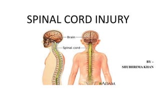
2. • The spinal cord is a collection of nerves that travels from the bottom
of the brain down to back.
• There are 31 pairs of nerves that leave the spinal cord and go to arms,
legs, chest and abdomen.
• These nerves allow brain to give commands to the muscles and cause
movements of the arms and legs.
• The spinal cord is very sensitive to injury. Unlike other parts of body,
the it does not have the ability to repair itself if it is damaged.
INTRODUCTION
3.
4. A spinal cord injury refers to any damage to
the spinal cord either from trauma, loss of its
normal blood supply, or compression from
tumor or infection.
DEFINITION
5. • Trauma: automobile or motor cycle accidents, gunshot or knife
wounds, falls and sports mishaps
• Weakened spine: rheumatoid arthritis or osteoporosis
• Spinal stenosis: spinal canal protecting the spinal cord has become too
narrow due to the normal aging process.
• Vertebrae most commonly involved are the 5th, 6th and 7th cervical
vertebrae, 12th thoracic vertebrae and 1st lumbar vertebrae.
CAUSES AND RISK FACTORS
7. Flexion injuries
• Occurs when the head strikes the steering wheel,
the spine is forced into acute hyper flexion
• Rupture of posterior ligaments results in forward
dislocation of the vertebrae
• Cervical spine usually affected are the C5 to C6
level
Mechanism of injury
8. Hyper extension injuries
• Results after a fall in which the chin hits an object
and the head is thrown back
• Anterior ligament is ruptured with fracture of the
posterior elements of the vertebral body
• Greatest area of stress is at the C4 and C5
CONT..
9.
10. Compression injuries
• Caused by falls or jumps in which the person
lands directly on the head, sacrum or feet
• Force of impact fractures the vertebrae and
the fragments compress the cord
• Lumbar and lower thoracic vertebrae are
usually affected
CONT..
11. Flexion rotation injuries
• Most unstable because ligaments that stabilize
the spine are torn.
• Rupture of the posterior ligament with
displacement of the vertebral body.
• This injury most often contributes to the
several neurologic deficits.
CONT..
12.
13. Skeletal level
• Skeletal level of injury is the vertebral level with the most damage to
the vertebral bones and ligaments.
Neurologic level
• Lowest segment of the spinal cord with normal sensory or motor
function on both the side of the body.
Level of injury
14. • Level of injury may be cervical, thoracic, lumbar or sacral.
• Cervical and lumber injuries are most common because they are
associated with the greatest flexibility and movement.
• Involvement of cervical cord = paralysis of all four extremities
(quadriplegia).
• Involvement of thoracic, lumbar or sacral cord = paralysis of lower
extremities (paraplegia).
• The degree of impairment of arms depends on the level of injury, the
lower the level, more function is retained in the arms.
CONT..
15.
16. • Complete cord injury: Results in total loss of sensory and motor
function below the level of injury.
• Incomplete cord injury: mixed loss of voluntary motor activity and
sensation and leaves some tracts intact.
Central cord syndrome
Anterior cord syndrome
Brown sequard syndrome
Conus medullaris and cauda equina syndrome
Degree of injury
17. Central cord syndrome
• Damage to central spinal cord
• Occurs most commonly in the cervical region
• Motor weakness and sensory loss are present in
both upper and lower extremities
CONT..
18. Anterior cord syndrome
• Results from injury causing compression of
anterior portion of the spinal cord.
• Paralysis and loss of pain and temperature
sensation below the level of injury
• Sensation of touch, position and vibration
remains intact.
CONT..
19. Brown sequard syndrome
• Result of damage to one half of the spinal cord
(knife injury)
• Ipsilateral paralysis with ipsilateral loss of
motor function and pressure and contralateral
loss of pain and temperature.
CONT..
20. Conus medullaris and cauda equina syndrome
• Result from damage to the very lowest portion
of the spinal cord (conus) and the lumbar and
sacral nerve roots (cauda equina).
• Flaccid paralysis of the lower limbs and
areflexia (flaccid bladder and bowel).
CONT..
21. Respiratory system
• Injury below the level of C4 diaphragmatic breathing hypoventilation
• Cervical and thoracic injuries paralysis of abdominal and intercostal
muscles patient cannot cough effectively to remove secretions
atelectasis and pneumonia
• Neurogenic pulmonary edema.
CLINICAL MANIFESTATIONS
22. Cardio vascular system
• Injury above the level of T6 decreases the influence of sympathetic
nervous system bradycardia occurs peripheral vasodilation
reduces return of blood to the heart Decreases cardiac output
hypotension.
CONT..
23. Urinary system
• Urinary retention (loss of sensation and decreased reflexes) in acute
SCI or spinal shock.
• Neurogenic bladder (urgency, frequency, incontinence, inability to void,
increase bladder pressure, reflux urine into kidney).
CONT..
24. Gastrointestinal system
• Injury above the level of T5 decreased gastro intestinal motility
development of paralytic ileus and gastric distension
• Development of stress ulcers
• Intra abdominal bleeding
• Less voluntary control over the bowel neurogenic bowel (bowel is
areflexic and sphincter tone is decreased)
CONT..
25. Problems with thermoregulation
• Poikilothermism is lost in spinal cord injuries
• Decreased ability to sweat or shiver below the level of the lesion
• Patients with high cervical injury have a greater loss of ability to
regulate temperature
CONT..
26. Peripheral vascular problems
• Deep vein thrombosis (during the first 3
months), if remain unrecognized , leading
to pulmonary embolism which is the
leading cause od death.
CONT..
27. Spinal shock
• Temporary loss of neurologic function characterized by decreased reflexes,
loss of sensation and flaccid paralysis below the level of injury.
• Syndrome lasts days to months
Neurogenic shock
• Effects are associated with cervical or high thoracic injury
• Due to loss of vasomotor tone caused by injury and is characterized by
hypotension and bradycardia.
• Peripheral vasodilation decreased cardiac output
CONT..
28. DIAGNOSTIC EVALUATION
History and physical examination
X ray spine
CT scan
MRI scan
Vertebral angiography: an x ray study of blood vessels suppling spine.
29. MANAGEMENT
Initial care
• Neck should be stabilized in a neutral
position without flexion or extension
• Place the affected person on a spine
board and secure the spine with a hard
collar around the neck
30. CONT..
• Log rolling technique
• Maintain a patent airway
• Mechanically assisted ventilation
• patients with severe cervical injury,
placed in skeletal traction
31. CONT..
Drug therapy
• Methyl prednisolone (effective if given within 8 hours of injury)
• Loading dose of 30mg/kg given within 3 hours of injury followed by
24 hours of 5.4mg/kg IV methyl prednisolone drip.
• Vasopressor agents (dopamine)
• Histamine 2 receptor blocking agents
32. CONT..
Managing respiratory dysfunction
• If the injury is at or above C3 endotracheal intubation and
mechanical ventilation.
• Chest physiotherapy, adequate oxygenation and pain management
• Use of incentive spirometry.
33. CONT..
Managing cardiovascular instability
In case of bradycardia, administer anticholinergic (atropine)
Hypotension managed with dopamine infusion
Compression gradient stockings to prevent DVT
If severe blood loss has occurred, blood should be administered
according to protocol
34. CONT..
Fluid and nutritional balance
• First 48 to 72 hours after SCI, GI tract may stop functioning (paralytic
ileus)
• NG tube insertion for gastric decompression
• Introduce oral foods and fluids once the bowel sounds returns
• In patients with high cervical injuries swallowing capacity must be
evaluated
• Increased dietary fiber
35. CONT..
Temperature control
Monitor body temperature
Monitor the environment closely to maintain appropriate temperature
Patient should not be overloaded with covers or unduly exposed
36. CONT..
Managing stress ulcers
Stool and gastric contents are tested daily for blood.
Give corticosteroids along with antacids
H2 receptor blockers or proton pump inhibitors
37. CONT..
Bladder and bowel management
Insertion of indwelling catheter
After patient is stabilized, start intermittent catheterization
Suppository should be inserted daily
Increased fiber intake
38. NURSING DIAGNOSIS
• Ineffective breathing pattern related to weakness or paralysis of
abdominal and intercostal muscles
• Impaired physical mobility related to motor and sensory impairments
• Disturbed sensory perception related to motor and sensory impairment
• Impaired urinary elimination related to inability to void spontaneously
• Constipation related to presence of atonic bowel
• Risk for impaired skin integrity related to immobility
39. COMPLICATIONS
Neurologic deterioration
Pressure sores
Pulmonary complications
- Atelectasis
- Increased work of breathing
- Decrease cough retained secretions Pneumonia
- Muscle fatigue
Loss of circulatory control
Muscle tone problems
- Spastic and flaccid muscles
depression.