2. 18 H. Ulrich and C. Wrenger
cells, and even whole organisms (4–6). Since these RNA and DNA
molecules bind their targets with similar affinities and specificities
as antibodies, and are able to distinguish between isotypes of an
enzyme, aptamers have been also called synthetic antibodies (7).
Recently, the use of in vitro selection methods has been extended
to complex targets, such as receptors that are only functional in
their membrane-bound form (8), and living organisms such as bac-
teria (9), trypanosomes (10), and Plasmodium falciparum-infected
erythrocytes (11). Moreover, since it became apparent that aptam-
ers are capable to distinguish between little differences in cell-sur-
face marker proteins expressed by, i.e., cancer cells, parasite-infected
cells, or stem-cells in comparison to normal somatic cell (5), they
have been developed into promising agents for diagnostic and
therapeutic applications.
Aptamers are ideal candidates for studying protein interactions
in vitro and in vivo and for developing novel molecules of pharma-
ceutical importance for the following reasons: aptamers isolated
from combinatorial library have high dissociation constants, rang-
ing from nanomolar to femtomolar, similar to the best affinity
interactions between monoclonal antibodies and antigens. Most
importantly, the aptamers can be chemically altered depending on
their desired purpose. Chemical modifications that enhance
aptamer stability for in vivo applications and possible therapy
include substitutions of the 2¢OH group of the ribose backbone of
pyrimidines by amino, fluoride, or O-methylene functions. With
such modifications, the half-life of RNA aptamers in biological
fluids such as human serum or urine increases from approximately
8 s for unmodified RNA to 86 h for fluoro- or amino-modified
RNA (12). Modifications at the 5¢ position of pyrimidines expand
the possibilities for chemically decorating oligonucleotide libraries
including iodide or bromide substitutions or the introduction of
SH-groups for chemical and photocross-linking of aptamers with
their targets. Aptamers can be attached to radioactive or fluorescent
molecules and can be used this way for in vitro and in vivo imaging
of target proteins.
Aptamers can be selected against any proteins within a complex
mixture of potential targets and then be used for ligand-mediated
target purification. Larry Gold and coworkers developed aptamers
against erythrocyte membranes that bind specifically to prior
unidentified target proteins (4). The same strategy, also denomi-
nated as deconvolution SELEX, was used to stain rat brain tumor
vessels and to identify the endothelial regulatory protein pigpen as
aptamer-target protein (13). These studies indicate the possibility
of purifying any protein target for which a high-affinity nucleic acid
ligand has been identified. In order to generate covalent cross-links
of selected aptamers with their targets, pyrimidines of the oligonu-
cleotides are modified at the C5 position of the ribose. The aptam-
ers can either be selected from oligonucleotide libraries that contain
3. 192 Aptamers as Specific Receptor Ligands
photo-cross linkable pyrimidines or already selected aptamers can
be modified posterior to the SELEX process. These modified
oligonucleotides attached to a reporter molecule generate reactive
groups that, when irradiated by UV light, can form covalent link-
age with another molecule in close proximity. For ligand-mediated
target purification, aptamers are selected that recognize specifically
an unknown target protein, i.e., in a cell membrane. Binding
specificity is based on the fact that a given aptamer species (i.e.,
radiolabeled) can be eluted by a competitor (molar excess of unla-
beled vs. labeled aptamer) from the cell membrane. In a second
step, the 5¢-biotin-labeled aptamer is immobilized on a solid sur-
face, such as streptavidin-coated magnetic beads, and then used as
an affinity matrix to purify and identify the bound target protein.
The binding proteins retained on aptamer-coated magnetic beads
are separated from the remaining protein mixture by applying a
magnetic field. Alternatively, aptamers can be covalently photo-
cross-linked to their targets and used as a tracer in order to separate
the target protein from remaining proteins in a cell extract.
Aptamers are capable to identify individual epitopes on cell
surfaces as it has been demonstrated for human membranes. Since
aptamers differentiate in molecular signature of cell-surface anti-
gens, this technology has been recognized as a powerful tool for
biomarker discovery and targeting cancer cells. Aptamer probes
were developed recognizing leukemia, prostate, or glioblastoma
cancer cells (14–16). Molecular recognition is a key mechanism for
the diagnosis of cancer which has been exploited by applying mul-
tipleanticancercellaptamersconjugatedonnanorods.Up-regulated
ErbB2 expression in breast cancer cells was targeted by fluorescence-
tagged RNA aptamers in surface plasmon resonance measurements
(17). Aptamer-based therapeutic approaches are based on the
specific delivery of si-RNAs or toxic loads to tumor cells (18, 19)
or by inhibition of cancer cell-surface receptors, such as the EGF
receptors involved in protection of tumor cells against apoptosis
(20, 21).
Moreover, aptamers have been developed as specific ligands
and/or inhibitors of neurotransmitter receptors, including GABA
receptors, AMPA-glutamate receptors, as well as neuronal and nic-
otinic acetylcholine receptors (nAChRs) (22–25, 8). In addition to
acting as inhibitors, such as anesthetics, anticonvulsives, or the
abused drug cocaine, on nAChRs, aptamers can be developed as
specific ligands of defined conformational sites of neurotransmitter
receptors in order to reestablish dysfunctions in channel-open and
-closing equilibrium. Such approaches aim at reestablishment of
decreased GABA A-receptor activity in epilepsy involving mutation
of this receptor (22), or at restoring of normal nicotinic receptor
activity following inhibition by abused drugs such as cocaine and
phencyclidine (23).
4. 20 H. Ulrich and C. Wrenger
A simple protocol detailed in this chapter was established to
develop RNA aptamers that bind to the electric organ nAChR
and that are displaced by cocaine (8) (see Fig. 1 for a scheme).
This protocol can be easily transferred to SELEX applications
with other receptors or cell-surface epitopes, given that these are
enriched in membrane preparations.
The aptamers obtained against the nAChR either are bio-
logically active by themselves and inhibit the muscle-type
nAChR activity as cocaine does or are by them biologically inac-
tive and, therefore, protect the receptor against inhibition by
cocaine (25).
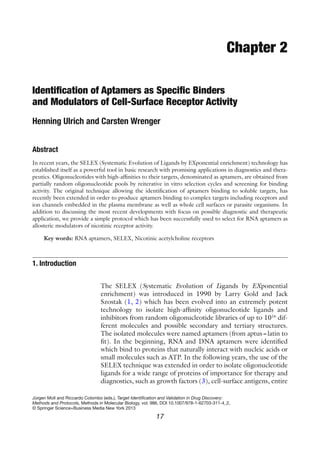
Obtaining RNA aptamers by using the SELEX technology is a
conceptually straightforward process. In the beginning, an oligo-
nucleotide is synthesized by a DNA synthesizer that consists of a
random sequence of typically 16–75 oligonucleotides flanked by
two constant regions. A T7-promoter site is incorporated in one of
the constant sequences by taking advantage of the introduced
EcoRI and HindIII sites, which allows removal of the constant
sites and to insert the random regions into the multi-cloning site of
a bacterial vector. Random sequences are created by premixing all
four nucleotides during DNA synthesis. The double-stranded
DNA template is enzymatically generated and amplified by primer
extension and error-prone PCR. The random nucleotide sequence
of the double-stranded DNA template is verified by cloning and
sequencing of around 40 individual RNA molecules. In the ran-
dom sequence of these RNA molecules, sequence motifs (i.e., AA,
AC, AG, AT) should be equally distributed.
For the in vitro selection process, the RNA pool containing
1013
different sequences and structural motifs is generated by
an in vitro transcription reaction. Folding of the RNA mole-
cules is induced by heat denaturation and renaturation at room
temperature (26).
The RNA molecules are made nuclease resistant either by
incorporating modified bases, i.e., 2¢-fluoro- or by employing 2¢-
amino-modified pyrimidines during the in vitro transcription step
(12), or by protection during the selection step using a dithiothre-
itol (DTT)-independent RNAse inhibitor (27). The RNA pool is
exposed to its target in a 10–1,000 ratio of RNA:target-binding
sites, and ligands are separated from nonfunctional molecules by
filtration or by migration on polyacrylamide gel where target-
bound RNA molecules migrate more slowly than unbound RNA
which allows gel purification as indicated in Fig. 2 (28).
The recovered RNA is reverse transcribed to cDNA and
amplified. The next-generation RNA pool, generated again by
in vitro transcription, is already enriched in RNA molecules that
bind to their target. The procedure is repeated with rising strin-
gency until the random RNA pool is purified to a fraction of RNA
5. 212 Aptamers as Specific Receptor Ligands
molecules with the desired binding properties and no further
improvement of binding can be achieved. The final RNA pool is
reverse transcribed into cDNA, amplified by PCR, and cloned into
a bacterial vector. Clones containing the aptamer insert are picked
and about 40 individual aptamers are identified by sequencing.
The previously random regions are aligned and analyzed for con-
sensus motifs, and the structures of RNA aptamers containing con-
sensus motifs are predicted on the basis of free energy minimization
(29). Aptamers containing consensus motifs are characterized
regarding their binding affinity for their target and are screened for
their biological activity by patch-clamping (30), using the whole-cell
current-recording technique (31) in combination with rapid ligand
delivery within 10 ms (called the cell-flow technique) (32, 33)
(see Fig. 2.1 for a general scheme of RNA aptamer selection).
The protocol described here for the nAChR can be used with
modification for the selection of aptamers that bind to other pro-
teins expressed on a cell surface. In addition to the electrophysio-
logical assay described here, screening for biological activity can
include the quantification of ion flux into cells using fluorescent
reporters (34, 35). For imaging purposes, fluorescence-tagged
nucleotides can be enzymatically incorporated into the aptamer
sequence (i.e., biotin-CTP or fluorescein-CTP). Alternatively, the
aptamer can be modified at its 5¢-end in order to attach a biotin
moiety by substituting 5¢-GTP for 5¢-GDP-b-S and followed by
covalent coupling of biotin molecule to this group and addition of
a streptavidin-fluorophore (12, 36).
Fig. 1. Scheme for RNA aptamer identification targeting cocaine-binding sites on the
nicotinic acetylcholine receptor (nAchR).
6. 22 H. Ulrich and C. Wrenger
1. Motor-driven blade homogenizer, low-speed refrigerated centri-
fuge, ultracentrifuge, spectrophotometer, and gamma counter.
2. Frozen electric organ from Torpedo californica (Aquatic Research
Organisms, Inc., Hampton, NH): Maintain at −80°C.
3. Buffer H: 10 mM sodium phosphate, 400 mM sodium chlo-
ride, 5 mM Na2
EDTA, 10 mM sodium azide, at pH 7.4. Per
liter: 2.68 g Na2
HPO4
×7H2
O, 112.88 g NaCl, 1.86 g
Na2
EDTA, 0.65 g NaN3
. Adjust to pH 7.4 with 1 M HCl.
4. Buffer A (buffer H without NaCl).
5. Phenylmethylsulfonylfluoride (PMSF) 0.1 M stock solution:
0.174 g PMSF in 10 ml isopropanol.
6. 60% (w/w) sucrose solution. Per 100 ml: 77.2 g D-glucose in
buffer A.
7. 36% (w/w) sucrose solution. Per 100 ml: 60 ml 60% sucrose
solution diluted to 100 ml with buffer A.
8. Bovine serum albumin and Lowry protein determination
reagents.
9. Buffer B: 10 mM sodium phosphate, 50 mM NaCl, 0.1%
(w/v) Triton X-100 at pH 7.5. Per liter: 2.68 g
Na2
HPO4
×7H2
O, 14.11 g NaCl, 1.0 ml Triton X-100. Adjust
to pH 7.5 with 1 M HCl.
10. Unlabeled a-bungarotoxin (BGT) solution: Prepare a
10 mM BGT stock solution from lyophilized BGT (Sigma,
St. Louis, MO).
11. 125
I-BGT solution: Prepare a 200 nM 125
I-BGT stock solution
from lyophilized 125
I-BGT (Perkin Elmer/New England
Nuclear, Boston, MA).
12. DE-81 filters (Whatman, Kent, UK).
1. Equipment: PCR machine, scintillation counter, tabletop cen-
trifuge, temperature-controlled water baths, equipment for
horizontal and vertical electrophoresis, UV-illuminator, phos-
phor imager, automatic DNA sequencer, vacuum dot-blot
manifold (Schleicher and Schuell). PCR 0.5 ml hot-start tubes,
aerosol resistant pipette tips, autoclaved Eppendorf tubes (all
from Fischer Scientific, Brightwaters, NY) and glassware,
diethyl pyrocarbonate (DEPC, Sigma)-treated solutions.
2. Sterile disposable Petri dishes and sterile inoculating loops for
bacterial culture.
3. BA-85 nitrocellulose and gel-blotting paper (GB002, both
102×133 mm, Schleicher and Schuell, Keene, NH).
2. Materials
2.1. Target Preparation
2.2. SELEX Procedure
7. 232 Aptamers as Specific Receptor Ligands
4. GF/F glass fiber filters, 1.3 cm diameter (Whatman, Kent, UK).
5. Sterile syringe filters (0.45 mm; 25 mm diameter) (Whatman).
6. Sigmacote (Sigma).
7. Kodak X-Omat K Diagnostic Film for autoradiography or
imaging plate for phospho imager.
8. Incubation buffer: 25 mM HEPES, 145 mM sodium chloride,
5.3 mM potassium chloride, 1.8 mM calcium chloride dihy-
drate, 1.7 mM magnesium chloride hexahydrate, pH 7.4. Per
1 l: 59.5 g HEPES, 8.47 g NaCl, 3.95 g KCl, 2.65 g
CaCl2
×2H2
O, 3.46 g MgCl2
×6H2
O. Adjust to pH 7.4 with
1 N NaCl.
9. pGEM-3Z vector (cloning vector that allows highly efficient
synthesis of RNA in vitro) (Promega, Madison, WI) and for
transformation of E. coli competent cells (strains JM 29 or
DH5a) (Promega).
10. pCR4-TOPO vector with One-shot cells (for fast cloning)
(Invitrogen, Carlsbad, CA).
11. LB medium and agar: Per liter LB medium dissolve in distilled
water (dd H2
O) and autoclave: 10 g tryptone, 5 g yeast extract,
5 g NaCl, 1 ml 1 N NaOH. Per liter LB agar dissolve in dd
H2
O and autoclave: 10 g tryptone, 5 g yeast extract, 5 g NaCl,
1 ml 1 N NaOH, 15 g agar.
12. Partial random DNA template (108 bp) (synthesized by
Biosource International, Foster City, CA).
5¢-ACC-GAG-TCC-AGA-AGC-TTG-TAG-TAC-TNN-
NNN-NNN-NNN-NNN-NNN-NNN-NNN-NNN-NNN-
NNN-NNN-NNN-NNG-CCT-AGA-TGG-CAG-TTG-AAT-
TCT-CCC-TAT-AGT-GAG-TCG-TAT-TAC-3¢ (N=A, C, G,
or T are incorporated with equal probabilities). Restriction
sites (HindIII and EcoRI) are underlined.
13. Primer region 1 (P-40, 40 bp, forward primer with T7 pro-
moter site)
5¢-GTA-ATA-CGA-CTC-ACT-ATA-GGG-AGA-ATT-CAA-
CTG-CCA-TCT-A-3¢.
14. Primer region 2 (P-22, 22 bp, reverse primer).
5¢-ACC-GAG-TCC-AGA-AGC-TTG-TAG-T-3¢
Reverse primer for aptamer amplification in pGEM-3Z
vector (P-22 pGEM, 22 bp)
5¢-GAA-TAC-TCA-AGC-TTG-TAG-TAC-T-3¢ (forward
primer is the same as above).
15. Kitsandenzymes:Superscriptreversetranscriptase(Invitrogen),
Maxiscript and Megascript in vitro transcription kits (Ambion,
Austin, TX). Taq-DNA polymerase, T4-polynucleotide kinase,
EcoRI and HindIII restriction enzymes (Invitrogen).
8. 24 H. Ulrich and C. Wrenger
16. Wizard Miniprep kit (Promega).
17. Big Dye sequencing kit (Applied Biosystems, Foster City, CA).
18. DTT-dependentand-independentRNAseinhibitors(Ambion),
NTPs, dNTPs, MgCl2
, MnCl2
, yeast t-RNA, DNA, and RNA
molecular weight standards.
19. Acrylamide/bisacrylamide, low-melting agarose (Sea Plaque
GTG, Biocompare, Inc, South San Francisco, CA).
20. Ethanol, saturated phenol (pH 7 and 5.2), chloroform, iso-
amylalcohol (molecular biology grade, Sigma).
21. Tris–borate–EDTA (TBE) buffer (10× solution): 0.89 M
TRIZMA base, 0.89 M H3
BO3
, 20 mM EDTA. Per liter: 108 g
TRIZMA base, 55 g boric acid, 40 ml 0.5 M ethylenedi-
aminetetraacetate (EDTA) solution.
22. Tris–EDTA (TE) buffer: 10 mM TRIZMA hydrochloride,
1 mM EDTA.
10 mM Tris–HCl, 1 mM EDTA, pH 7.5. Per liter: 1.57 g
Tris–HCl, 0.29 g EDTA. Adjust to pH 7.5.
23. Formamide-loading buffer for RNA gel: Per 10 ml add 0.2 ml
0.5 M EDTA, pH 8.0, 10 mg bromophenol blue, 10 mg
xylene cyanol, 10 ml formamide (deionized).
24. Loading buffer for DNA gel: Per 10 ml: 1 ml 10× TBE, 10 mg
bromophenol blue, 10 mg xylene cyanol, 2 g glycerol.
25. Urea, ammonium acetate, sodium acetate.
26. Spin columns S-30 (cut-off 30 nucleotides) (Sigma).
27. Cocaine hydrochloride (1 mg/ml in methanol) (Sigma):
Evaporate the methanol and resuspend in incubation buffer at
a concentration of 10 mM.
28. MK-801 (RBI Biochemicals, Natick, MA): Dissolve at a con-
centration of 10 mM in incubation buffer.
29. (a-32
P)-UTP (3,000 Ci/mmol), (g-32
P)-ATP (5,000 Ci/
mmol), both from Amersham Biosciences, Uppsala, Sweden.
1. Equipment: CO2
-cell culture incubator, laminar flow cabinet,
vibration–isolation table (TMC, Peabody, MA), Faraday cage
(TMC), Axopatch 200B integrated patch-clamp amplifier with
Digidata 1322A interface and pClamp program (Axon
Instruments, Union City, CA), inverted microscope, one micro
manipulator and two coarse manipulators (Narishige
International, East Meadow, NY), computer-operated 12 V
solenoid valves (Lee Company, Westbrook, CT), two peristal-
tic pumps for ligand and inhibitor delivery (Rainin, Oakland,
CA), Kendall tubing diameter: 0.05, 0.16, and 0.42 cm (VWR
International, Westchester, PA), U-shaped stainless steel capil-
lary tubes (250 mM inner diameter) with a circular porthole of
2.3. Cell Culture and
Electrophysiology
9. 252 Aptamers as Specific Receptor Ligands
150 mm at the base of the U (37) for ligand and inhibitor
application made from Hamilton HPLC-tubing (Hamilton
Corp., NV), patch-pipette puller (Sutter, Novato, CA), pipette
polisher (Microforge, Narishige International, USA).
2. BC3
H1 cells (American Type Culture Collection, Manassas,
VA).
3. DMEM high glucose, trypsin–EDTA solution, fetal bovine
serum (FBS), heat inactivated (all from Invitrogen).
4. 25 cm2
cell culture flasks (Costar, Corning, Acton, MA).
5. 15 ml screw capped vials (Greiner, Fischer Scientific).
6. 35 mm cell culture dishes (Corning, Acton, MA).
7. Incubation buffer (extracellular buffer): 25 mM HEPES,
145 mM NaCl, 5.3 mM KCl, 1.8 mM CaCl2
×2H2
O, 1.7 mM
MgCl2
×6H2
O, pH 7.4.
8. Pipette solution (intracellular buffer): 25 mM HEPES,
140 mM potassium chloride, 10 mM sodium chloride, 2 mM
magnesium chloride hexahydrate, 1 mM EGTA, pH 7.4. Per
liter: 59.5 g HEPES, 10.44 g KCl, 0.58 g NaCl, 0.41 g
MgCl2
×6H2
O, 0.38 g EGTA. Adjust to pH 7.4 with 1 M
KCl.
9. Carbamoylcholine (Sigma).
10. Cocaine hydrochloride (Sigma).
11. Borosilicate glass capillaries for patch pipettes (World Precision
Instruments, Sarasota, FL).
1. Weigh 60–80 g of frozen electric organ and return it to the
freezer. Measure a volume of buffer H equal in ml to the tissue
weight in grams and add PMSF to a final concentration of
1 mM. Put the frozen pieces of organ in a sturdy cloth sack and
pulverize with a hammer. Transfer the still frozen powder into
a graduated cylinder, add the buffer containing PMSF, and
leave the tissue/buffer mixture for about 10 min at room tem-
perature to thaw. Then homogenize the tissue for 30 s four
times using a motor-driven blade homogenizer at maximal
speed, resting 10 s between each run. All subsequent steps
should be performed on ice.
2. Transfer the homogenate into precooled centrifuge bottles and
centrifuge for 10 min at 2,400×g, 4°C. Collect the superna-
tant and discard the pellet. Transfer the supernatant to a pre-
cooled ultracentrifuge bottle and ultracentrifuge for 90 min at
34,000×g, 4°C. Discard the supernatant and resuspend the
3. Methods
3.1. Preparation and
Evaluation of the
nAChR-Enriched
Plasma Membranes
as a Target for the
In Vitro Selection
Process
10. 26 H. Ulrich and C. Wrenger
pellet in 60 ml of buffer A containing 1 mM PMSF by
homogenizing the pellet three times for 10 s each using a
motor-driven blade homogenizer at low speed. Wash the pellet
again as above in buffer A and resuspend the pellet in 25 ml
of buffer A containing 1 mM PMSF.
3. This step employs a discontinuous gradient ultracentrifuga-
tion method (38). Add 44 ml of 60% sucrose solution to the
resuspended pellet, mix well, transfer to a 250 ml graduated
cylinder, and add buffer A until the total volume is 108 ml to
obtain a final sucrose concentration of 28%. Add 10 ml of a
36% sucrose solution to six swinging bucket rotor 30 ml ultra-
centrifuge tubes. Gently overlay the layers in each centrifuge
tube with 18 ml of the resuspended pellet. Top off each tube
with buffer A. Ultracentrifuge on a swinging bucket rotor for
4 h at 100,000×g, 4°C. Aspirate and discard the floating pad
at the buffer A/28% sucrose interface, together with most of
the 28% layer. Collect the visible membrane layer containing
nAChRs at the 28%/36% interface. Dilute the recovered
28%/36% interface with an equal volume of buffer A and col-
lect the pellet by ultracentrifugation for 30 min at 38,000×g,
4°C. Resuspend the pellet in 10 ml buffer A using a syringe
equipped with a #18 needle. Aliquot and store the membranes
frozen at −80°C.
4. The concentration of the membrane proteins is determined
according to the method of Lowry et al. (39) using bovine
serum albumin as the reference standard.
(125
I) BGT binding to the receptor is assayed using a filtration
method (40).
1. Dilute 50 ml of the membrane suspension 1:100 in buffer B
and then measure 50 ml aliquots of the diluted membranes into
six Eppendorf vials.
2. To each of the three vials add 50 ml buffer B and to each of the
other three vials add 50 ml unlabeled BGT solution.
3. Vortex and preincubate on a rotator at room temperature for
60 min. Then add 100 ml of 125
I-BGT to each vial.
4. Vortex and incubate on a rotator at room temperature for
60 min.
5. Load vacuum manifold ports with DE-81 filters (Whatman).
6. Prewash filters with 0.6 ml buffer B using vacuum.
7. Transfer 50 ml of each reaction mixture directly onto the filters
(do this in triplicate) and wash each filter with 1 ml buffer B.
8. Allow buffer to filter through and air-dry the filters for 1 min
using vacuum suction.
3.2. Determination of
nAChR-Binding Sites
in Torpedo Electric
Organ Membrane
Preparations
11. 272 Aptamers as Specific Receptor Ligands
9. Transfer filters into counting vials and determine filter-bound
radioactivity in a gamma counter. Subtract nonspecific binding
(binding with unlabeled BGT present) to determine the concen-
tration of specifically bound 125
I-BGT. The concentration of
nAChR in the membrane suspension is one-half this value (×400)
since there are two BGT-binding sites per nAChR molecule.
1. Check purity by resuspending 35 nmol of the pre-purified sin-
gle-stranded oligonucleotide in 100 ml TE buffer. End label
1 ml of the oligonucleotides with (g)32
P ATP using T4-kinase
and check for purity on 8% denaturing urea (6 M) PAGE (41).
If there is not a single clean band, but shorter oligonucleotides
are also present, the random oligonucleotides need to be
purified.
2. Prepare a purification gel by adding an equal amount of forma-
mide loading buffer to the resuspended DNA pool and a small
amount of 32
P-labeled DNA from step 1.
3. Denature the mixture at 65°C for 10 min and load into nine
lanes (each lane contains 500,000 cpm).
4. Expose gel using a phosphor imaging plate (15 min), and
match the obtained image with the gel to localize the bands
with the right size.
5. Cut out the bands and reimage the gel.
6. Crush the gel slices with a pipette tip and elute the DNA twice
with 400 ml sodium acetate (0.3 M, pH 5.2) and 1 mM EDTA
(for 12 h each).
7. Ethanol-precipitate the eluted purified DNA (see Note 1).
Use hot-start tubes and assemble the bottom and top part of the
reaction for second-strand synthesis and amplification of the DNA
template by error-prone PCR. Hot-start PCR is the PCR tech-
nique of assembling the reaction mixture at a temperature that is
greater than the annealing temperature. This procedure increases
precision, yield, and specificity. The pre-adhered wax bead assures
synchronous reaction start-up and eliminates the need for using
mineral oil.
1. Assemble the reaction as reported in Table 1. This mix is
enough for 30 PCR reactions of 120 ml volume each.
2. Second-strand synthesis: Execute the following program on a
PCR machine:
94°C 3 min, 42°C 2 min, and 72°C for 8 min. Then add
1.5 ml primer P-22 (100 pmol/ml) to each 120 ml PCR
and start the following program: 94°C 2 min, 42°C 1 min, and
72°C 1 min for one cycle and 94°C 2 min, 42°C 1 min,
and 72°C 1 min+0.5 min increase each round for 11 cycles.
3.3. Preparation of the
Random DNA Pool for
In Vitro Transcription
3.4. Second-Strand
Synthesis and
Amplification by
Error-Prone PCR
12. 28 H. Ulrich and C. Wrenger
3. Analyze on native PAGE (8% in 1× TBE buffer).
4. Repeat the protocol that is described above three times (so
that the total reaction volume is 10.8 ml).
5. Purify if necessary.
6. Ethanol precipitate and dissolve in 100 ml dd H2
O.
PCR amplification of the single-stranded (ss) DNA pool will result
in multiple copies of a double-stranded DNA pool. Chemical
lesions occurring during the chemical synthesis of the ss DNA
pool, and the possibility that some sequences are more amplified
than others during PCR procedures, may result in a limited pool
size and predominance of sequence motifs in the random sequence
(see below). Therefore it is necessary to sequence and analyze a
small number of individual sequences of the random pool (2).
1. Clone 1 ml of the purified PCR product into a pCR4-TOPO
vectorandtransformto“OneShot”competentcells(Invitrogen)
according to the manufacturer’s recommendation.
2. Isolate and sequence 40 clones.
3. Use a word processing program to analyze random sequences
for equal occurrence of AA, AC, AG, and AT motifs in the ran-
dom regions (repeat for C, G, and T). If one or more of these
motifs predominate or the base composition is not random in
the random sequences, the pool may not contain enough dif-
ferent sequences for a successful SELEX experiment.
3.5. Evaluation
of the Degree
of Randomness
of the Pool
Table 1
Second-strand synthesis and amplification by error-prone
PCR mix
Addition Bottom (ml) Top (ml)
H2
O 981 1,540
10× PCR buffer 180 180
dATP (100 mM) 12
dGTP (100 mM) 12
dCTP (100 mM) 30
dTTP (100 mM) 30
MgCl2
(50 mM) 504
P-40 (100 pM) 45
ss DNA pool (60 pM) 6
MnCl2
(1 M) 1.8
Taq DNA polymerase 30
13. 292 Aptamers as Specific Receptor Ligands
1. To perform in vitro transcription reaction: Save 20 ml of the
total volume of 100 ml (for 32
P-UTP transcription for binding
studies with nAChRs in Torpedo electric organ membranes,
and in case you have to amplify the DNA again) and use 80 ml
for transcription.
2. Assemble the following reaction: DNA solution (80 ml); H2
O
(DEPC-treated) (74 ml); 10× synthesis buffer (25 ml); ATP,
CTP, GTP, UDP 10 mM solutions (10 ml) each; DTT (100 mM)
(6 ml); and T7 RNA polymerase (50 U/ml) (25 ml).
3. Incubate the reaction at 37°C overnight.
4. Add 25 ml DNAse (1 U/ml) and incubate for 20 min.
5. Analyze 10 ml of the reaction product on a denaturing PAGE
(8%, 6 M urea) to verify the right size of the RNA (90 nt) and
to determine if smaller products are present.
6. If smaller reaction products are present, purify the full-length
RNA from the gel to avoid the selection and amplification of
shorter sequences. Alternatively, for generation of nuclease-
resistant transcripts substitute CTP and GTP by 2¢-fluoro- or
2¢-amino-pyrimidines (TriLink BioTechnologies, San Diego,
CA) at final concentrations of 1.2 mM.
7. For radiolabeling of the RNA pool, add 50 mCi (a-32
P)-ATP
(10 mCi/ml).
8. To purify RNA, extract the reaction product with 275 ml of a
saturated solution of phenol in 0.1 M NaOAc, pH 5.2.
9. The supernatant is then extracted with an equal volume of
chloroform in order to remove remaining phenol and then
ethanol-precipitated.
The first two rounds are performed under low stringency condi-
tions to enhance RNA–protein binding and to avoid early deple-
tion of sequences present in the SELEX RNA pool. For SELEX
cycles 1–3 a nitrocellulose-filter binding assay is used to separate
receptor-bound from free aptamers. Beginning from SELEX cycle
4, the nitrocellulose-filter binding and a gel-shift selection step are
employed as two consecutive selection processes (see Note 2).
1. The respective RNA pool is heated up for 10 min to 85°C
prior to selection and allowed to cool down to room tempera-
ture in order to allow for proper formation of secondary and
tertiary structures.
2. To perform SELEX experiment using nitrocellulose-filter bind-
ing, prepare filtration unit by preincubating a nitrocellulose
sheet in incubation buffer.
3. Mount nitrocellulose sheet in autoclaved filtration unit.
Assemble the following reaction mixtures for the SELEX
process: nAChR-enriched plasma membranes (800 mg/ml
3.6. In Vitro
Transcription and
Purification of the
SELEX RNA Pool
3.7. Selection of
Aptamers that Bind
to the nAChR and
Displace Cocaine
14. 30 H. Ulrich and C. Wrenger
protein; 1.6 mM receptor) (40 ml); incubation buffer (280 ml);
anti-RNAse (40 U/ml) (40 ml); RNA pool (50 mM) (40 ml).
4. Incubate for 40 min at room temperature.
5. Place 4×100 ml of the selection mixture into each of the four
wells of the filtration unit.
6. Wash each well twice with 200 ml incubation buffer.
7. Cut out the pieces of the nitrocellulose filters that cover the
used wells of the filtration unit.
8. For the elution of receptor-bound, cocaine-displaceable RNA
aptamers, incubate each of these filter pieces in 100 ml of 1 mM
cocaine (or 100 ml of 1 mM MK-801 that binds to the same
site as cocaine on the nAChR) in incubation buffer for 20 min
at room temperature.
9. For the recovery and purification of the eluted RNA, add 5 mg
t-RNA as a carrier, and phenol- and chloroform-extract the
eluate, precipitate with ethanol, and resuspend the pellet in
12 ml DEPC-treated H2
O.
10. Then perform reverse transcription (RT-PCR) assembling four
separate reactions as follows: 2.5 ml of the recovered RNA, 2 ml
P-22 (100 mM), and 6.5 ml DEPC-treated H2
O.
11. Incubate at 70°C for 10 min, and then place on ice for 1 min.
12. Add to the following RNA/P-22 solution: 14 ml RNA/P-22
solution, 2 ml 10× synthesis buffer, 2 ml dNTP mix (10 mM),
2 ml DTT (0.1 M), 1 ml MgCl2
(50 mM), and 1 ml superscript
RT (200 U/ml).
13. Incubate at room temperature for 10 min, then at 46°C for
50 min, and then at 70°C for 10 min.
14. Place the vial on ice for 5 min, add 1 ml RNAse H, and incu-
bate at 37°C for 15–20 min (volume is now 21 ml).
15. Transfer each reaction into a hot-start tube and prepare a PCR
mix as reported in Table 2 and perform the following program:
cycle 1 (94°C, 5 min; 60°C, 5 min; 72°C, 1 min), 5–10 cycles
(94°C, 1 min; 60°C, 1 min; 72°C, 1 min), and final cycle
(94°C, 1 min; 60°C, 1 min; 72°C, 10 min).
16. Analyze 10 ml aliquot of the PCR product on a native PAGE
(see Note 3).
17. Gel-purify PCR products if necessary.
18. Phenol- and chloroform-extract, ethanol-precipitate, and
resuspend in 100 ml DEPC-treated H2
O.
19. In vitro transcribe and purify RNA as already described.
20. To perform gel-shift selection experiments, prepare a native
gel, containing 3% acrylamide (stock: 38.5% acrylamide and
1.5% bis-acrylamide) and 1× TBE, adjusted to pH 7.4.
15. 312 Aptamers as Specific Receptor Ligands
21. The RNA pool unlabeled or radiolabeled - as detailed later - is
diluted in incubation buffer prior to the SELEX step, dena-
tured and renatured as detailed in Subheading 3.7.
22. Assemble the following reaction: 20 ml nAChR-enriched
plasma membranes (800 mg/ml protein; 1.6 mM receptor),
9 ml incubation buffer, 1 ml anti-RNAse (40 U/ml), and 10 ml
RNA (320 mM).
23. The reaction is carried out for 40 min at room temperature in
incubation buffer.
24. Add one part of 4× loading buffer (20% glycerol, 0.2% bro-
mophenol blue in 1× TBE) to three parts of reaction mixture.
25. Load the samples on the gel and electrophorese the gel at
10 V/cm for 3 h in 1× TBE, adjusted to pH 7.4.
26. Stain the gel with 0.5 mg/ml ethidium bromide.
27. Excise the band containing the RNA–protein complex.
28. Elute the bound RNA with 0.5 M NaOAc pH 5.2 containing
1 U/ml anti-RNAse.
29. Phenol- and chloroform-extract and ethanol precipitate the
recovered RNA (see Subheading 3.7). The RNA is reverse
transcribed, purified, and precipitated as detailed in
Subheadings 3.6 (25) and 3.7, and then RNA is used for the
nitrocellulose filter selection step and cocaine displacement of
nAChR-bound RNA molecules (Fig. 2).
Radiolabeled RNA can be generated either by incorporation of a
(a32
P) nucleoside triphosphate during an in vitro transcription
reaction or by the transfer of (g32
P)-ATP to the 5¢ terminus of a
dephosphorylated RNA molecule (41). The authors prefer the first
mentioned method, as it needs only a single enzymatic reaction.
3.8. Synthesis and
Purification of
32
P-Labeled RNA
Table 2
PCR mix
Addition Bottom (ml) Top (ml)
RT-reaction product 21
dd-H2
O 21 44
10× buffer 3 5
P-40 primer (50 mM) 2
MgCl2
(50 mM) 3
Taq polymerase (5 U/ml) 1
16. 32 H. Ulrich and C. Wrenger
1. Gel-purify DNA pools of various SELEX cycles.
2. Ethanol-precipitate the DNAs and resuspend at a concentra-
tion of 200 ng/ml for in vitro transcription reactions.
3. Make labelling cocktail: (Volume needs to be multiplied by the
number of reactions to be set up). Add 6 ml DEPC-treated
H2
O, 2 ml 10× transcription buffer, 1 ml ATP (0.5 mM), 1 ml
CTP (10 mM), 1 ml GTP (10 mM), and 1 ml UTP (10 mM).
4. Add to the 12 ml mix: 2 ml DNA (SELEX pools), 5 ml (a-32
P)-
ATP (10 mCi/ml), and 1 ml T7 RNA polymerase (50 U/ml) for
a total volume of 20 ml.
5. Incubate at 37°C for at least 2 h.
6. Add 2 ml DNAse I (1 U/ml) and incubate for 15 min at
37°C.
7. Add 3 ml 0.5-M EDTA and purify the 32
P-RNAs from unincor-
porated 32
P-nucleotides using a spin column 30 (Sigma).
8. Run an 8% denaturing gel (6 M urea) to check the size and the
purity of the (32
P)-RNA (see Note 4). (For nuclease-resistant
(32
P)-RNA aptamers set up transcription reactions in the pres-
ence of 1.5 mM 2¢-fluoro- or 2¢-amino-modified pyrimidines
instead of CTP and UTP).
9. Gel-purify the (32
P)-RNA if necessary.
10. For gel purification of 32
P-labeled RNA, pour denaturing gel
(8% acrylamide, 6 M urea) (see Note 3).
Fig. 2.Alternation of gel-shift and filter-binding selection steps:Target-bound and unbound
radiolabeled RNA aptamers are separated by polyacrylamide gel electrophoresis, visualized
by autoradiography, purified from the gel, and used for the subsequent nitrocellulose-filter
binding selection step.The experiments are carried out in the presence (+) and absence (−)
of target protein using the SELEX cycles 0 (control), 3, and 7. The figure illustrates the
increase of binding affinity of selected RNA pools, seen as augmented quantity of RNA
retained together with the receptor protein at the top of the gel (modified from ref. (8)).
17. 332 Aptamers as Specific Receptor Ligands
11. Load left and right outer lanes with loading dye, and pre-run
the gel at 250 V until the dye has migrated down approxi-
mately 75% of the gel.
12. Mix (32
P)-RNA reaction mixture with 15 ml formamide load-
ing buffer, and incubate at 85°C for 15 min.
13. Load samples and run the gel at 250 V until the rapidly migrat-
ing dye (bromophenol blue) has almost moved out of the gel.
14. When finished, visualize the location of the (32
P)-RNA on the
gel by autoradiography or phospho imaging, and cut out the
gel slices containing the full-length (32
P) RNA using a fresh,
sterile razor blade for each sample.
15. Transfer the gel slabs into a 2 ml Eppendorf tube, crush with a
pipette tip, and extract overnight with 1 ml 0.3 M NaOAc, pH
5.2, 10 ml DTT, and 5 ml RNAsin.
16. Pass the suspension through a syringe filter (0.45 mm sterile
filter) to remove the gel, add an equal volume of a saturated
solution of phenol in 0.3 M NaOAc pH 5.2, and vortex hard.
17. Centrifuge for 3 min at 12,000×g in a tabletop centrifuge.
18. Collect the supernatant and concentrate it by ethanol precipi-
tation (see Note 1).
1. Soak GF/F filters (diameter 1.3 cm, Whatman) and a sheet of
filter paper (GB002, 102×133 mm, Schleicher and Schuell)
for 1 h in incubation buffer containing 1% Sigmacote to reduce
unspecific binding of the RNA molecules to the filters.
2. The (32
P) RNAs are diluted in incubation buffer containing
10 mg/ml to a specific activity of 5×10⁵
cpm/ml and dena-
tured and renatured before the experiment.
3. The specific binding of the selected aptamers to their target is
determined as the difference between the total binding and the
unspecific binding in the presence of an excess of unlabeled
competitor. Assemble the following reactions for duplicate
estimations in a total volume of 100 ml (for determination of
total and unspecific binding each). The specific binding is the
difference between total and unspecific binding. Binding of
the selected RNA molecules to the nAChR reaction: 89 ml
incubation buffer, 10 ml nAChR-enriched plasma membranes
(800 mg/ml protein; 1.6 mM receptor), and 1 ml 32
P-RNA
dilution containing 10 mg/ml t-RNA anti-RNAse (40 U/ml).
Determination of unspecific binding reaction by competition of
the RNA aptamers with cocaine: 79 ml incubation buffer, 10 ml
nAChR-enriched plasma membranes (800 mg/ml protein;
1.6 mM receptor), 1 ml 32
P-RNA dilution containing 10 mg/ml
t-RNA anti-RNAse (40 U/ml), and 10 ml cocaine (10 mM).
The percentage of binding of the (32
P) RNA aptamers to the
3.9. Determination
of Specific Binding
to the nAChR
18. 34 H. Ulrich and C. Wrenger
filters (background binding) is determined in the absence of
receptor protein.
4. Incubate reactions for 40 min at room temperature.
5. In the meantime, mount the glass fiber filters on top of the
filter sheet in the dot-blot filtration unit (Schleicher and
Schuell). For duplicate determinations, two samples each of
45 ml solution are spotted on separate filters.
6. Gentle suction is applied to the filtration unit. The filters are
washed with 200 ml incubation buffer.
7. The filters are removed and transferred into 5 ml scintillation
fluid.
8. The filter-bound radioactivity is measured by scintillation
counting.
The IC50
value is the concentration at which a competitor (cocaine)
displaces 50% of the radioactive ligand (32
P-RNA) from the
nAChR. On the assumption that cocaine binds with an apparent
dissociation constant (Kd
) of 50 mM to the membrane-bound
nAChR (42), the Ki
of the RNA:nAChR complex is calculated from
this data, using the equation of Cheng and Prusoff (43) where
32
i 50 dIC / (1 [ P RNA] / )K K= + − . (32
P-RNA) is the radioligand
concentration used in the experiment.
1. For IC50
determinations, incubate 5×10⁵
cpm (32
P)-ATP-
labeled RNA in the presence of 20 nM unlabeled RNA with
constant concentrations of nAChR protein.
2. Increase cocaine concentration from 0 to 10 mM and separate
the reaction mixtures as described in Subheading 3.9.
1. For cloning and sequencing of individual RNA aptamers, nine
SELEX cycles are necessary to obtain high-affinity RNA ligands
for the nAChR.
2. Reverse transcribe the final RNA pool, and amplify and gel-
purify the resulting cDNA (see Subheading 3.7).
3. Cut the constant regions using the restriction enzymes EcoRI
and HindIII.
4. Purify the 69-bp reaction product on a gel made of low-melting
agarose (Sea Plaque GTG).
5. Cut pGEM-3Z vector (Promega) using EcoRI and HindIII
and gel-purify the linearized vector.
6. Ligate the purified DNA into the linearized pGEM-3Z vector.
7. Transform E. coli JM109 or DH5a cells with the vector accord-
ing to the instructions of Sambrook and Russell (40).
8. After streaking the transformed cells on ampicillin-selective
LB-agar plates, grow the cells overnight.
3.10. Determination
of the Binding
Affinities Using IC50
Determinations
3.11. Identification
and Characterization
of Individual Aptamers
19. 352 Aptamers as Specific Receptor Ligands
9. Isolate individual white colonies, each containing one aptamer
sequence.
10. Grow the isolated cells to the desired optical density (1.8–
2.0 O.D. at 600 nm).
11. Purify the plasmids using the Wizard DNA Miniprep kit
(Promega), and sequence the inserts according to the proto-
col supplied with the Big Dye sequencing kit (Applied
Biosystems).
12. Then identify the consensus sequences and predict the aptamer
secondary structure. The previous random sequences of the
cloned RNA molecules are aligned and compared for consen-
sus motifs found in almost all RNA molecules by simply align-
ing the sequences in a word processing program and
visualizing sequence similarities by eye or the “find” function
of the program, or by using a sequence alignment computer
program. A thorough analysis of identifying aptamer consen-
sus motifs was published (44). The corresponding aptamer
secondary structures are predicted using the multifold com-
puter program (41).
13. Amplify plasmid inserts coding for individual RNA aptamers
by PCR using the primers P-40 and P-22pGEM and purify the
PCR products on a low-melting agarose gel.
14. The purified DNAs are used as templates for in vitro transcrip-
tion reactions. Forward primer (P-40, same sequence as primer
used during SELEX process) and reverse primer (p-22 pGEM).
1. Screening for binding affinity: Sequences containing consen-
sus regions are in vitro transcribed in the presence of (32
P)
UTP and tested for their affinity towards membrane prepara-
tions from Torpedo organs containing nAChRs (see
Subheading 3.9).
2. Screening for biological activity: The biological activity of the
selected RNA aptamers are determined in vitro, as to whether
they inhibit the nAChR function as cocaine does or whether
they compete with cocaine but do not have any biological
activity by themselves and, therefore, protect the receptor
against inhibition by cocaine (25).
3. For the electrophysiological assay, BC3
H1 cells that express the
muscle-type nAChR are plated in cell culture dishes.
4. A patch-clamp (recording) pipette is attached to an individual
cell and a gigaohm seal is formed between the cell and the
patch-clamp pipette by applying suction through the pipette.
5. The whole-cell configuration is achieved by breaking the cell
membrane; the buffer in the recording pipette is now in free
exchange with the cytosol of the cells.
3.12. Screening for
Binding Affinity and
Biological Activity
20. 36 H. Ulrich and C. Wrenger
6. The flow of ions across the plasma membrane is recorded as
current. An extensive description of the patch-clamp technique
is given in ref. (30).
7. Currents induced by carbamoylcholine in the absence or the
presence of cocaine and/or RNA aptamers are recorded by
using the whole-cell recording technique (31) and the cell-
flow method for rapid solution exchange and correction of the
current for desensitization that occurs during the rising phase
of the current (32). The recordings are made with a cell bathed
in incubation buffer at a holding potential of −60 mV, pH 7.4,
and 22°C. The recorded currents are amplified by using a
patch-clamp amplifier and low-pass Bessel filtered at 1–2 kHz.
The filtered signal is digitized on a 1322A-Digidata interface
controlled by the Axon pCLAMP software. The recorded cur-
rents are corrected for receptor desensitization as has been
described (32).
8. In vitro transcribe the large amounts of aptamers needed for
electrophysiological experiments using the Megascript kit
(Ambion).
9. Prepare BC3
H1 cells by plating them on 35-mm cell culture
dishes and culture them for 14 days in subconfluent conditions
in DMEM medium (high glucose) containing 1% FBS at 37°C
and 7.5% CO2
.
10. Use filtered incubation buffer as the extracellular buffer and
pipette solution (intracellular buffer), to fill the recording
pipette.
11. Pull and fire polish patch pipettes when filled with buffer solu-
tion show a resistance of 3–5 MW and a series resistance of
5–6 MW.
12. Use the cell-flow technique (32) to determine the inhibition of
the receptor. In these experiments, cells containing the nAChR
are used. The maximum current amplitude, a measure of the
concentration of open receptor-channels, is determined. The
experiments are done in the presence of 100 mM carbamoyl-
choline (a stable analog of acetylcholine) and in the absence
and presence of 150 mM cocaine.
13. Repeat the experiment with 100 mM carbamoylcholine in the
presence of increasing concentrations of the RNA aptamer to
be tested, in order to determine if the RNA aptamer inhibits
receptor function. If an RNA aptamer does not inhibit recep-
tor function, experiments are performed in the presence of a
constant concentration of carbamoylcholine and cocaine with
increasing concentrations of the RNA aptamer to determine
whether the aptamer alleviates inhibition (25).
21. 372 Aptamers as Specific Receptor Ligands
1. For precipitation of DNA and RNA, add 10% of the volume
sodium acetate, pH 7 or 5.2, respectively. Add four parts of
ice-cold 98% ethanol and mix well. Add 1 ml of linear acrylam-
ide (3%). Incubate for 2 h at −20°C and centrifuge at 12,000×g
for 30 min in a tabletop centrifuge. Carefully remove the
supernatant—the DNA or RNA should be visible against the
light as one or several small shiny specks, usually attached to
the tube, but they may be loose—and wash the pellet with
200 ml 80% ethanol. Remove the supernatant, and dry the
DNA or RNA (3 min in a speed vacuum concentrator) until all
visible liquid has gone, but avoid overdrying of the pellet
because the DNA or RNA will then be difficult to redissolve.
Dissolve the DNA in 100 ml dd H2
O.
2. In order to avoid the selection and amplification of RNA mole-
cules that do not bind to the target site, two assays for in vitro
selection are both employed after SELEX cycle 3. In addition a
negative preselection step can be used at which unspecific bind-
ers to nitrocellulose (used for separation of receptor-bound from
unbound RNA molecules) are discarded (see Subheading 3.7).
3. Too many PCR cycles will result in the occurrence of multim-
ers of denatured DNA molecules that will migrate in the high
molecular weight range in an analyzing PAGE. These sequences
cannot be renatured to double-stranded DNA molecules and
are lost, as they cannot be in vitro transcribed.
4. For denaturing PAGE for RNA: Prepare a 40% stock solution
weigh out 38.5 g acrylamide ultrapure (pay attention is carci-
nogenic) and 25 g N,N¢ methylenebisacrylamide ultrapure.
Dissolve in 100 ml DEPC-treated H2
O, microwave the solu-
tion for 40s, filter it and warrant protection from light. Prepare
10× TBE and 10 M urea solution in DEPC-treated H2
O (8 ml
40% acrylamide solution, 4 ml 10× TBE buffer, 28 ml dd
H2
O). Fill between the glass plates and start polymerization.
Add 30 ml N,N,N’,N’-tetramethyl-ethylenediamine (TEMED)
and 300 ml ammonium persulfate (APS) (10% solution).
Acknowledgments
The authors would like to thank FAPESP (Fundação de Amparo à
Pesquisa do Estado de São Paulo), Brazil, for financial support via
the respective grants (Project No. 06/61285-9 to H.U.; Project
No. 2009/54325-2 to C.W.). H.U. also acknowledges financial
support from CNPq (Conselho Nacional de Desenvolvimento
Científico e Tecnológico), Brazil.
4. Notes
22. 38 H. Ulrich and C. Wrenger
References
1. Tuerk C, Gold L (1990) Systematic evolution
of ligands by exponential enrichment: RNA
ligands to bacteriophage T4 DNA polymerase.
Science 249:505–510
2. Ellington AD, Szostak JW (1990) In vitro
selection of RNA molecules that bind specific
ligands. Nature 346:818–822
3. Ng EW, Shima DT, Calias P et al (2006)
Pegaptanib, a targeted anti-VEGF aptamer for
ocular vascular disease. Nat Rev Drug Discov
5:123–132
4. Morris KN, Jensen KB, Julin CM et al (1998)
High affinity ligands from in vitro selection:
complex targets. Proc Natl Acad Sci USA
95:2902–2907
5. Ulrich H, Wrenger C (2009) Disease-specific
biomarker discovery by aptamers. Cytometry A
75:727–733
6. Homann M, Goringer HU (1999)
Combinatorial selection of high affinity RNA
ligands to live African trypanosomes. Nucleic
Acids Res 27:2006–2014
7. Jayasena SD (1999) Aptamers: an emerging
class of molecules that rival antibodies in diag-
nostics. Clin Chem 45:1628–1650
8. Ulrich H, Ippolito JE, Pagan OR et al (1998)
In vitro selection of RNA molecules that dis-
place cocaine from the membrane-bound nico-
tinic acetylcholine receptor. Proc Natl Acad Sci
USA 95:14051–14056
9. Li H, Ding X, Peng Z et al (2011) Aptamer
selection for the detection of Escherichia coli
K88. Can J Microbiol 57:453–459
10. Ulrich H, Magdesian MH, Alves MJ et al
(2002) In vitro selection of RNA aptamers that
bind to cell adhesion receptors of Trypanosoma
cruzi and inhibit cell invasion. J Biol Chem
277:20756–20762
11. Barfod A, Persson T, Lindh J (2009) In vitro
selection of RNA aptamers against a conserved
region of the Plasmodium falciparum erythro-
cyte membrane protein. Parasitol Res 105:
1557–1566
12. Ulrich H, Martins AH, Pesquero JB (2004)
RNA and DNA aptamers in cytomics analysis.
Cytometry A 59:220–231
13. Blank M, Weinschenk T, Priemer M et al
(2001) Systematic evolution of a DNA aptamer
binding to rat brain tumor microvessels.
Selective targeting of endothelial regulatory
protein pigpen. J Biol Chem 276:
16464–16468
14. Shangguan D, Li Y, Tang Z et al (2006)
Aptamers evolved from live cells as effective
molecular probes for cancer study. Proc Natl
Acad Sci USA 103:11838–11843
15. Lupold SE, Hicke BJ, Lin Y et al (2002)
Identification and characterization of nuclease-
stabilized RNA molecules that bind human
prostate cancer cells via the prostate-specific
membrane antigen. Cancer Res 62:
4029–4033
16. Bayrac AT, Sefah K, Parekh P et al (2011) In
vitro selection of DNA aptamers to glioblas-
toma multiforme. ACS Chem Neurosci 2:
175–181
17. Kim MY, Jeong S (2011) In vitro selection of
RNA aptamer and specific targeting of ErbB2
in breast cancer cells. Nucleic Acid Ther 21:
173–178
18. Dassie JP, Liu XY, Thomas GS et al (2009)
Systemic administration of optimized aptamer-
siRNA chimeras promotes regression of PSMA-
expressing tumors. Nat Biotechnol 27:
839–849
19. Yu C, Hu Y, Duan J et al (2011) Novel aptamer-
nanoparticle bioconjugates enhances delivery
of anticancer drug to MUC1-positive cancer
cells in vitro. PLoS One 6:e24077
20. Li N, Nguyen HH, Byrom M et al (2011)
Inhibition of cell proliferation by an anti-EGFR
aptamer. PLoS One 6:e20299
21. Cerchia L, de Franciscis V (2011) Nucleic acid
aptamers against protein kinases. Curr Med
Chem 18:4152–4158
22. Cui Y, Rajasethupathy P, Hess GP (2004)
Selection of stable RNA molecules that can
regulate the channel-opening equilibrium of
the membrane-bound gamma-aminobutyric
acid receptor. Biochemistry 43:16442–16449
23. Du M, Ulrich H, Zhao X, Aronowski J et al
(2007) Water soluble RNA based antagonist of
AMPA receptors. Neuropharmacology 53:
242–251
24. Huang Z, Han Y, Wang C et al (2010) Potent
and selective inhibition of the open-channel
conformation of AMPA receptors by an RNA
aptamer. Biochemistry 49:790–798
25. Hess GP, Ulrich H, Breitinger HG et al (2000)
Mechanism-based discovery of ligands that
prevent inhibition of the nicotinic acetylcholine
receptor. Proc Natl Acad Sci USA 97:
13895–13900
26. Marshall KA, Ellington AD (2000) In vitro
selection of RNA aptamers. Methods Enzymol
318:193–214
27. Kusser W (2000) Chemically modified nucleic
acid aptamers for in vitro selections: evolving
evolution. J Biotechnol 74:27–38
28. Blackwell TK (1995) Selection of protein bind-
ing sites from random nucleic acid sequences.
Methods Enzymol 254:604–618
23. 392 Aptamers as Specific Receptor Ligands
29. Zuker M, Mathews DH, Turner DH (1999)
Algorithms and thermodynamics for RNA sec-
ondary structure prediction: a practical guide.
In: Barciszewski J, Clark BFC (eds) RNA bio-
chemistry and biotechnology. NATO ASI
series. Kluwer Academic Publishers, Dordrecht,
pp 11–43
30. Sakmann B, Neher H (1995) Single channel
recording, 2nd edn. Plenum, New York
31. Hamill OP, Marty A, Neher E et al (1981)
Improved patch clamp techniques for high-
resolution current recording from cells and
cell-free membrane patches. Pflugers Arch
391:85–100
32. Udgaonkar JB, Hess GP (1987) Chemical
kinetic measurements of a mammalian acetyl-
choline receptor using a fast reaction technique.
Proc Natl Acad Sci USA 84:8758–8762
33. Hess GP (2002) Rapid chemical reaction tech-
niques developed for use in investigations of
membrane-bound proteins (neurotransmitter
receptors). Biophys Chem 100:493–506
34. Moreton RB (1997) Optical methods for imag-
ing ionic activities. Scanning Microsc Suppl
8:371–390
35. Sabri S, Richelme F, Pierres A et al (1997)
Interest of image processing in cell biology and
immunology. J Immunol Methods 208:1–27
36. Davis KA, Lin Y, Abrams B et al (1998) Staining
of a cell surface human CD4 with
2´F-pyrimidine-containing RNA aptamers for
flow cytometry. Nucleic Acids Res 26:
3915–3924
37. Milburn T, Matsubara N, Billington AP et al
(1989) Synthesis, photochemistry, and biologi-
cal activity of a caged photolabile acetylcholine
receptor ligand. Biochemistry 28:49–55
38. Sczczawinska K, Ferchmin PA, Hann RM et al
(1992) Electric organ polyamines and their
effect on the acetylcholine receptor. Cell Mol
Neurobiol 12:95–106
39. Lowry OH, Roseborough NJ, Farr AL et al
(1951) Protein measurement with the Folin
phenol reagent. J Biol Chem 193:265–275
40. Schmidt J, Raftery MA (1973) A simple assay
for the study of solubilized acetylcholine recep-
tors. Anal Biochem 52:349–354
41. Sambrook J, Russell DW (2001) Molecular
cloning: a laboratory manual, 3rd edn. Cold
Spring Harbor Laboratory, Cold Spring
Harbor, New York
42. Niu L, Abood LG, Hess GP (1995) Cocaine:
mechanism of inhibition of a muscle acetylcho-
line receptor studied by a laser-pulse photolysis
technique. Proc Natl Acad Sci USA 92:
12008–12012
43. Cheng CY, Prusoff WH (1973) Relationship
between the inhibition constant (K1) and the
concentration of inhibitor which causes 50 per
cent inhibition (IC50) of an enzymatic reac-
tion. Biochem Pharmacol 22:3099–3108
44. Bailey TL, Boden M, Buske FA et al (2009)
MEME SUITE: tools for motif discovery and
searching. Nucleic Acids Res 7:202–208