DOC1 - National Toxicology Program - Toxicology and Carcinogenesis 4 MI

Este es el único estudio realizado en seres vivos para evaluar el potencial toxicológico y cancerígeno del compuesto 4-Metilidimadazol que se encuentra en el colorante Caramelo IV usado por Coca Cola, las bebidas de Cola y otros productos. Fue realizado por el Programa Nacional de Toxicología del Servicio de Salud Pública de los Estados Unidos en 2007. Para evaluar el riesgo que pueden presentar para la salud humana diversos compuestos, se realizan estudios en animales, considerando que si hay un efecto negativo en su salud, estos compuestos no deberían ser consumidos por las personas. El estudio se realizó en ratas y ratones durante dos años y su conclusión fue: "Concluimos que el 4-Metilidimadazol causa cáncer de pulmón en ratones machos y hembras. El 4-Metilidimazol se puede asociar también con el desarrollo de leucemia en ratas hembras".

Recomendados
Recomendados
Más contenido relacionado
La actualidad más candente
La actualidad más candente (20)
Destacado
Destacado (20)
Similar a DOC1 - National Toxicology Program - Toxicology and Carcinogenesis 4 MI
Similar a DOC1 - National Toxicology Program - Toxicology and Carcinogenesis 4 MI (20)
Más de ToxiColaOrg
Más de ToxiColaOrg (8)
Último
Último (20)
DOC1 - National Toxicology Program - Toxicology and Carcinogenesis 4 MI
- 1. NTP TECHNICAL REPORT ON THE TOXICOLOGY AND CARCINOGENESIS STUDIES OF 4-METHYLIMIDAZOLE (CAS NO. 822-36-6) IN F344/N RATS AND B6C3F1 MICE (FEED STUDIES) NATIONAL TOXICOLOGY PROGRAM P.O. Box 12233 Research Triangle Park, NC 27709 January 2007 NTP TR 535 NIH Publication No. 07-4471 National Institutes of Health Public Health Service U.S. DEPARTMENT OF HEALTH AND HUMAN SERVICES
- 2. FOREWORD The National Toxicology Program (NTP) is an interagency program within the Public Health Service (PHS) of the Department of Health and Human Services (HHS) and is headquartered at the National Institute of Environmental Health Sciences of the National Institutes of Health (NIEHS/NIH). Three agencies contribute resources to the program: NIEHS/NIH, the National Institute for Occupational Safety and Health of the Centers for Disease Control and Prevention (NIOSH/CDC), and the National Center for Toxicological Research of the Food and Drug Administration (NCTR/FDA). Established in 1978, the NTP is charged with coordinating toxicological testing activities, strengthening the science base in toxicology, developing and validating improved testing methods, and providing information about potentially toxic substances to health regulatory and research agencies, scientific and medical communities, and the public. The Technical Report series began in 1976 with carcinogenesis studies conducted by the National Cancer Institute. In 1981, this bioassay program was transferred to the NTP. The studies described in the Technical Report series are designed and conducted to characterize and evaluate the toxicologic potential, including carcinogenic activity, of selected substances in laboratory animals (usually two species, rats and mice). Substances selected for NTP toxicity and carcinogenicity studies are chosen primarily on the basis of human exposure, level of production, and chemical structure. The interpretive conclusions presented in NTP Technical Reports are based only on the results of these NTP studies. Extrapolation of these results to other species, including characterization of hazards and risks to humans, requires analyses beyond the intent of these reports. Selection per se is not an indicator of a substance’s carcinogenic potential. The NTP conducts its studies in compliance with its laboratory health and safety guidelines and FDA Good Laboratory Practice Regulations and must meet or exceed all applicable federal, state, and local health and safety regulations. Animal care and use are in accordance with the Public Health Service Policy on Humane Care and Use of Animals. Studies are subjected to retrospective quality assurance audits before being presented for public review. NTP Technical Reports are indexed in the NIH/NLM PubMed database and are available free of charge electronically on the NTP website (http://ntp.niehs.nih.gov) or in hardcopy upon request from the NTP Central Data Management group at cdm@niehs.nih.gov or (919) 541-3419.
- 3. NTP TECHNICAL REPORT ON THE TOXICOLOGY AND CARCINOGENESIS STUDIES OF 4-METHYLIMIDAZOLE (CAS NO. 822-36-6) IN F344/N RATS AND B6C3F1 MICE (FEED STUDIES) NATIONAL TOXICOLOGY PROGRAM P.O. Box 12233 Research Triangle Park, NC 27709 January 2007 NTP TR 535 NIH Publication No. 07-4471 National Institutes of Health Public Health Service U.S. DEPARTMENT OF HEALTH AND HUMAN SERVICES
- 4. 2 CONTRIBUTORS National Toxicology Program NTP Pathology Working Group Evaluated and interpreted results and reported findings Evaluated slides and prepared pathology report on rats (October 20, 2004) P.C. Chan, Ph.D., Study Scientist G.D. Hill, D.V.M., Ph.D., Study Pathologist J.T. Boyce, D.V.M., Ph.D., Chairperson Pathology Associates International, A Charles River Company D.W. Bristol, Ph.D. N. Allison, D.V.M. J.R. Bucher, Ph.D. Experimental Pathology Laboratories, Inc. L.T. Burka, Ph.D. S.A. Elmore, D.V.M., Observer R.S. Chhabra, Ph.D. National Toxicology Program R.A. Herbert, D.V.M., Ph.D. G.P. Flake, M.D. A.P. King-Herbert, D.V.M. National Toxicology Program G.E. Kissling, Ph.D. R.A. Herbert, D.V.M., Ph.D. D.E. Malarkey, D.V.M., Ph.D. National Toxicology Program R.R. Maronpot, D.V.M. G.D. Hill, D.V.M., Ph.D. National Toxicology Program J.C. Peckham, D.V.M., M.S., Ph.D. K. Karli, D.V.M., Observer S.D. Peddada, Ph.D. National Toxicology Program J.H. Roycroft, Ph.D. G. Pearse, B.V.M.&S. R.C. Sills, D.V.M., Ph.D. National Toxicology Program C.S. Smith, Ph.D. J.C. Peckham, D.V.M., M.S., Ph.D. G.S. Travlos, D.V.M. National Toxicology Program K.L. Witt, M.S. H. Satoh, D.V.M., Ph.D., Observer National Toxicology Program Y. Tani, D.V.M., Ph.D. Southern Research Institute National Toxicology Program Conducted studies and evaluated pathology findings H.G. Wall, D.V.M., Ph.D. GlaxoSmithKline C.D. Hébert, Ph.D., Principal Investigator K. Yoshizawa, D.V.M., Ph.D., Observer J.E. Heath, D.V.M. National Toxicology Program D.R. Farnell, D.V.M., M.S., Ph.D. Evaluated slides and prepared pathology report on mice (November 17, 2003) Experimental Pathology Laboratories, Inc. Provided pathology review M.P. Jokinen, D.V.M., Chairperson Pathology Associates International, A Charles River Company M.H. Hamlin, II, D.V.M., Principal Investigator G.P. Flake, M.D. N. Allison, D.V.M. National Toxicology Program J.C. Peckham, D.V.M., M.S., Ph.D. R.A. Herbert, D.V.M., Ph.D. National Toxicology Program Dynamac Corporation G.D. Hill, D.V.M., Ph.D. Prepared quality assurance audits National Toxicology Program A. Nyska, D.V.M. S. Brecher, Ph.D., Principal Investigator National Toxicology Program G. Pearse, B.V.M.&S. National Toxicology Program J.C. Peckham, D.V.M., M.S., Ph.D. Experimental Pathology Laboratories, Inc. R.C. Sills, D.V.M., Ph.D. National Toxicology Program Y. Tani, D.V.M., Ph.D. National Toxicology Program
- 5. 4-Methylimidazole, NTP TR 535 3 Constella Group, Inc. Biotechnical Services, Inc. Provided statistical analyses Prepared Technical Report P.W. Crockett, Ph.D., Principal Investigator S.R. Gunnels, M.A., Principal Investigator L.J. Betz, M.S. P.A. Gideon, B.A. M.R. Easterling, Ph.D. L.M. Harper, B.S. K.P. McGowan, M.B.A. D.C. Serbus, Ph.D. J. Matthews, M.S.
- 6. 4 CONTENTS ABSTRACT . . . . . . . . . . . . . . . . . . . . . . . . . . . . . . . . . . . . . . . . . . . . . . . . . . . . . . . . . . . . . . . . . . . . . . . . . . . . . 7 EXPLANATION OF LEVELS OF EVIDENCE OF CARCINOGENIC ACTIVITY . . . . . . . . . . . . . . . . 10 TECHNICAL REPORTS REVIEW SUBCOMMITTEE . . . . . . . . . . . . . . . . . . . . . . . . . . . . . . . . . . . . . . . 11 SUMMARY OF TECHNICAL REPORTS REVIEW SUBCOMMITTEE COMMENTS . . . . . . . . . . . . 12 INTRODUCTION . . . . . . . . . . . . . . . . . . . . . . . . . . . . . . . . . . . . . . . . . . . . . . . . . . . . . . . . . . . . . . . . . . . . . . . . 13 MATERIALS AND METHODS . . . . . . . . . . . . . . . . . . . . . . . . . . . . . . . . . . . . . . . . . . . . . . . . . . . . . . . . . . . . 19 RESULTS . . . . . . . . . . . . . . . . . . . . . . . . . . . . . . . . . . . . . . . . . . . . . . . . . . . . . . . . . . . . . . . . . . . . . . . . . . . . . . . 25 DISCUSSION AND CONCLUSIONS . . . . . . . . . . . . . . . . . . . . . . . . . . . . . . . . . . . . . . . . . . . . . . . . . . . . . . . 47 REFERENCES . . . . . . . . . . . . . . . . . . . . . . . . . . . . . . . . . . . . . . . . . . . . . . . . . . . . . . . . . . . . . . . . . . . . . . . . . . 51 APPENDIX A Summary of Lesions in Male Rats in the 2-Year Feed Study of 4-Methylimidazole . . . . . . . . . . . . . . . . . . . . . . . . . . . . . . . . . . . . . . . . . . . . . . . . . . . . . . . 55 APPENDIX B Summary of Lesions in Female Rats in the 2-Year Feed Study of 4-Methylimidazole . . . . . . . . . . . . . . . . . . . . . . . . . . . . . . . . . . . . . . . . . . . . . . . . . . . . . . . 95 APPENDIX C Summary of Lesions in Male Mice in the 2-Year Feed Study of 4-Methylimidazole . . . . . . . . . . . . . . . . . . . . . . . . . . . . . . . . . . . . . . . . . . . . . . . . . . . . . . . 133 APPENDIX D Summary of Lesions in Female Mice in the 2-Year Feed Study of 4-Methylimidazole . . . . . . . . . . . . . . . . . . . . . . . . . . . . . . . . . . . . . . . . . . . . . . . . . . . . . . . 169 APPENDIX E Genetic Toxicology . . . . . . . . . . . . . . . . . . . . . . . . . . . . . . . . . . . . . . . . . . . . . . . . . . . . . . . . . 207 APPENDIX F Chemical Characterization and Dose Formulation Studies . . . . . . . . . . . . . . . . . . . . . . . . 215 APPENDIX G Feed and Compound Consumption in the 2-Year Feed Studies of 4-Methylimidazole . . . . . . . . . . . . . . . . . . . . . . . . . . . . . . . . . . . . . . . . . . . . . . . . . . . . . . . 229 APPENDIX H Ingredients, Nutrient Composition, and Contaminant Levels in NTP-2000 Rat and Mouse Ration . . . . . . . . . . . . . . . . . . . . . . . . . . . . . . . . . . . . . . . . . . 235 APPENDIX I Sentinel Animal Program . . . . . . . . . . . . . . . . . . . . . . . . . . . . . . . . . . . . . . . . . . . . . . . . . . . 239 APPENDIX J Single-Dose Toxicokinetic Studies in F344/N Rats and B6C3F1 Mice . . . . . . . . . . . . . . . 243 APPENDIX K Physiologically Based Pharmacokinetic Model . . . . . . . . . . . . . . . . . . . . . . . . . . . . . . . . . 249
- 7. 4-Methylimidazole, NTP TR 535 5 SUMMARY Background 4-Methylimidazole is an ingredient in a variety of chemical products including pharmaceuticals, photographic chemicals, dyes and pigments, and rubber. We studied the effects of 4-methylimidazole on male and female rats and mice to identify potential toxic or carcinogenic hazards to humans. Methods We gave feed containing 4-methylimidazole to groups of 50 animals for 2 years. Male and female rats received 625, 1,250, or 2,500 parts per million (ppm) 4-methylimidazole in their feed (the highest concentration corresponding to 0.25%). Male and female mice received feed containing 312, 625, or 1,250 ppm 4-methylimidazole. Groups of animals receiving untreated feed served as controls. Tissues from more than 40 sites were examined for every animal. Results Survival by animals exposed to 4-methylimidazole was the same as for the controls, but animals exposed to the higher concentrations weighed less than the controls. Female rats receiving 4-methylimidazole had a slightly higher rate of leukemia than the controls. Male and female mice had increased rates of adenomas and carcinomas of the lung. Conclusions We conclude that 4-methylimidazole caused lung cancer in male and female mice. 4-Methylimidazole may also have been associated with development of leukemia in female rats.
- 8. 6 4-Methylimidazole, NTP TR 535
- 9. 7 ABSTRACT H3C H3C C N C NH HC CH HC CH N N H 4-METHYLIMIDAZOLE CAS No. 822-36-6 Chemical Formula: C4H6N2 Molecular Weight: 82.11 Synonyms: 1H-Imidazole, 4-methyl (9Cl); imidazole, 4-methyl; 4(5)-methylglyoxaline; 4(5),4(5)-methylimidazole; 5-methylimidazole Trade name: 4-MeI 4-Methylimidazole is used in the manufacture of phar- of male and female rats was similar to that of the control maceuticals, photographic chemicals, dyes and pig- groups. Mean body weights of males in the 1,250 and ments, cleaning and agricultural chemicals, and rubber. 2,500 ppm groups and females in the 2,500 and It has been identified as a by-product of fermentation in 5,000 ppm groups were less than those of the control foods and has been detected in mainstream and side- groups throughout the study; mean body weights of stream tobacco smoke. 4-Methylimidazole was nomi- 1,250 ppm females were less after week 41. Feed con- nated by the National Cancer Institute for a long-term sumption by 5,000 ppm females was less than that by the study because of the high potential for human exposure. controls. Clonic seizures, excitability, hyperactivity, and Male and female F344/N rats and B6C3F1 mice were impaired gait were observed primarily in 2,500 and exposed to 4-methylimidazole (99.5% pure) in feed for 5,000 ppm females. 2 years. Fifteen-day and 14-week toxicity studies of 4-methylimidazole in F344/N rats and B6C3F1 mice are The incidence of mononuclear cell leukemia in reported in NTP Toxicity Report No. 67. Genetic toxi- 5,000 ppm females was significantly greater than that in cology studies were conducted in Salmonella the controls, and the incidence exceeded the historical typhimurium, rat and mouse bone marrow cells, and range in feed study controls. The incidences of hepatic mouse peripheral blood. histiocytosis, chronic inflammation, and focal fatty change were generally significantly increased in all exposed groups of male and female rats. The incidences 2-YEAR STUDY IN RATS of hepatocellular eosinophilic and mixed cell focus were Groups of 50 male and 50 female rats were fed diets con- significantly increased in 2,500 ppm males and taining 0, 625, 1,250, or 2,500 ppm 4-methylimidazole 5,000 ppm females. (males) or 0, 1,250, 2,500, or 5,000 ppm 4-methylimid- azole (females) (equivalent to average daily doses of approximately 30, 55, and 115 mg 4-methylimidazole/kg 2-YEAR STUDY IN MICE body weight to males and 60, 120, and 260 mg/kg to Groups of 50 male and 50 female mice were fed diets females) for 106 weeks. Survival of all exposed groups containing 0, 312, 625, or 1,250 ppm 4-methylimidazole
- 10. 8 4-Methylimidazole, NTP TR 535 (equivalent to average daily doses of approximately TA98, TA100, or TA1535, with and without hamster or 40, 80, and 170 mg 4-methylimidazole/kg body weight rat liver metabolic activation enzymes. No consistent or to males and females) for 106 weeks. Survival of all significant increases in the frequencies of micronucle- exposed groups of male and female mice was similar ated erythrocytes were seen in the bone marrow of male to that of the control groups. Mean body weights of rats or mice treated with 4-methylimidazole by intraperi- males and females in the 1,250 ppm groups were less toneal injection, or in peripheral blood samples from than those of the control groups after weeks 17 and male and female mice administered the compound in 12, respectively. Mean body weights of 312 and dosed feed for 14 weeks. 625 ppm females were less after weeks 85 and 65, respectively. Feed consumption by exposed groups of male and female mice was generally similar to that by CONCLUSIONS the controls. Under the conditions of these 2-year feed studies, there was no evidence of carcinogenic activity* of The incidences of alveolar/bronchiolar adenoma in all 4-methylimidazole in male F344/N rats exposed to 625, exposed groups of females, alveolar/bronchiolar carci- 1,250, or 2,500 ppm. There was equivocal evidence of noma in 1,250 ppm males, and alveolar/bronchiolar ade- carcinogenic activity of 4-methylimidazole in female noma or carcinoma (combined) in 1,250 ppm males and F344/N rats based on increased incidences of mononu- 625 and 1,250 ppm females were significantly greater clear cell leukemia. There was clear evidence of car- than those in the control groups. The incidence of alve- cinogenic activity of 4-methylimidazole in male and olar epithelium hyperplasia was significantly increased female B6C3F1 mice based on increased incidences of in 1,250 ppm females. alveolar/bronchiolar neoplasms. Exposure to 4-methylimidazole resulted in nonneoplas- GENETIC TOXICOLOGY tic lesions in the liver of male and female rats and the 4-Methylimidazole was not mutagenic in the S. typhi- lung of female mice and in clinical findings of neurotox- murium mutation assay when tested in strains TA97, icity in female rats. __________ * Explanation of Levels of Evidence of Carcinogenic Activity is on page 10. A summary of the Technical Reports Subcommittee comments and the public discussion on this Technical Report appears on page 12.
- 11. 4-Methylimidazole, NTP TR 535 9 Summary of the 2-Year Carcinogenesis and Genetic Toxicology Studies of 4-Methylimidazole Male Female Male Female F344/N Rats F344/N Rats B6C3F1 Mice B6C3F1 Mice Concentrations in feed 0, 625, 1,250, or 0, 1,250, 2,500, or 0, 312, 625, or 1,250 ppm 0, 312, 625, or 1,250 ppm 2,500 ppm 5,000 ppm Body weights 1,250 and 2,500 ppm 1,250, 2,500, and 1,250 ppm group less 625 and 1,250 ppm groups less than the 5,000 ppm groups less than the control group groups less than the control group than the control group control group Survival rates 31/50, 34/50, 33/50, 43/50, 39/50, 34/50, 45/50, 44/50, 42/50, 43/50, 40/50, 43/50, 32/50 35/50 46/50 40/50 Nonneoplastic effects Liver: histiocytosis Liver: histiocytosis None Lung: alveolar (38/50, 45/50, 50/50, (40/50, 50/50, 48/48, epithelium hyperplasia 50/50); chronic 50/50); chronic (3/50, 2/50, 3/50, 11/50) inflammation (18/50, inflammation (17/50, 32/50, 31/50, 36/50); 28/50, 34/48, 35/50); hepatocyte, focal fatty hepatocyte, focal fatty change (21/50, 24/50, change (16/50, 29/50, 37/50, 33/50); 29/48, 32/50); eosinophilic focus (4/50, eosinophilic focus (1/50, 3/50, 7/50, 12/50); mixed 2/50, 5/48, 11/50); mixed cell focus (5/50, 7/50, cell focus (10/50, 7/50, 11/50, 27/50) 6/48, 18/50) Neoplastic effects None None Lung: Lung: alveolar/bronchiolar alveolar/bronchiolar carcinoma (2/50, 4/50, adenoma (0/50, 8/50, 4/50, 8/50); 16/50, 8/50); alveolar/bronchiolar alveolar/bronchiolar adenoma or carcinoma carcinoma (3/50, 0/50, (combined) (9/50, 13/50, 2/50, 7/50); 16/50, 22/50) alveolar/bronchiolar adenoma or carcinoma (combined) (3/50, 8/50, 17/50, 14/50) Equivocal findings None Mononuclear cell None None leukemia: (9/50, 7/50, 16/50, 20/50) Level of evidence of No evidence Equivocal evidence Clear evidence Clear evidence carcinogenic activity Genetic toxicology Salmonella typhimurium gene mutations: Negative in strains TA97, TA98, TA100, and TA1535 with and without S9 Micronucleated erythrocytes Rat bone marrow in vivo: Negative when administered by intraperitoneal injection Mouse bone marrow in vivo: Negative when administered by intraperitoneal injection Mouse peripheral blood in vivo: Negative in males and females
- 12. 10 4-Methylimidazole, NTP TR 535 EXPLANATION OF LEVELS OF EVIDENCE OF CARCINOGENIC ACTIVITY The National Toxicology Program describes the results of individual experiments on a chemical agent and notes the strength of the evidence for conclusions regarding each study. Negative results, in which the study animals do not have a greater incidence of neoplasia than control animals, do not necessarily mean that a chemical is not a carcinogen, inasmuch as the experiments are conducted under a limited set of conditions. Positive results demonstrate that a chemical is carcinogenic for laboratory animals under the conditions of the study and indicate that exposure to the chemical has the potential for hazard to humans. Other organizations, such as the International Agency for Research on Cancer, assign a strength of evidence for conclusions based on an examination of all available evidence, including animal studies such as those conducted by the NTP, epidemiologic studies, and estimates of exposure. Thus, the actual determination of risk to humans from chemicals found to be carcinogenic in laboratory animals requires a wider analysis that extends beyond the purview of these studies. Five categories of evidence of carcinogenic activity are used in the Technical Report series to summarize the strength of the evidence observed in each experiment: two categories for positive results (clear evidence and some evidence); one category for uncertain findings (equivocal evidence); one category for no observable effects (no evidence); and one category for experiments that cannot be evaluated because of major flaws (inadequate study). These categories of interpretative conclusions were first adopted in June 1983 and then revised in March 1986 for use in the Technical Report series to incorporate more specifically the concept of actual weight of evidence of carcinogenic activity. For each separate experiment (male rats, female rats, male mice, female mice), one of the following five categories is selected to describe the findings. These categories refer to the strength of the experimental evidence and not to potency or mechanism. • Clear evidence of carcinogenic activity is demonstrated by studies that are interpreted as showing a dose-related (i) increase of malignant neoplasms, (ii) increase of a combination of malignant and benign neoplasms, or (iii) marked increase of benign neoplasms if there is an indication from this or other studies of the ability of such tumors to progress to malignancy. • Some evidence of carcinogenic activity is demonstrated by studies that are interpreted as showing a chemical-related increased incidence of neoplasms (malignant, benign, or combined) in which the strength of the response is less than that required for clear evidence. • Equivocal evidence of carcinogenic activity is demonstrated by studies that are interpreted as showing a marginal increase of neoplasms that may be chemical related. • No evidence of carcinogenic activity is demonstrated by studies that are interpreted as showing no chemical-related increases in malignant or benign neoplasms. • Inadequate study of carcinogenic activity is demonstrated by studies that, because of major qualitative or quantitative limitations, cannot be interpreted as valid for showing either the presence or absence of carcinogenic activity. For studies showing multiple chemical-related neoplastic effects that if considered individually would be assigned to different levels of evidence categories, the following convention has been adopted to convey completely the study results. In a study with clear evidence of carcinogenic activity at some tissue sites, other responses that alone might be deemed some evidence are indicated as “were also related” to chemical exposure. In studies with clear or some evidence of carcinogenic activity, other responses that alone might be termed equivocal evidence are indicated as “may have been” related to chemical exposure. When a conclusion statement for a particular experiment is selected, consideration must be given to key factors that would extend the actual boundary of an individual category of evidence. Such consideration should allow for incorporation of scientific experience and current understanding of long-term carcinogenesis studies in laboratory animals, especially for those evaluations that may be on the borderline between two adjacent levels. These considerations should include: • adequacy of the experimental design and conduct; • occurrence of common versus uncommon neoplasia; • progression (or lack thereof) from benign to malignant neoplasia as well as from preneoplastic to neoplastic lesions; • some benign neoplasms have the capacity to regress but others (of the same morphologic type) progress. At present, it is impossible to identify the difference. Therefore, where progression is known to be a possibility, the most prudent course is to assume that benign neoplasms of those types have the potential to become malignant; • combining benign and malignant tumor incidence known or thought to represent stages of progression in the same organ or tissue; • latency in tumor induction; • multiplicity in site-specific neoplasia; • metastases; • supporting information from proliferative lesions (hyperplasia) in the same site of neoplasia or in other experiments (same lesion in another sex or species); • presence or absence of dose relationships; • statistical significance of the observed tumor increase; • concurrent control tumor incidence as well as the historical control rate and variability for a specific neoplasm; • survival-adjusted analyses and false positive or false negative concerns; • structure-activity correlations; and • in some cases, genetic toxicology.
- 13. 4-Methylimidazole, NTP TR 535 11 NATIONAL TOXICOLOGY PROGRAM BOARD OF SCIENTIFIC COUNSELORS TECHNICAL REPORTS REVIEW SUBCOMMITTEE The members of the Technical Reports Review Subcommittee who evaluated the draft NTP Technical Report on 4-methylimidazole on September 28, 2005, are listed below. Subcommittee members serve as independent scientists, not as representatives of any institution, company, or governmental agency. In this capacity, subcommittee members have five major responsibilities in reviewing the NTP studies: • to ascertain that all relevant literature data have been adequately cited and interpreted, • to determine if the design and conditions of the NTP studies were appropriate, • to ensure that the Technical Report presents the experimental results and conclusions fully and clearly, • to judge the significance of the experimental results by scientific criteria, and • to assess the evaluation of the evidence of carcinogenic activity and other observed toxic responses. Charlene A. McQueen, Ph.D., Chairperson Special Ad Hoc Reviewers College of Pharmacy University of Arizona Kenny Crump, Ph.D. Tucson, AZ Environ International * Ruston, LA Diane F. Birt, Ph.D. Department of Food Science & Human Nutrition Prescott Deininger, Ph.D.* Iowa State University Tulane University Medical Center Ames, IA New Orleans, LA Michael R. Elwell, D.V.M., Ph.D. Harish Sikka, Ph.D. Pathology, Drug Safety Evaluation Environmental Toxicology and Chemistry Laboratory Pfizer Global Research and Development State University of New York College at Buffalo Groton, CT Buffalo, NY Thomas A. Gasiewicz, Ph.D., Principal Reviewer Keith Soper, Ph.D. Department of Environmental Medicine Merck Research Laboratories Environmental Health Sciences Center West Point, PA University of Rochester School of Medicine Rochester, NY Vernon Walker, Ph.D.* Lovelace Respiratory Institute John P. Giesy, Jr., Ph.D. Albuquerque, NM Department of Zoology Michigan State University East Lansing, MI Shuk-Mei Ho, Ph.D.* Department of Surgery, Division of Urology University of Massachusetts Medical School Worcester, MA Stephen M. Roberts, Ph.D., Principal Reviewer Center for Environmental & Human Toxicology University of Florida Gainesville, FL Mary Vore, Ph.D. Graduate Center for Toxicology University of Kentucky Lexington, KY __________ * Did not attend
- 14. 12 4-Methylimidazole, NTP TR 535 SUMMARY OF TECHNICAL REPORTS REVIEW SUBCOMMITTEE COMMENTS On September 28, 2005, the draft Technical Report Dr. Chan explained that because the increased incidence on the toxicology and carcinogenesis studies of of mononuclear cell leukemia in female rats was statisti- 4-methylimidazole received public review by the cally significant but not strong, it was considered equiv- National Toxicology Program’s Board of Scientific ocal evidence. He noted also that there was an earlier Counselors’ Technical Reports Review Subcommittee. onset in exposed females. Dr. Chan said there was a dif- The review meeting was held at the National Institute of ference in metabolism between rats and ruminants and Environmental Health Sciences (NIEHS), Research that metabolism may be limited in rats and absorption Triangle Park, NC. faster in gavage studies. Dr. J.R. Bucher, NIEHS, agreed this version of the model did not fit the data well and it Dr. P.C. Chan, NIEHS, described the nomination, would have to be revised. Dr. C.J. Portier, NIEHS, said design, and results of the toxicology and carcinogenesis the main discrepancy was with the chronic exposure studies of 4-methylimidazole. The proposed conclu- plasma concentrations. sions were no evidence of carcinogenic activity of 4-methylimidazole in male F344/N rats exposed to 625, Dr. Crump noted this was the third study reviewed 1,250, or 2,500 ppm, equivocal evidence of carcinogenic with rather similar increases in the incidences of activity in female F344/N rats based on increased mononuclear cell leukemia in female rats and suggested incidences of mononuclear cell leukemia, and clear the conclusions should have been similar in all the cases, evidence of carcinogenic activity in male and female perhaps equivocal evidence. Dr. Soper agreed. B6C3F1 mice based on increased incidences of Dr. Bucher explained that staff debated between some alveolar/bronchiolar neoplasms. evidence and equivocal evidence and also that inhalation studies, which consistently have higher background rates for this leukemia, are considered somewhat differently Dr. Gasiewicz, the first principal reviewer, noted this from studies using other routes. Dr. Elwell said that he was another study where the occurrence of mononuclear considered the pattern in this study at least as strong as cell leukemia in female rats entered into the conclusions, any of the others reviewed and felt equivocal evidence and he urged explanation of how the incidences in other was appropriate. studies and the comparison with historical rates would ensure that conclusions were consistent. He agreed with Dr. Gasiewicz moved, and Dr. Roberts seconded, that the all the other conclusions and appreciated the inclusion of conclusions be accepted as written with the addition of toxicokinetics. the word “marginally” added before the increased inci- dences of mononuclear cell leukemia. Dr. Crump sug- Dr. Roberts, the second reviewer, also called for consis- gested that word may not have been appropriate. tency between studies in formulating conclusions. He Drs. Gasiewicz and Roberts agreed that the response felt the description of the kinetics was contradictory and itself was not marginal, but the basis of the equivocal thought the kinetics in rats were Michaelis-Menten conclusion seemed more about the highly variable back- rather than first-order. He found several problems with ground frequency of this lesion. Dr. Roberts moved, and the toxicokinetic model, possibly related to assumptions Dr. Vore seconded, to remove the word marginal. The made about elimination, saturation, and the time-course vote to accept the conclusions as originally drafted was of absorption. approved unanimously with five votes.
- 15. 13 INTRODUCTION H3C H3C C N C NH HC CH HC CH N N H 4-METHYLIMIDAZOLE CAS No. 822-36-6 Chemical Formula: C4H6N2 Molecular Weight: 82.11 Synonyms: 1H-Imidazole, 4-methyl (9Cl); imidazole, 4-methyl; 4(5)-methylglyoxaline; 4(5),4(5)-methylimidazole; 5-methylimidazole Trade name: 4-MeI CHEMICAL AND PHYSICAL PROPERTIES pharmaceuticals, photographic and photothermographic 4-Methylimidazole is a light yellow, crystalline solid chemicals, dyes and pigments, agricultural chemicals, that is soluble in water and alcohol. It has a melting and rubber. In addition, 4-methylimidazole has been point range from 46° to 48° C and a boiling point of investigated for use as a starting material in the synthesis 263° C (MSDS, 1996). of cardiovascular stimulants, epoxy resin anticholes- teremics, neurotransmitter antagonists, disinfectants/ antiprotozoal antiseptic agents, and aromatase inhibitors. The chemical is also used as a component in imidazole- PRODUCTION, USE, phenoxyalkanal oven cleaners, a crosslinking agent for AND HUMAN EXPOSURE epoxy resin hardeners, a corrosion inhibitor for cooling Preparation of 4-methylimidazole involves cyclocon- water in heat exchange apparatus, a component of densation of an aldehyde and ammonia with methylgly- absorbent to remove acid gases from hydrocarbon or oxal. Variations include the use of ammonium carbonate synthesis gas, and a starting material for inks and paper or ammonium oxalate as the ammonia source and cyclo- dyes (Chemical Dynamics, Corp., 1989; NCI, 1991). condensation of ammonia and formamide with hydroxy- acetone. Another method to synthesize the compound is 4-Methylimidazole, formed by interaction of ammonia by catalytic dehydrogenation of imidazoline derivatives. with reducing sugars, has been identified as a toxic 4-Methylimidazole may be synthesized from propanol by-product of fermentation in ammoniated hay forage and formamide, by catalytic cyclization of bisfor- for livestock animals (Ray et al., 1984). Ammoniation mamidipropane, or by photolysis of alkenyltetrazole of carbohydrate-containing material including hay to derived from alkenes by sequential epoxidation, ring increase nonprotein nitrogen content is a common farm opening, and dehydration. Production figures for the practice. Ammonia treatment also increases digestibility compound are not available (Chemical Dynamics, Corp., of fiber components (Waagepetersen and Vestergaard, 1989; NCI, 1991). 1977). Neurologic signs had been reported in sheep and calves of nursing cows fed ammoniated hay (Weiss 4-Methylimidazole is used as a chemical intermediate, et al., 1986; Motoi et al., 1997). The disorder included starting material, or component in the manufacture of febrile, hyperexcitability, abnormal circling behavior,
- 16. 14 4-Methylimidazole, NTP TR 535 and epileptoid seizures. The compound responsible for In ewes, the absorption and elimination of a single oral causing abnormal neurologic behavior in calves was dose of 4-methylimidazole followed first-order kinetics. identified as 4-methylimidazole (Motoi et al., 1997). One half of an oral dose (20 mg/kg) of 4-methylimid- azole was absorbed in about 27 minutes, and the maxi- Humans may be exposed to low levels of 4-methylimid- mum plasma level was reached 5 hours after oral azole in food and tobacco smoke. Muller et al. (1998a,b) administration (Karangwa et al., 1990). The bioavail- reported alkylimidazoles including 4-methylimidazole ability calculated using plasma data from three ewes was in milk, plasma, and urine in sheep and cattle fed ammo- 69%, and the biological half-life was 9.03 hours. Only niated forage. 4-Methylimidazole has also been identi- 0.07 mg/kg of the oral dose was recovered in urine fied as an undesirable by-product of fermentation in unchanged. Metabolites of 4-methylimidazole were not several food products including caramel coloring, soy detected by high-performance liquid chromatography sauce, Worcestershire sauce, wine, ammoniated (HPLC). molasses, and caramel-colored syrups (Yoshikawa and Fujiwara, 1981; Huang et al., 1983; Matyasovszky In goats and heifers, the mean residence time of and Jeszenszky, 1985; Wong and Bernhard, 1988). 4-methylimidazole administered orally or intravenously However, only caramel colors (caramel color III and IV) was about 5 hours, and the volume of distribution was manufactured with ammonia or its salts contain measur- 0.9 L/kg body weight in both goats and heifers (Nielsen able levels of 4-methylimidazole (Chappel and Howell, et al., 1993). 4-Methylimidazole and its metabolites 1992). Two batches of caramel color IV used in bever- were excreted mainly in urine, but also in milk and feces. ages reportedly contained 110 mg and 164 mg Metabolites identified included 5-methyl hydantoin and 4-methylimidazole per kilogram (MacKenzie et al., 2-methylhydantoic acid, an unidentified metabolite, and 1992). 4-Methylimidazole has also been detected in urea. The administered 4-methylimidazole was distrib- mainstream and sidestream smoke (Moree-Testa et al., uted mainly to the liver, kidney, and lung. In pregnant 1984; Sakuma et al., 1984). No quantitative data on and postpartum cows and mice, 4-methylimidazole was human exposure were found in the literature. found in milk following oral administration (Morgan and Edwards, 1986). The United States Food and Drug Administration lists caramel color as “generally recognized as safe” Following gavage administration of 5, 50, or 150 mg/kg (Chappel and Howell, 1992). A Danish law, enacted 4-methylimidazole to F344/N rats, peak plasma concen- in 1976, restricted the use of caramel coloring in food tration was reached between 0.5, 1.0, and 3.0 hours, and beverages, citing a cancer risk. No standards or respectively (Yuan and Burka, 1995). At 150 mg/kg, the 14 guidelines have been set for occupational exposures or plasma concentration of [ C]-4-methylimidazole was environmental levels of 4-methylimidazole in the almost constant during the first 5 hours after gavage; at United States. lower doses, the decline was more rapid. The estimated terminal half-life was dose dependent. The results suggest that the elimination of parent 4-methylimidazole was saturable. Using the total urinary recovery of parent ABSORPTION, DISTRIBUTION, 4-methylimidazole, the estimated bioavailability was METABOLISM, AND EXCRETION approximately 60% to 70%. Little or no metabolism of 4-methylimidazole was found. Only one minor Experimental Animals hydrophilic metabolite was present in urine and plasma. There appears to have been a species difference in Fecal, biliary, or respirated elimination of radioactivity 4-methylimidazole disposition in previous studies. was negligible. In rats, the uptake at 5 minutes after a single 216 mg/kg Urinary metabolites from Long-Evans rats given intraperitoneal injection of 4-methylimidazole was high- 490 mg/kg by intraperitoneal injection were isolated and est in the intestines, followed by blood, liver, stomach, characterized (Cowgill, 1955). 4-Hydroxymethylimid- and kidney (Hidaka, 1976a). The compound was azole and 4-imidazolecarboxylic acid were identified by excreted unchanged in urine, beginning approximately comparison to authentic standards (Figure 1). The alde- 30 minutes after injection, and reached approximately hyde was inferred as an intermediate because the same 90% within 8 hours. metabolites were observed when it was injected.
- 17. 4-Methylimidazole, NTP TR 535 15 O O H3C HO H HO N N N N [O] [O] [O] N N N N H H H H FIGURE 1 Metabolism of 4-Methylimidazole (Cowgill, 1955) Information on absorption, distribution, metabolism, and intraperitoneally for rabbits; and 590 mg/kg orally and excretion in dosed feed studies are not available. 210 mg/kg intraperitoneally for chickens (Nishie et al., 1969). The LD50 value of 4-methylimidazole orally Humans administered in rats was 173 mg/kg (Hidaka, 1976b). 4-Methylimidazole selectively inhibits thromboxane synthetase but shows no inhibition of arachidonic acid- 4-Methylimidazole has been associated with acute toxi- induced platelet-fibrin clot retraction in vitro (Di Minno city to foraging animals fed commercially ammoniated et al., 1982). Neither 2- nor 4-methylimidazole signifi- grasses or grains. Animals fed ammoniated feed exhib- cantly affected human platelet aggregation in vitro, ited convulsant activity including restlessness, bellow- whereas imidazole and 1-methylimidazole did (Horton ing, frothing at the mouth, and paralysis (Wiggins, et al., 1983). In a study of antioxidant activity in a 1956). Ewes fed ammoniated hay showed facial twitch- 2,2N-azobis 2-amidinopropane dihydrochloride-induced ing and general body tremors initially, followed by lipid oxidation system, 2- and 4-methylimidazole opisthotonos (tetanic spasms in which the spine is fixed reduced the rate of phosphatidylcholine oxidation by in an extended position) and convulsion. Death may 28% and 50%, respectively; imidazole produced a 39% ensue (Weiss et al., 1986). Neurologic signs and con- reduction, and 1-methylimidazole had little antioxidant vulsant activity have been observed in cattle fed ammo- activity (Kohen et al., 1988). niated molasses (Nishie et al., 1970; Morgan and Edwards, 1986). Calves nursing from cows fed ammo- 4-Methylimidazole is a strong inhibitor of cytochrome niated hay would run in circles and into walls and were P450-mediated drug oxidation. The inhibitory effects easily excited by noise and touch (Weiss et al., 1986; can be demonstrated by hepatic metabolism of tolbu- Perdok and Leng, 1987). 4-Methylimidazole was impli- tamide (measuring plasma hydroxytolbutamide concen- cated, but not identified, for the toxicosis (Weiss et al., tration by HPLC) in vivo in adult male Wistar rats or 1986). However, in goats and heifers, intravenous in vitro with human liver microsomes. In contrast, administration of 20 mg/kg 4-methylimidazole induced 2-methylimidazole does not inhibit microsomal oxida- coughing, salivation, urination, or defecation within tion (Back and Tjia, 1985; Back et al., 1988). 30 minutes; 40 to 60 mg/kg induced convulsions or 4-Methylimidazole also stimulated the phosphorylation clonic seizure (Nielsen et al., 1993). + + of rabbit kidney (Na and K )-ATPase, while 2-methyl- imidazole did not (Schuurmans Stekhoven et al., 1988). In mice, 4-methylimidazole has been shown to induce similar toxic neurologic effects (e.g., tremor, restless- ness, running, sialorrhea, opisthotonos, Straub tail, and tonic extensor seizure) terminating in death at high doses TOXICITY and loss of balance at lower doses. The convulsant doses Experimental Animals (CD50) were 360 mg/kg orally and 155 mg/kg intraperi- Reported LD50 values are 370 mg/kg orally and toneally (Nishie et al., 1970). Considering the oral LD50 165 mg/kg intraperitoneally for mice; 120 mg/kg of 370 mg/kg and the intraperitoneal LD50 of 165 mg/kg,
- 18. 16 4-Methylimidazole, NTP TR 535 the convulsions were probably agonal rather than related relative weights were observed in the kidney and liver of to specific neurological activity. At subconvulsant doses males and in the liver of females. Hepatocytic vacuola- (50 to 100 mg/kg intraperitoneally), 4-methylimidazole tion indicating lipid accumulation was observed in males decreased spontaneous motor activity measured with a exposed to 1,250 ppm or greater and in females exposed Woodard animal activity cage with six photocells and a to 5,000 or 10,000 ppm. The incidences of degeneration circular raceway (Nishie et al., 1969). Convulsions were of the seminiferous tubules of the testes were increased also induced in rabbits and day old chicks by in males exposed to 2,500 ppm or greater. Atrophy of 4-methylimidazole (Nishie et al., 1969). The results the prostate gland was noted in males exposed to from mice, rabbits, and chicks suggested that 625 ppm or greater. The incidences of prostate gland 4-methylimidazole was at least partly responsible for the inflammation and epididymal hypospermia were signifi- signs of toxicity observed in cattle fed ammoniated cantly increased in the 10,000 ppm males. The esti- feeds. mated NOAEL level of 4-methylimidazole was 1,250 ppm for males and 5,000 ppm for females. 4-Methylimidazole at 150 mg/kg (1,827 µmol/kg) in mice induced convulsions, hyperactivity, tremor, opis- Male and female mice in the 14-week toxicity study thotonos, and Straub tail (Nishie et al., 1969). Liver were administered 4-methylimidazole in dosed feed at 0, hypertrophy in mice following intraperitoneal adminis- 625, 1,250, 2,500, 5,000, or 10,000 ppm (NTP, 2004a). tration of 4-methylimidazole has been reported (Hidaka, One of 10 males and seven of 10 females from the 1976c). 4-Methylimidazole given intraperitoneally 10,000 ppm groups died early. Body weight gains of induced aggressive behavior in male Wistar rats treated mice exposed to 1,250 ppm or greater were significantly with lisuride; 4-methylimidazole was more potent than reduced compared to the controls. Exposure concentra- 2-methylimidazole (Ferrari et al., 1987). MacKenzie tion-related increases in relative liver weights were et al. (1992) administered caramel color IV, which con- observed in exposed mice. Relative testis weights in tained 110 mg 4-methylimidazole/kg, in drinking water males and relative kidney weights in females were to male and female F344 rats and B6C3F1 mice at 0, 2.5, higher in groups exposed to 2,500 ppm or greater. A 5.0, or 10.0 g/kg for 24 months. No differences in over- minimal microcytic, normochromic, nonresponsive ane- all survival, body weights, or tumor incidences were mia was observed in females at all exposure concentra- observed. The authors concluded that the no-observed- tions. In males, there were transient increases in serum adverse-effect level (NOAEL) was 10.0 g caramel color T4 levels in the 5,000 ppm group at days 29 and 86 and IV/kg for rats and mice. Hargreaves et al. (1994) exposure concentration-related increases in serum T3 reported that 4-methylimidazole inhibited rat liver P450 levels at days 8 and 29. In females, serum T4 levels were 2E1 activities. lower at day 86 in the exposed groups; the levels were also significantly lower at days 8 and 29 in the The NTP conducted 15-day and 14-week dose-finding 10,000 ppm group. Exposure concentration-related and toxicity studies of 4-methylimidazole in F344/N rats increases in serum T3 levels were observed at days 29 and B6C3F1 mice (NTP, 2004a). In the 14-week toxic- and 86 in females. TSH levels were not assayed. ity study, male and female rats were administered Microscopic evaluation of tissues showed that no lesions 4-methylimidazole in dosed feed at 0, 625, 1,250, 2,500, were related to 4-methylimidazole exposure. No signif- 5,000, or 10,000 ppm. Survival of the exposed groups of icant differences occurred in sperm motility or vaginal rats was not different from that of the controls. cytology parameters between exposed and control Abnormal breathing, nasal/eye discharge, ruffled fur, groups. The estimated NOAEL of 4-methylimidazole tremors, and ataxia were observed in the 5,000 and was 10,000 ppm for mice. 10,000 ppm groups. Final body weights were signifi- cantly lower in the 5,000 ppm males (85% of the con- Humans trols) and in the 10,000 ppm males (70% of the controls) No information on the toxicity of 4-methylimidazole in and females (63% of the controls). Feed consumption humans was found in a review of the literature. was reduced in an exposure concentration-related manner. A microcytic, normochromic, nonresponsive anemia was observed in rats exposed to 2,500 ppm or greater. 4-Methylimidazole administration affected REPRODUCTIVE TOXICITY serum triiodothyronine (T3), total thyroxine (T4), and Experimental Animals thyroid stimulating hormone (TSH) with no apparent Results from a 14-week toxicity study of 4-methyl- pattern. Exposure concentration-related increases in imidazole (625 to 10,000 ppm in feed) in F344/N rats
- 19. 4-Methylimidazole, NTP TR 535 17 showed an exposure concentration-dependent degenera- CARCINOGENICITY tion of seminiferous tubules of the testes, atrophy of No information on the carcinogenicity of 4-methylimid- the prostate gland, and decreased epididymal sperm azole in animals or humans was found in a search of the motility (NTP, 2004a). For the 2,500 ppm group, in available literature. However, in 2-year feed studies of which there were three animals with testicular degenera- 2-methylimidazole in male F344/N rats at exposure con- tion and five without, both the relative and the absolute centrations up to 3,000 ppm, in female F344/N rats at testis weights and sperm densities were correlated exposure concentrations up to 5,000 ppm, and in male with this degeneration. Although decreased testis and female B6C3F1 mice at exposure concentrations up weight was associated with decreased body weight, no to 2,500 ppm, the incidences of thyroid gland follicular instances of testicular degeneration resulting from cell neoplasms were increased in the highest exposure reduced body weight alone are known; therefore, the concentration groups of male and female rats and male NTP concluded that 4-methylimidazole is a reproductive mice at the end of the studies (NTP, 2004b). toxicant in male rats based on the exposure concentra- tion-dependent testicular degeneration. Further, Adams et al. (1998) reported that a high dose of 4-methylimid- azole (50 to 100 mg/kg) injected subcutaneously into GENETIC TOXICITY male Sprague-Dawley rats caused decreases in luteiniz- No information on the mutagenicity of 4-methylimid- ing hormone secretion. 4-Methylimidazole administra- azole was found in a search of the available literature. tion also caused decreases in serum testosterone, testicular interstitial fluid testosterone concentration, and testicular interstitial fluid formation in a dose-dependent STUDY RATIONALE manner 2 hours after treatment. 4-Methylimidazole The National Cancer Institute nominated 2- and inhibited male fertility through suppression of testos- 4-methylimidazole for study. The nomination was based terone secretion and testicular interstitial fluid forma- on the chemical’s widespread use in electronic and phar- tion. These results suggest that 4-methylimidazole maceutical industries, potential for widespread human disrupts pituitary luteinizing hormone secretion regula- exposure as contaminants in food products and in the tory mechanisms. environment, neurotoxicity in various animal species, the lack of chronic toxicity data, and a suspicion of car- Humans cinogenicity from a structure-activity standpoint. The No information on the reproductive toxicity of 2-year study of 2-methylimidazole showed exposure 4-methylimidazole in humans was found in the concentration-related increases in thyroid gland follicu- literature. lar cell neoplasms in rats and mice.
- 20. 18 4-Methylimidazole, NTP TR 535
- 21. 19 MATERIALS AND METHODS PROCUREMENT studies using GC; no degradation of the bulk chemical was detected. AND CHARACTERIZATION OF 4-METHYLIMIDAZOLE 4-Methylimidazole was obtained from Sigma Chemical PREPARATION AND ANALYSIS Company (St. Louis, MO) in one lot (116H0901). OF DOSE FORMULATIONS Identity and purity analyses were conducted by the ana- lytical chemistry laboratory, Battelle Columbus The dose formulations were prepared every 2 weeks Operations (Columbus, OH), Galbraith Laboratories, by mixing 4-methylimidazole with feed (Table F2). Inc. (Knoxville, TN), and the study laboratory, Southern Homogeneity studies of 100 and 2,400 ppm or 300 Research Institute (Birmingham, AL; Appendix F). and 5,000 ppm dose formulations were performed by Stability analyses were performed by the analytical the analytical chemistry laboratory and the study labora- chemistry laboratory. Reports on analyses performed in tory, respectively, using HPLC. Stability studies of support of the 4-methylimidazole studies are on file at a 100 ppm dose formulation were performed by the ana- the National Institute of Environmental Health Sciences. lytical chemistry laboratory using HPLC. Homogeneity was confirmed, and stability of the dose formulations The chemical, a white powder, was identified as was confirmed for at least 36 days for dose formulations 4-methylimidazole by infrared, ultraviolet/visible, and stored in sealed plastic containers in a refrigerator. proton and carbon-13 nuclear magnetic resonance spec- troscopy and melting point determination. The purity of Periodic analyses of the dose formulations of lot 116H0901 was determined by elemental analyses, 4-methylimidazole were conducted by the study labora- functional group titration, gas chromatography (GC), tory using HPLC. The dose formulations were analyzed and high-performance liquid chromatography (HPLC). at least every 12 weeks (Table F3). Of the dose Karl Fischer titration indicated 0.12% water. Elemental formulations analyzed and used, 140 of 141 for rats and analyses for carbon, hydrogen, and nitrogen were in all 74 for mice were within 10% of the target agreement with the theoretical values for 4-methylim- concentrations. idazole. Functional group titration indicated a purity of approximately 100%. GC indicated one major peak and two volatile impurities with a combined relative area of 2-YEAR STUDIES 0.7%; these impurities were not identified. HPLC detected two impurities with a combined relative area of Study Design 0.5% of the major peak. The overall purity of lot Groups of 50 male rats were fed diets containing 0, 625, 116H0901 was determined to be greater than 99%. 1,250, or 2,500 ppm 4-methylimidazole for 106 weeks. Groups of 50 female rats were fed diets containing 0, Stability studies of the bulk chemical were performed by 1,250, 2,500, or 5,000 ppm 4-methylimidazole for the analytical chemistry laboratory using GC. These 106 weeks. Groups of 50 male and 50 female mice were studies indicated that 4-methylimidazole was stable as a fed diets containing 0, 312, 625, or 1,250 ppm bulk chemical for at least 14 days when stored in 4-methylimidazole for 106 weeks. ® Teflon -sealed amber glass vials at temperatures up to 60° C. To ensure stability, the bulk chemical was stored For rats in the 2-year study, the top exposure concentra- ® at 5° C in Teflon -sealed containers, protected from light tions selected for males and females were 2,500 and and moisture. Stability was monitored during the 2-year 5,000 ppm, respectively. In 2,500 ppm male rats in the
- 22. 20 4-Methylimidazole, NTP TR 535 14-week toxicity study, body weights were 95% of the every 4 weeks thereafter, and at the end of the studies. controls’, changes in hematology and clinical chemistry Clinical findings were recorded every 4 weeks. parameters were slight, absolute and relative liver weights were increased, and vacuolization was observed Complete necropsies and microscopic examinations in hepatocytes; even though the no-observed-adverse- were performed on all rats and mice. At necropsy, all effect level was at 1,250 ppm, the hepatic histopathology organs and tissues were examined for grossly visible at 2,500 ppm was not considered detrimental for a 2-year lesions, and all major tissues were fixed and preserved in study (NTP, 2004a). In 5,000 ppm female rats in the 10% neutral buffered formalin (except eyes initially 14-week toxicity study, body weights were 94% of the fixed in Davidson’s solution), processed and trimmed, control group’s, changes in hematology and clinical embedded in paraffin, sectioned to a thickness of 5 µm, chemistry parameters were slight, and absolute and rela- and stained with hematoxylin and eosin for micro- tive spleen weights were reduced compared to controls; scopic examination. For all paired organs (e.g., adrenal there were no other organ weight or histopathologic gland, kidney, ovary), samples from each organ were changes. examined. Tissues examined microscopically are listed in Table 1. Based on the reduced body weights observed in the 14-week toxicity study (NTP, 2004a), 1,250 ppm was Microscopic evaluations were completed by the study selected as the highest exposure concentration for the laboratory pathologist, and the pathology data were 2-year study in mice. The top dose level was selected entered into the Toxicology Data Management System. based on body weights in the 14-week toxicity study The slides, paraffin blocks, and residual wet tissues were (NTP, 2004a). In the 1,250 ppm males and females, sent to the NTP Archives for inventory, slide/block body weights were 93% and 88% of the controls’, match, and wet tissue audit. The slides, individual ani- respectively. There were no changes in hematology, mal data records, and pathology tables were evaluated clinical chemistry, organ weights, or histopathology. by an independent quality assessment laboratory. The individual animal records and tables were compared for accuracy; the slide and tissue counts were verified, and Source and Specification of Animals the histotechnique was evaluated. For the 2-year studies, Male and female F344/N rats and B6C3F1 mice were a quality assessment pathologist evaluated slides from obtained from Taconic Farms, Inc. (Germantown, NY), all tumors and all potential target organs, which included for use in the 2-year studies. Rats and mice were quar- the bone marrow, liver, lung, pituitary gland, spleen, and antined for 15 days before the beginning of the studies. thyroid gland of male and female rats; the kidney and Five male and five female rats and mice were randomly prostate gland of male rats; the eye, heart, ovary, pan- selected for parasite evaluation and gross observation of creas, and uterus of female rats; the lung, mesenteric disease. Rats and mice were approximately 6 weeks old lymph node, and thyroid gland of male and female mice; at the beginning of the studies. The health of the animals the adrenal gland, kidney, and preputial gland of male was monitored during the studies according to the proto- mice; and the mandibular lymph node and mammary cols of the NTP Sentinel Animal Program (Appendix I). gland of female mice. In addition, selected brain slides corresponding to neurological clinical signs exhibited by Animal Maintenance female rats were evaluated during the peer review Male rats were housed three per cage; male mice were process. housed individually, and female rats and mice were housed five per cage. Feed and water were available The quality assessment report and the reviewed slides ad libitum. Feed consumption was measured by cage were submitted to the NTP Pathology Working Group weekly for the first 13 weeks and every 4 weeks there- (PWG) chairperson, who reviewed the selected tissues after for rats and mice. Cages and racks were rotated and addressed any inconsistencies in the diagnoses made every two weeks. Further details of animal maintenance by the laboratory and quality assessment pathologists. are given in Table 1. Information on feed composition Representative histopathology slides containing exam- and contaminants is provided in Appendix H. ples of lesions related to chemical administration, examples of disagreements in diagnoses between the laboratory and quality assessment pathologists, or Clinical Examinations and Pathology lesions of general interest were presented by the chair- All animals were observed twice daily. Body weights person to the PWG for review. The PWG consisted of were recorded initially, weekly for the first 13 weeks, the quality assessment pathologist and other pathologists
- 23. 4-Methylimidazole, NTP TR 535 21 experienced in rodent toxicologic pathology. This group gist(s), and the PWG. Details of these review proce- examined the tissues without any knowledge of dose dures have been described, in part, by Maronpot and groups or previously rendered diagnoses. When the Boorman (1982) and Boorman et al. (1985). For subse- PWG consensus differed from the opinion of the labora- quent analyses of the pathology data, the decision of tory pathologist, the diagnosis was changed. Final diag- whether to evaluate the diagnosed lesions for each tissue noses for reviewed lesions represent a consensus type separately or combined was generally based on the between the laboratory pathologist, reviewing patholo- guidelines of McConnell et al. (1986). TABLE 1 Experimental Design and Materials and Methods in the 2-Year Feed Studies of 4-Methylimidazole Study Laboratory Southern Research Institute (Birmingham, AL) Strain and Species F344/N rats B6C3F1 mice Animal Source Taconic Farms, Inc. (Germantown, NY) Time Held Before Studies 15 days Average Age When Studies Began 6 weeks Date of First Exposure Rats: January 20, 2000 Mice: February 3, 2000 Duration of Exposure 106 weeks Date of Last Exposure Rats: January 25, 2002 Mice: February 8, 2002 Necropsy Dates Rats: January 17 to 25, 2002 Mice: January 31 to February 8, 2002 Average Age at Necropsy 111 weeks Size of Study Groups 50 males and 50 females Method of Distribution Animals were distributed randomly into groups of approximately equal initial mean body weights.
- 24. 22 4-Methylimidazole, NTP TR 535 TABLE 1 Experimental Design and Materials and Methods in the 2-Year Feed Studies of 4-Methylimidazole Animals per Cage Rats: 3 (males) or 5 (females) Mice: 1 (males) or 5 (females) Method of Animal Identification Tail tattoo Diet Irradiated NTP-2000 open formula meal (Zeigler Brothers, Inc., Gardners, PA), available ad libitum Water Tap water (Birmingham, AL, municipal supply) via automatic watering system (Edstrom Industries, Inc., Waterford, WI), available ad libitum Cages Solid bottomed polycarbonate (Lab Products, Inc., Maywood, NJ), changed once (male mice) or twice weekly Bedding Heat-treated, irradiated hardwood chips (P.J. Murphy Forest Products Corp., Montville, NJ), changed once (male mice) or twice weekly Cage Filters Reemay® spun-bonded polyester (Andico, Birmingham, AL), changed every 2 weeks Racks Stainless steel (Lab Products, Inc., Maywood, NJ), changed every 2 weeks Animal Room Environment Temperature: 72° ± 3° F Relative humidity: 50% ± 15% Room fluorescent light: 12 hours/day Room air changes: 15-30/hour Exposure Concentrations Rats: 0, 625, 1,250, or 2,500 ppm (males) or 0, 1,250, 2,500, or 5,000 ppm (females) in feed, available ad libitum Mice: 0, 312, 625, or 1,250 ppm in feed, available ad libitum Type and Frequency of Observation Observed twice daily; animals were weighed initially, weekly for 13 weeks then every 4 weeks, and at the end of the studies; clinical findings were recorded every 4 weeks. Feed consumption was recorded weekly for 13 weeks then every 4 weeks for a 7-day period. Method of Sacrifice CO2 asphyxiation Necropsy Necropsies were performed on all animals. Histopathology Complete histopathology was performed on all rats and mice at the end of the studies. In addition to gross lesions and tissue masses, the following tissues were examined: adrenal gland, bone with marrow, brain, clitoral gland, esophagus, eye, gallbladder (mice only), harderian gland, heart, large intestine (cecum, colon, rectum), small intestine (duodenum, jejunum, ileum), kidney, liver, lung with mainstem bronchi, lymph nodes (mandibular and mesenteric), mammary gland, nose, ovary, pancreas, parathyroid gland, pituitary gland, preputial gland, prostate gland, salivary gland, skin, spleen, stomach (forestomach and glandular), testis (with epididymis and seminal vesicle), thymus, thyroid gland, trachea, urinary bladder, and uterus. In addition, spinal cord and sciatic nerve were examined in exposed female rats that displayed clinical signs of possible neurotoxicity and corresponding tissue samples for comparison from five terminally sacrificed control female rats.
- 25. 4-Methylimidazole, NTP TR 535 23 STATISTICAL METHODS For analysis of a given site, each animal is assigned a risk weight. This value is one if the animal had a lesion Survival Analyses at that site or if it survived until terminal sacrifice; if the The probability of survival was estimated by the prod- animal died prior to terminal sacrifice and did not have a uct-limit procedure of Kaplan and Meier (1958) and is lesion at that site, its risk weight is the fraction of the presented in the form of graphs. Animals found dead of entire study time that it survived, raised to the kth power. other than natural causes or missing were censored from the survival analyses; animals dying from natural causes This method yields a lesion prevalence rate that depends were not censored. Statistical analyses for possible only on the choice of a shape parameter for a Weibull dose-related effects on survival used Cox’s (1972) hazard function describing cumulative lesion incidence method for testing two groups for equality and Tarone’s over time (Bailer and Portier, 1988). Unless otherwise (1975) life table test to identify dose-related trends. All specified, a value of k=3 was used in the analysis of site- reported P values for the survival analyses are two sided. specific lesions. This value was recommended by Bailer and Portier (1988) following an evaluation of neoplasm Calculation of Incidence onset time distributions for a variety of site-specific neo- The incidences of neoplasms or nonneoplastic lesions plasms in control F344 rats and B6C3F1 mice (Portier are presented in Tables A1, A4, B1, B5, C1, C5, D1, and et al., 1986). Bailer and Portier (1988) showed that the D5 as the numbers of animals bearing such lesions at a Poly-3 test gave valid results if the true value of k was specific anatomic site and the numbers of animals with anywhere in the range from 1 to 5. A further advantage that site examined microscopically. For calculation of of the Poly-3 method is that it does not require lesion statistical significance, the incidences of most neoplasms lethality assumptions. Variation introduced by the use of (Tables A3, B3, C3, and D3) and all nonneoplastic risk weights, which reflect differential mortality, was lesions are given as the numbers of animals affected at accommodated by adjusting the variance of the Poly-3 each site examined microscopically. However, when statistic as recommended by Bieler and Williams (1993). macroscopic examination was required to detect neo- plasms in certain tissues (e.g., harderian gland, intestine, Tests of significance included pairwise comparisons of mammary gland, and skin) before microscopic evalua- each exposed group with controls and a test for an over- tion, or when neoplasms had multiple potential sites of all exposure-related trend. Continuity-corrected Poly-3 occurrence (e.g., leukemia or lymphoma), the denomina- tests were used in the analysis of lesion incidence, and tors consist of the number of animals on which a reported P values are one sided. The significance of necropsy was performed. Tables A3, B3, C3, and D3 lower incidences or decreasing trends in lesions is repre- also give the survival-adjusted neoplasm rate for each sented as 1–P with the letter N added (e.g., P=0.99 is group and each site-specific neoplasm. This survival- presented as P=0.01N). adjusted rate (based on the Poly-3 method described below) accounts for differential mortality by assigning a Analysis of Continuous Variables reduced risk of neoplasm, proportional to the third Average severity values were analyzed for significance power of the fraction of time on study, only to site- with the Mann-Whitney U test (Hollander and Wolfe, specific, lesion-free animals that do not reach terminal 1973). sacrifice. Historical Control Data Analysis of Neoplasm The concurrent control group represents the most valid and Nonneoplastic Lesion Incidences comparison to the treated groups and is the only control The Poly-k test (Bailer and Portier, 1988; Portier and group analyzed statistically in NTP bioassays. However, Bailer, 1989; Piegorsch and Bailer, 1997) was used to historical control data are often helpful in interpreting assess neoplasm and nonneoplastic lesion prevalence. potential treatment-related effects, particularly for This test is a survival-adjusted quantal-response proce- uncommon or rare neoplasm types. For meaningful dure that modifies the Cochran-Armitage linear trend comparisons, the conditions for studies in the historical test to take survival differences into account. More database must be generally similar. One significant specifically, this method modifies the denominator in the factor affecting the background incidence of neoplasms quantal estimate of lesion incidence to approximate at a variety of sites is diet. In 1995, the NTP incorpo- more closely the total number of animal years at risk. rated a new diet (NTP-2000) that contains less protein
- 26. 24 4-Methylimidazole, NTP TR 535 and more fiber and fat than the NIH-07 diet previously permitting a critical anticipation of a chemical’s carcino- used in toxicity and carcinogenicity studies (Rao, 1996, genicity in experimental animals based on numerous 1997). The current NTP historical database contains all considerations, including the molecular structure of studies that use the NTP-2000 diet with histopathology the chemical and its observed effects in short-term findings completed up to the present. A second potential in vitro and in vivo genetic toxicity tests (structure- source of variability is route of administration. In gen- activity relationships). The short-term tests were origi- eral, the historical database for a given study will include nally developed to clarify proposed mechanisms of studies using the same route of administration, and the chemical-induced DNA damage based on the relation- overall incidences of neoplasms for all routes of admin- ship between electrophilicity and mutagenicity (Miller istration are included for comparison, including the pres- and Miller, 1977) and the somatic mutation theory of ent study. cancer (Straus, 1981; Crawford, 1985). However, it should be noted that not all cancers arise through geno- toxic mechanisms. QUALITY ASSURANCE METHODS DNA reactivity combined with Salmonella mutagenicity The 2-year studies were conducted in compliance with is highly correlated with induction of carcinogenicity in Food and Drug Administration Good Laboratory multiple species/sexes of rodents and at multiple tissue Practice Regulations (21 CFR, Part 58). In addition, as sites (Ashby and Tennant, 1991). A positive response in records from the 2-year studies were submitted to the the Salmonella test was shown to be the most predictive NTP Archives, these studies were audited retrospec- in vitro indicator for rodent carcinogenicity (89% of the tively by an independent quality assurance contractor. Salmonella mutagens are rodent carcinogens) (Tennant Separate audits covered completeness and accuracy of et al., 1987; Zeiger et al., 1990). Additionally, no bat- the pathology data, pathology specimens, final pathol- tery of tests that included the Salmonella test improved ogy tables, and a draft of this NTP Technical Report. the predictivity of the Salmonella test alone. However, Audit procedures and findings are presented in the these other tests can provide useful information on the reports and are on file at NIEHS. The audit findings types of DNA and chromosomal damage induced by the were reviewed and assessed by NTP staff, and all com- chemical under investigation. ments were resolved or otherwise addressed during the preparation of this Technical Report. The predictivity for carcinogenicity of a positive response in acute in vivo bone marrow chromosome aberration or micronucleus tests appears to be less than that in the Salmonella test (Shelby et al., 1993; Shelby GENETIC TOXICOLOGY and Witt, 1995). However, clearly positive results in The genetic toxicity of 4-methylimidazole was assessed long-term peripheral blood micronucleus tests have high by testing the ability of the chemical to induce mutations predictivity for rodent carcinogenicity (Witt et al., in various strains of Salmonella typhimurium, micronu- 2000); negative results in this assay do not correlate well cleated erythrocytes in rat and mouse bone marrow, and with either negative or positive results in rodent carcino- increases in the frequency of micronucleated erythro- genicity studies. Because of the theoretical and cytes in mouse peripheral blood. Micronuclei (literally observed associations between induced genetic damage “small nuclei” or Howell-Jolly bodies) are biomarkers of and adverse effects in somatic and germ cells, the deter- induced structural or numerical chromosomal alterations mination of in vivo genetic effects is important to the and are formed when acentric fragments or whole chro- overall understanding of the risks associated with expo- mosomes fail to incorporate into either of two daughter sure to a particular chemical. Most organic chemicals nuclei during cell division (Schmid, 1975; Heddle et al., that are identified by the International Agency for 1983). The protocols for these studies and the results are Research on Cancer as human carcinogens, other than given in Appendix E. hormones, are genotoxic. The vast majority of these are detected by both the Salmonella assay and rodent bone The genetic toxicity studies have evolved from an earlier marrow cytogenetics tests (Shelby, 1988; Shelby and effort by the NTP to develop a comprehensive database Zeiger, 1990).
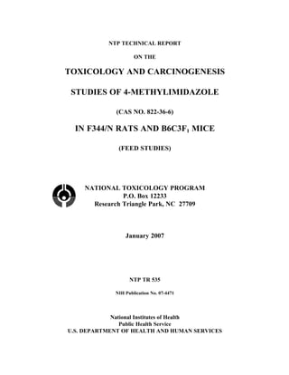
- 27. 25 RESULTS RATS Survival Estimates of 2-year survival probabilities for male and Clonic seizures, excitability, hyperactivity, and impaired female rats are shown in Table 2 and in the Kaplan- gait were observed in 5,000 ppm females; some of these Meier survival curves (Figure 2). Survival of all clinical findings were also observed in the lower exposed groups of male and female rats was similar to exposed groups at greater frequencies than in the con- that of the control groups. trols (Table 5). The study laboratory performed a histopathologic examination of brain, spinal cord, and sciatic nerve from 82 females; 77 displayed clinical Body Weights, Feed and Compound signs of possible neurotoxicity (two from the 1,250 ppm Consumption, and Clinical Findings group, 25 from the 2,500 ppm group, and 50 from the Mean body weights of males in the 1,250 and 2,500 ppm 5,000 ppm group), and five control animals were evalu- groups and females in the 2,500 and 5,000 ppm groups ated for comparison. As part of the pathology peer were less than those of the control groups throughout the review, a neuropathologic evaluation of 10 5,000 ppm study; mean body weights of 1,250 ppm females were and 10 control females was performed. FluoroJade B less after week 41 (Tables 3 and 4; Figure 3). Feed con- (a fluorescent marker for neuronal degeneration) stain- sumption by exposed groups of males was generally ing was conducted on five 5,000 ppm and five control similar to that by the controls (Table G1). However, feed females to identify any subtle antemortem neuronal consumption by 5,000 ppm females was less than that by changes that may not have been readily apparent during the controls (Table G2). Dietary concentrations of 625, evaluation of the standard sections stained with H&E. 1,250, or 2,500 ppm for males and 1,250, 2,500, or Positive controls used for comparison were from a pre- 5,000 ppm for females resulted in average daily doses of vious study in which neuronal necrosis was detected approximately 30, 55, and 115 mg 4-methylimidazole/kg (Morgan et al., 2004). H&E and FluoroJade B staining body weight to males and 60, 120, and 260 mg/kg to did not confirm morphologic neural correlates for the females. neurologic signs exhibited.
- 28. 26 4-Methylimidazole, NTP TR 535 TABLE 2 Survival of Rats in the 2-Year Feed Study of 4-Methylimidazole 0 ppm 625 ppm 1,250 ppm 2,500 ppm Male Animals initially in study 50 50 50 50 Moribund 13 11 12 10 Natural deaths 6 5 5 8 Animals surviving to study termination 31 34 33 32 a Percent probability of survival at end of study 62 68 66 64 b Mean survival (days) 701 681 695 689 c Survival analysis P=0.964 P=0.842N P=0.926N P=1.000 0 ppm 1,250 ppm 2,500 ppm 5,000 ppm Female Animals initially in study 50 50 50 50 Moribund 5 5 5 9 Natural deaths 2 6 11 6 d e Animals surviving to study termination 43 39 34 35 Percent probability of survival at end of study 86 78 68 70 Mean survival (days) 697 701 684 691 Survival analysis P=0.078 P=0.449 P=0.060 P=0.099 a Kaplan-Meier determinations b Mean of all deaths (uncensored, censored, and terminal sacrifice) c The result of the life table trend test (Tarone, 1975) is in the control column, and the results of the life table pairwise comparisons (Cox, 1972) with the controls are in the exposed group columns. A lower mortality in an exposed group is indicated by N. d Includes three animals that died during the last week of the study e Includes two animals that died during the last week of the study
- 29. 4-Methylimidazole, NTP TR 535 27 FIGURE 2 Kaplan-Meier Survival Curves for Male and Female Rats Exposed to 4-Methylimidazole in Feed for 2 Years