
Recomendados
Recomendados
Más contenido relacionado
La actualidad más candente
La actualidad más candente (14)
Similar a Cardiovascular system
Similar a Cardiovascular system (20)
Más de Arosek Padhi
Más de Arosek Padhi (20)
Último
Último (20)
Cardiovascular system
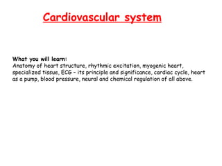
- 1. Cardiovascular system What you will learn: Anatomy of heart structure, rhythmic excitation, myogenic heart, specialized tissue, ECG – its principle and significance, cardiac cycle, heart as a pump, blood pressure, neural and chemical regulation of all above.
- 2. They are self-contracting, autonomically regulated and must continue to contract in rythmic fashion for the whole life of the organism. Hence they have special features. The cells are Y shaped and are shorter and wider than skeletal muscle cells. They are predominatly mononucleated. The arrangement of actin and myosin is similar to skeletal striated muscle. Some of the cardiac muscle cells are auto-rhythmic, i.e they contract even in the absence of neuronal innervation (known as pacemaker cells). Intercalated disks are located between cardiac muscles cells. These contain gap junctions which provide communicating channels between cells.The intercalated disks allows waves of depolarisations to sweep across the cells thus synchronising muscle contraction. Cardiac muscles Depolarisation of cardiac muscle cells differs from that of other muscle cells.Repolarisation takes much longer to occur and thus cells cannot be stimulated at high frequency. The advantage is that cardiac muscle are prevented from going into tetanus.
- 4. Skeletal muscle vs. Cardiac muscle
- 5. Skeletal muscle vs. Cardiac muscle Transmission electron microscopy
- 6. Heart muscle is a SYNCYTIUM of many Cardiac Myocytes
- 8. Differences in concentration of ions on opposite sides of a cellular membrane leads to a voltage called membrane potential
- 9. • Na+-K+ ATPase pump used for maintaining the large excess of Na+ outside the cell and the large excess of K+ ions on the inside. • This unbalanced charge transfer contributes to the separation of charge across the membrane. The sodium-potassium pump is an important contributor to action potential produced by nerve cells. This establishes two concentration gradients Leak of more potassium ions (more permeable; Na+-K+ leak channels) to diffuse across the membrane, down the concentration gradient that was established by the ATPase, creating a charge separation, and thus a voltage, across the membrane.
- 10. + + + + + + + + + + + + + + + + + + + K+ - - - - - - - - - - - - - - - - - - - - Na+ 3Na+ 2K+ More positive Positive charge less positive Negative charge All membranes have Na+-K+ channels or pumps (AT) ATP Na+ K+ Na+-K+ leak channels Deficit of positive on inside: gives negative potential to inside of cell membranes -90mV: resting membrane potential
- 11. Relatively static membrane potential of quiescent cells is called the resting membrane potential (or resting voltage), as opposed to the specific dynamic electrochemical phenomena called action potential and graded membrane potential. RESTING MEMBRANE POTENTIAL -90mV on the inside of the fibre AT of Na+ and K+ thro the membrane: SODIUM POTASSIUM PUMP OUT IN 3Na+ 2K+ Electrogenic Pump: more +ve out and -ve in leaving a deficit of +ve on inside Hence -ve Potential inside the membrane
- 13. In physiology, an action potential is a short-lasting event in which the electrical membrane potential of a cell rapidly rises and falls, following a consistent trajectory. Action potentials occur in several types of animal cells, called excitable cells, which include neurons, muscle cells, and endocrine cells, as well as in some plant cells. In neurons, they play a central role in cell-to-cell communication. In other types of cells, their main function is to activate intracellular processes. In muscle cells, for example, an action potential is the first step in the chain of events leading to contraction. In beta cells of the pancreas, they provoke release of insulin. Action potentials in neurons are also known as "nerve impulses" or "spikes", and the temporal sequence of action potentials generated by a neuron is called its "spike train". A neuron that emits an action potential is often said to "fire". Action Potential
- 14. Action potentials are generated by special types of voltage-gated ion channels embedded in a cell's plasma membrane. These channels are shut when the membrane potential is near the resting potential of the cell, but they rapidly begin to open if the membrane potential increases to a precisely defined threshold value. When the channels open, they allow an inward flow of sodium ions, which changes the electrochemical gradient, which in turn produces a further rise in the membrane potential. This then causes more channels to open, producing a greater electric current, and so on. The process proceeds explosively until all of the available ion channels are open, resulting in a large upswing in the membrane potential. The rapid influx of sodium ions causes the polarity of the plasma membrane to reverse, and the ion channels then rapidly inactivate. As the sodium channels close, sodium ions can no longer enter the neuron, and they are actively transported out of the plasma membrane. Potassium channels are then activated, and there is an outward current of potassium ions, returning the electrochemical gradient to the resting state. After an action potential has occurred, there is a transient negative shift, called the after hyperpolarization or refractory period, due to additional potassium currents. This is the mechanism which prevents an action potential traveling back the way it just came. Action Potential
- 15. NERVE ACTION POTENTIAL Transmission of nerve signals ----- action potential (AP) AP: rapid changes in membrane potential, which spreads long the nerve fibre -90mV (Negative potential) ---------> positive potential Resting Action 0 +35 overshoot -90 polarized Membrane permeable to Na+ Na+ channels close K+ channels open Diffusion of K+
- 16. Depolarisation of cardiac muscle cells differs from that of other muscle cells. Repolarisation takes much longer to occur and thus cells cannot be stimulated at high frequency. The advantage is that cardiac muscle are prevented from going into tetanus. Action Potential of Cardiac Muscle Spontaneous depolarization
- 17. Action potential causes ventricular contraction to last 15times longer than skeletal muscle -85mV to +20mV
- 18. 4: resting membrane potential; diastole 0: rapid depolarization (opening of fast Na+ channels and inward movt. of Na+) 1: inactivation of fast Na+ channels (K+, Cl- out) 2: “ plateau ” balance b/w inward Ca++ and outward K+ (slow) 3: “ rapid repolarization ” Ca++ channels close; K+ perm. incr.; K+ out, net outward positive current (loss of positive charge); 4: cells repolarize (-85-90mV) and resting membrane potential is achieved
- 19. Long Action Potential of Cardiac Muscle and Plateau AP is caused by opening of fast sodium channels; Na+ enters skeletal muscle from ECF (FAST). Channels close and repolarization occurs Permeability of K+ does not decr Skeletal Muscle Cardiac Muscle 1. Fast Na+ channels 2. Slow Ca++ channels (Ca-Na channels); remain open for 1/10th of a sec; slow; large Ca-Na+ flows into cardiac muscle; prolonged depolarization and hence plateau 3. Ca ions activate muscle to enter into contractile phase 4. Permeability of K+ ions decr (5X);outflux prevented; prevents early return of AP plateau 5. Channels close; influx of Ca and Na+ cease, K+ permeability incr and rapid loss of K from cardiac muscle returns membrane pot----end of AP 0.3-0.5m/sec: velocity of signal conduction of excitatory AP along A-V muscle fibre
- 21. Refractory Period of cardiac Muscle Cardiac muscle is refractory to restimulation RP: that interval of time during which a normal cardiac impulse cannot re excite an already excited area of cardiac muscle 1 2 3 sec contraction Normal refractory period of ventricle is 0.25-0.30 sec Relative RP: (0.05sec) muscle is difficult to excite but can be with a strong excitatory signal Early premature contraction Late premature contraction
- 22. Refractory Period of cardiac Muscle
- 23. Automaticity of Cardiac Muscle It means that it is self-exciting. (You could also call it "myogenic" tissue. Meaning a tissue able of creating its own excitement.) This is in contrast with skeletal muscle, which requires either conscious or reflex nervous stimuli for excitation. The heart's rhythmic contractions occur spontaneously, although the rate of contraction can be changed by nervous or hormonal influences, exercise and emotions. For example, the sympathetic nerves to accelerate heart rate and the vagus nerve decelerates heart rate.
- 24. The rhythmic sequence of contractions is coordinated by the sinoatrial (SA) and atrioventricular (AV) nodes. The sinoatrial node, often known as the cardiac pacemaker, is located in the upper wall of the right atrium and is responsible for the wave of electrical stimulation that initiates atrial contraction by creating an action potential. Once the wave reaches the AV node, situated in the lower right atrium, it is delayed there before being conducted through the bundles of His and back up the Purkinje fibers, leading to a contraction of the ventricles. The delay at the AV node allows enough time for all of the blood in the atria to fill their respective ventricles. In the event of severe pathology, the AV node can also act as a pacemaker; this is usually not the case because their rate of spontaneous firing is considerably lower than that of the pacemaker cells in the SA node and hence is overridden. Automaticity of Cardiac Muscle
- 27. Aa Aa
- 29. Cyt SERCA Sarcolemma ICa Ca Ca 3Na SR RyR 3Na Ca T-Tubule Na Na Na Na Ca NCX NCX ATP 2K ATP PLB/SLN AP (Em) [Ca]i Contraction 3Na 2K ATP by D.M. Bers Ca2+ Ion transport in Muscle SERCA: Sarco/Endoplasmic Reticulum Ca2+ ATPase SLN: Sarcolipin; PLB: Phospholamban
- 30. Cyt Ca RyR T-Tubule 3Na Ca Na Sarcolemma NCX AP (Em) [Ca]i Contraction SERCA ATP NCX Na 3Na Na 3Na 2K ATP PLB/SLN ICa SR by D.M. Bers Ca Na Ca Ca2+ Ion transport in Muscle SERCA: Sarco/Endoplasmic Reticulum Ca2+ ATPase SLN: Sarcolipin; PLB: Phospholamban
- 32. Heart Valves Produce One-way Blood Flow Atrioventricular(AV) valves –prevent backflow of blood into the atria when the ventricles contract. 1. bicuspid valve –between left atrium and left ventricle 2. tricuspid valve –between the right atrium and right ventricle
- 33. Heart Valves Produce One-way Blood Flow Semi lunar Valves – prevent backflow of blood into the ventricles when the ventricles relax. 1. aortic valve –at entrance to aorta 2. pulmonary valve –at entrance to pulmonary trunk The semilunar valves also have cusps, which catch blood as it flows back toward the ventricle during ventricular systole. “dubb”
- 34. The heart pumps by squeezing, compressing and pressurizing the blood which then flows down the pressure gradient. The heart’s valves force the blood to go in one direction and prevent (when working properly) backward flow.
- 36. 3D reconstruction of the heart as viewed from the apex towards the valves, image flipped 180° relative to illustration above. Pulmonary valve not visible, leaflets of the tricuspid and aortic valves only partly visible. To the left two images in 2D from the same dataset, showing tricuspid and mitral valves (above) and aortal and mitral valve (below). Tricuspid and mitral valve aortic valves aortal and mitral valve Tricuspid
- 37. Terms: DIASTOLE-the relaxation phase; unless otherwise specified refers to left ventricle, but each chamber has its own diastole; refers to filling of blood SYSTOLE-the contraction phase; unless otherwise specified refers to left ventricle, but each chamber has its own systole. CARDIAC CYCLE the sequence of events from the beginning of one heartbeat to the beginning of the next Each cycle is initiated by the spontaneous generation of AP in the SINO ATRIAL NODE. AP travels from here to atria and AV bundle into the ventricles (delay of 0.1 sec) The frequency of the cardiac cycle is described by the heart rate.
- 38. Each beat of the heart involves five major stages The first two stages, often considered together as the "ventricular filling" stage, involve the movement of blood from atria into ventricles. Action of valves relating to them The next three stages involve the movement of blood from the ventricles to the pulmonary artery (in the case of the right ventricle) and the aorta (in the case of the left ventricle). Action of valves relating to them CARDIAC CYCLE
- 39. The first, "late diastole", is when the semi lunar valves close, the atrioventricular (AV) valves open, and the whole heart is relaxed. The second, "atrial systole", is when the atrium (R and L) contracts, the AV valves open, and blood flows from atrium to the ventricle. The third, "isovolumic ventricular contraction", is when the ventricles begin to contract, the AV and semilunar valves close, and there is no change in volume. The fourth, "ventricular ejection", is when the ventricles are empty and contracting, and the semilunar valves are open. During the fifth stage, "Isovolumic ventricular relaxation", pressure decreases, no blood enters the ventricles, the ventricles stop contracting and begin to relax, and the semi lunar valves close due to the pressure of blood in the aorta. CARDIAC CYCLE
- 40. Throughout the cardiac cycle, blood pressure increases and decreases. The cardiac cycle is coordinated by a series of electrical impulses that are produced by specialized heart cells found within the sinoatrial node and the atrioventricular node. CARDIAC CYCLE The sino-atrial node sends out electrical waves of excitation to both atria, and it is prevented from flowing into the ventricles by strands of non-conducting fibrous tissue situated laterally from the tricuspid/bicuspid valves to the septum. These waves of excitation travel towards the septum and into the atrio- ventricular node, where they are held for roughly 0.1 seconds. They are then discharged down the bundle of his, then down the purkinje tissue, which are both situated inside the septum. The waves flow down towards the apex of the heart and are then discharged into the ventricles, causing them to contract (ventricular systole) This creates the well known beat of the heart. The cardiac muscle is composed of myocytes which initiate their own contraction without help of external nerves (with the exception of modifying the heart rate due to metabolic demand). Under normal circumstances, each cycle takes approximately one second.
- 42. Major Events in The Cardiac Cycle 1) quiescent period- period when all chambers are at rest and filling. 70% of ventricular filling occurs during this period. The AV valves are open, the semi-lunar valves are closed. 2) atrial systole- pushes the last 30% of blood into the ventricle. BP in atria rises and pushes blood into ventricles (atrial kick) [P wave: depolarization; due to SA node] 3) atrial diastole- atria begin filling. This occurs nearly simultaneously with the next event… 4) ventricular systole- ventricles contract, first closing the AV valves and causing the first heart sound then the semi lunarvalves open permitting ventricular ejection of blood into the arteries. 5) ventricular diastole- As the ventricles relax the semi lunarvalves close first producing the second heart sound, then the AV valves open allowing ventricular filling.
- 43. Heart diastole Atrial systole Heart systole Ventricular systole Atrial Pressure a wave: atrial contraction;; AV valves open; RAP incr 4-6mm Hg LAP incr 7-8 mm Hg c wave: ventricles contract (V systole); backflow of blood in atria; incr in ventricular pressure; AV valves closed, SLV open v wave: end of contraction; slow flow of blood into atria from SVC/IVC (diastole); AV valves open Heart diastole Atrial systole
- 44. V V V middle stage of diastole during the cycle of a heartbeat
- 46. Electrocardiogram P: spread of depolarization through atria Atrial contraction Rise in atrial pressure after P wave After 0.016sec QRS wave: electrical depolarization of ventricles Initiation of contraction Ventricular pressure rises QRS rises slightly before ventricular systole T wave stage of repolarization of ventricles (ventricular muscles relax; occurs slightly before end of ventricular contraction) P Q R S T
- 48. Cardiac Diastole CD is the period of time when the heart relaxes after ventricular contraction in preparation for refilling with circulating blood. Ventricular diastole is when the ventricles are relaxing, while atrial diastole is when the atria are relaxing. Together they are known as complete cardiac diastole. During ventricular diastole, the pressure in the (left and right) ventricles drops from the peak that it reaches in systole. When the pressure in the left ventricle drops to below the pressure in the left atrium, the mitral valve opens, and the left ventricle fills with blood that was accumulating in the left atrium. The isovolumic relaxation time (IVRT) is the interval from the aortic component of the second heart sound, that is, closure of the aortic valve, to onset of filling by opening of the mitral valve. Likewise, when the pressure in the right ventricle drops below that in the right atrium, the tricuspid valve opens, and the right ventricle fills with blood that was accumulating in the right atrium. During diastole the pressure within the myocardium is lower than that in aorta, allowing blood to circulate in the heart itself via the coronary arteries.
- 52. AV open SL close
- 53. AV open SL close
- 54. Cardiac Cycle
- 55. Cardiac Output The volume of blood per minute that the left ventricle pumps into the systemic circulation Depends on two factors: 1. Heart rate: rate of contraction i.e. number of beats per minute 2. Stroke volume: amount of blood pumped by left ventricle in each contraction. Average SV is 75ml in humans; heart rate is 70 beats per minute cardiac output is 5.25 L/min (= to total vol of blood in body) CO incrs during heavy excersise
- 56. Cardiac Output Minute Volume= Heart Rate X Stroke Volume Heart rate, HR at rest = 65 to 85 bpm (widest range usually quoted) Each heartbeat at rest takes about 0.8 sec. of which 0.4 sec. is quiescent period. Stroke volume, SV at rest = 60 to 70 ml. C.O. at rest = 70 bpmX 70 ml/beat = 4900 ml/min/vent. The heart can increase both rate and volume with exercise. Rate increase is limited due to necessity of minimum ventricular diastolic period for filling. Upper limit is usually put at about 220 bpm. Maximum heart rate calculations are usually below 200.
- 57. SV / EDV = Ejection Fraction Normally around 50% at rest and will increase during strenuous exercise in a healthy heart. Well trained athletes may have ejection fractions approaching 70% in the most strenuous exercise. Preload-This is the pressure at the end of ventricular diastole, the beginning of ventricular systole. It is roughly proportional to the End Diastolic Volume (EDV), i.e. as the EDV increases so does the preload of the heart. Afterload- This is the pressure at the end of ventricular systole and is related to the resistance to blood flow. It is roughly proportional to the End Systolic Volume (ESV). When the peripheral resistance increases so does the ESV and the afterload of the heart. The difference between preload and afterload is a measure of the hearts efficiency. ADDITIONAL TERMS
- 58. Blood Flow ~ Δblood pressure Resistance to blood flow Blood flow is directly proportional to the pressure gradient over a section of a blood vessel, and inversely proportional to the resistance to flow. Resistance is produced by friction along the vascular wall, and is increased with vasoconstriction, atherosclerosis, and hypertension.
- 59. Arteries have blood flowing at high velocity and pressure and maintain BP even when heart is relaxed; thick and elastic walls Vein have thin walls and blood flows at low velocity and low pressure and flows as a result of skeletal muscle action; contain valves to move blood towards heart