
Recomendados
Recomendados
Más contenido relacionado
La actualidad más candente
La actualidad más candente (20)
Destacado
Similar a Paper dmzbb nqo1
Similar a Paper dmzbb nqo1 (20)
Más de delaine_marie
Más de delaine_marie (20)
Último
Último (20)
Paper dmzbb nqo1
- 1. NQO1, NAD(P)H dependent cytosolic oxidoreductase, modulates redox status and H2O2 levels in pancreatic β-cells Delaine M. Zayas-Bazán Burgos1 and Joshua Gray2, Emma Heart3 1 University of Puerto Rico at Cayey; 2US Coast Guard Academy, New London, CT; 3Marine Biological Laboratory, Woods Hole, MA Abstract: Diabetes is the illness in which either the β-cells do not secrete enough insulin or secrete insulin that is not efficient. Type 2 diabetes is marked by a reduction in the ability of beta cells of the pancreas to secrete insulin. Redox status, defined as the ratio of the reduced-to-oxidized forms of redox couples (such as NADH-to-NAD+ and NADPH-to-NADP+), plays an important role in overall cell health and in the in glucose-stimulated insulin secretion (GSIS) from pancreatic β-cells. Here we have investigated the role of the cytosolic NAD(P)H-dependent oxidoreductase NQO1 on β-cell redox status and quinone-dependent production of reactive oxygen intermediates (ROI). In both clonal insulin secreting β-cells (INS-1 832/13) and isolated rodent islets, NQO1 over-expression blunted quinone-dependent ROI production, while islets from NQO1 knockout mice had enhanced ROI formation. Furthermore, NQO1 has been found to decrease NAD(P)H-to-NAD(P)+ ratio, consistent with the NQO1-dependent utilization of NAD(P)H for the intrinsic Plasma Membrane Electron Transport activity in β-cells. Together, these data show that NQO1 plays an important role in maintaining proper redox status and maintains insulin secretion in the face of oxidative stress. endocrine gland that, in most species, arises Introduction from ventral and dorsal buds which Background subsequently merge to form the pancreas.” This J definition has been conserved and further orgen Jonsson and his team in detailed to give everyone a better 1994 stated that: “The understanding of the pancreas. The pancreas mammalian pancreas is a mixed exocrine and serves both digestive and endocrine functions
- 2. and it is composed of two main parts. The first All of these cells can be directly or part is known as the Pancreatic Acini and indirectly linked to many of the metabolism corresponds to the exocrine area of the diseases we know. Metabolism can be defined pancreas. The second main part and our area of as the essential chemical processes involved in interest is the endocrine area known as the converting nutrients into chemical energy and Islets of Langerhans. The Islets of Langerhans molecular are cluster of cells that produce various maintaining the living system. (Hickman 2007). hormones. There are five types of cells These chemical processes include digestion, composing the Islets of Langerhans and their acquisition of energy, respiration and synthesis type dictate the type of hormone they produce, of molecules and structures. Diabetes mellitus, release and under what stimuli. The cells and exocrine their function are illustrated in the following pseudocyst among others are diseases that table. affect the metabolism by affecting one or components pancreatic for building insufficiency and and various steps of the digestion and nutrient Table 1.1 Islet Composition absorbance of the organism. Type of Cell Function α cells Produce Glucagon In 1990 Lind C et al identified NQO1, an NAD(P)H-dependent cytosolic oxidoreductase, was for its ability to reduce β cells Produce Insulin δ cells Produce Somatosin (Growth quinones such as menadione. Quinones are a class of organic compound that could either hormone-inhibiting Hormone) be exogenous or endogenous. Haefeli in 2011stated the features of quinones: “quiones ε cells Produce Gherlin PP cells Produce Pancreatic Polypeptide feature a quinoid conjugated double-bond
- 3. system, which is responsible for their the formation of relatively stable quinols electrophilic nature” (hydroquinones), which undergo less redox cycling due to the greater stability of the hydroquinone versus the semiquinone and further detoxification via phase 2 metabolismThis concept is consistent with the increased susceptibility of the NQO1 knockout model mouse to high and toxic doses of redox cycling compounds and xenobiotics evidenced in 2000 by Joseph and his team. This supports Figure 1 NQO1 reduces H2O2 production by quinones. Quinones such as menadione (1) are reduced by NAD(P)H-dependent oxidoreductase enzymes to either quinols (2) or semiquinols (3). the protective role of NQO1 against oxidative stress in a variety of tissues. However, there are Semiquinols, generated by 1-electron reducation of quinones, are highly reactive and reduce molecular oxygen to generate reactive oxygen intermediates (ROIs), while regenerating the parent quinone no studies on the protective role of NQO1 in pancreatic islet β-cells, which otherwise compound (1). Complete 2-electron to the quinol (2), however, facilitiates further metabolism and elimination of the quinone. NQO1 catalyzes complete 2-electron reduction of quinones to relatively stable contain relatively low levels of classical antioxidant enzymes (Tiedge M et al 1997). quinols, which results in a lower level of ROI production. Inside the cell, quinones undergo either one- or two electron NAD(P)H-dependent reduction: 1-electron reduction leads to the formation of unstable semiquinones, which can readily reduce molecular oxygen to superoxide, regenerating the parent quinone compound via redox cycling (Fig. 2). In contrast, complete 2 electron reductions mediated by NQO1 lead to In parallel to its role as a detoxification enzyme, NQO1 has more recently been implicated in the regulation of intermediary metabolism. NQO1 expression is correlated with fasting insulin levels evidenced in Palming J et al work in 2007 and an association between reduced activity of NQO1 due to polymorphism and dysregulated blood
- 4. glucose levels has been reported in humans 832/13 cells as well as primary rodent islets, we (Kim 2009). Gaiwkad et al evidenced in 2001 have demonstrated that NQO1 regulates the NQO1’s role in cellular metabolism with level of the NAD(P)H/NAD(P)+ ratio, which studies using global NQO1 knock-out mice, can explain its effect on the GSIS. Furthermore, which found that these mice suffer from several using over-expression or knockout strategies, metabolic defects, including insulin resistance we have demonstrated that NQO1 reduces the in the periphery, and fail to increase insulin degree of quinone-dependent redox cycling and output in the face of their insulin resistance in superoxide production in β-cells, thus acting to contrast to the normal β-cell compensatory prevent toxicity under enhanced pro-oxidant hyperinsulinemic response (Pi 2007). We have load. We believe that NQO1 reduces oxidative previously demonstrated that NQO1 regulates stress in β-cells by lowering quinone-dependent glucose-stimulated insulin secretion (GSIS) and H2O2 production, through the complete two Plasma Membrane Electron Transport (PMET) electron reduction. We also believe that at the activity in pancreatic β-cells and our lab metabolic level NQO1 modulates NAD(P)H- continues to investigate the mechanisms of this to-NAD(P)+ ratio in β-cells, as it does in other pathway. tissues. Problem and hypotheses Materials and Methods In this study we investigated the role of In order to conduct the experiments the NQO1 in β-cell metabolic pathways, namely first procedures were to over express NQO1 in the role of NQO1 in modifying the redox our cell line. This was achieved through the status. This is one of the key determinants of infection with the adenovirus that either was an the β-cell health and a coupling factor in empty vector, control, or the over expressing glucose-stimulated insulin secretion (GSIS). gene. Then the NQO1 activity was measured. Using both clonal insulin secreting INS-1 Whole cell lysates were prepared by sonication
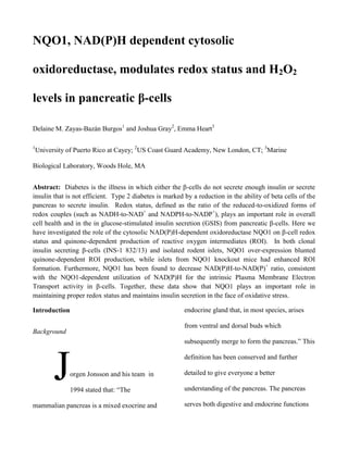
- 5. followed by centrifugation at 12 kg for 5 min at Then the level of released hydrogen peroxide 4 C. Equal concentrations of cell lysate (H2O2) was quantified using Amplex protein were tested for NQO1 activity, which Red/horseradish peroxidase. Fluorescence (540 was quantified by the decrease in absorbance of excitation, 595 emission) was monitored using dichlorophenolindophenol (DPI) (600 nm) over a SpectraMax M5 multi-mode microplate a period of one minute. The difference in reader (Molecular Devices, Sunnyvale, CA). activity in the absence and presence of This was evaluated with both rodent islets and dicoumarol (20 µM) are expressed as NQO1 INS-1 832/13 cells. The rodent islets were activity. Figure 2 isolated from normal mice and the global knock out utilizing the corresponding procedures. NQO1 over-expression in INS-1 832/13 cells. Adenoviral-mediated over-expression resulted in the increase of NQO1 protein (adapted Results from[10]) and enzyme activity, measured as the The effects of NQO1 over-expression reduction of DCPIP (dichlorophenolindophenol) on menadione-dependent hydrogen peroxide production in INS-1 832/13 cells were measured and analyzed (Figure 3). Dicoumarol (DIC), an inhibitor of NQO1, blocks NQO1 Control NQO1+ inhibitory action on redox cycling and H2O2 production. On the first column with no addition of Dicoumarol NQO1’s protection can be clearly seen. Under high glucose conditions NQO1 lowers statistically hydrogen peroxide production. Legend: 3G is 3 mM glucose, 16
- 6. is 16 mM glucose. Data are means ± SE from 2 NQO1 KO. The levels of hydrogen production experiments performed in quadruplicate are clearly lowered by the presence of NQO1. measurement.*P 0.05 Control vs. NQO1+ . Figure 3. Effects of NQO1 on H2O2 production in INS-1 Figure 4. Effects of NQO1 on H2O2 production in rodent islets 832/13 cells The effects of NQO1 over-expression (panel A) and knock-down (panel B) on menadionedependent H2O2 production in isolated rodent islets (Figure 4). The over-expressing islets came from normal mice and after isolation were infected with adenovirus. The NQO1 knock-out islets came from the global knockout mice. Data are means ± SE from 2 Lastly in order to evidence the effects of experiments performed in quadruplicate NQO1 in the β-cells metabolism the NAD(H) measurement. *P 0.05 Control vs. NQO1+ or –to- NAD+ ratio was quantified. NQO1 regulates NADH-to-NAD+ ratio in INS-1
- 7. 832/13 cells and islets. INS-1 832/13 (A), Conclusions infected with control adenovirus (Ad-control) or NQO1 over-expressing adenovirus (Ad- All of these results lead to the various conclusions about NQO1 role in β-cell health NQO1) or isolated NQO1 Wild Type and and metabolism. In terms of the health of the βNQO1 Knock-Out islets (B) were exposed to cell NQO1 protects from oxidant stress, 4mM or 16 mM glucose and NADH and NAD+ therefore enhances β-cell health. In terms of were measured by LC/MS/MS. Data are means metabolism NQO1 modulates β-cell redox ± SE from 2-3 experiments performed in status by lowering the NADH-to-NAD+ ratio as duplicate measurement.*P 0.05 Ad-control vs. it does in other tissues such as liver and Ad-NQO1, and KO vs. WT. adipose. This effect on redox cycle has a direct Figure 5. NQO1 mediates redox cylcing increasing role in glucose metabolism and glucose stimulated insulin secretion. Future experiments may include exploring NQO1’s role in the physiological state. Under normal conditions almost every eukaryotic cell posses the endogenous quinine ubiquinone. Another great follow-up would be to determine the role of NOQ1 under glucotoxicity. Glucotoxicity is high glucose and high fatty acids.
- 8. Acknowledgements Palming J et al (2007) J Clin Endocrinol Metab 92: 2346-52 This project was conducted thanks to Pi J et al (2007) Diabetes 56, 1783-91. the collaboration and help of many individuals. Tiedge M et al (1997) Diabetes 46: 1733-42 This work was supported by the National Science Foundation through the Biological Discovery in Woods Hole DBI 1005378, the American Diabetes Association 7-12-BS-073 and National Institute of Health through both Dr. Emma Heart’s grant IH R56DK088093, and through the Research Initiative for Scientific Enhancement Program in the University of Puerto Rico at Cayey R25 GM059429. We extend our gratitude towards the REU directors Dr. Mensinger and Dr. Malchow. References Gaikwad A et al (2001) JBC 276: 22559-64 Gray JP et al (2011) AJP Endo Metab 301: E113021. Haefeli RH (2011) PLos One 6: e17963. Joseph P et al (2000) Biochem Pharmacol 60: 207-14 Kim D (2009) Korean Diabetes J 33: 24-30 Lind C et al (1990) Method Enzymol, 186:287-301.