Mycobacterium tuberculosis seminar
•Descargar como PPT, PDF•
212 recomendaciones•55,107 vistas

Basics of tuberculosis
![KOCH’S DISEASE : TUBERCULOSIS ,[object Object],[object Object],[object Object],[object Object],[object Object],[object Object],[object Object],[object Object]](data:image/gif;base64,R0lGODlhAQABAIAAAAAAAP///yH5BAEAAAAALAAAAAABAAEAAAIBRAA7)
Recomendados
Más contenido relacionado
La actualidad más candente
La actualidad más candente (20)
Destacado
Destacado (20)
Similar a Mycobacterium tuberculosis seminar
Similar a Mycobacterium tuberculosis seminar (20)
Más de deepak deshkar
Más de deepak deshkar (20)
Último
Último (20)
Mycobacterium tuberculosis seminar
- 3. “ NO ONE IS SAFE FROM TUBERCULOSIS UNTIL EVERY ONE IS SAFE”.
- 6. Organisms belonging to the genus Mycobacterium are---- 1.Very Thin 2.Rod shaped 3.0.2 to 0.4 X 2 to 10 µ m 4.Non motile 5.Sometimes showing filamentous branching like fungus. 6.Forming mould like pellicle in liquid culture. HENCE CALLED MYCOBACTERIA
- 8. Mycobacterium tuberculosis Mycobacterium bovis Mycobacterium bovis BCG Mycobacterium africanum Mycobacterium microti (Vole) MYCOBACTERIUM TUBERCULOSIS COMPLEX
- 9. MYCOBACTERIUM TUBERCULOSIS Mycobacterium tuberculosis Mycobacterium tuberculosis Scientific classification Kingdom: Bacteria Phylum: Actinobacteria Order: Actinomycetales Suborder: Corynebacterineae Family: Mycobacteriaceae Genus: Mycobacterium Species: M. tuberculosis Binomial name
- 13. Mycobacterium tuberculosis: Ziehl-Neelsen stain.
- 14. Mycobacterium Tuberculosis Stained with Fluorescent Dye
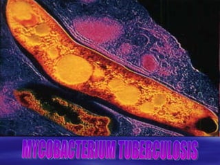
- 16. Mycobacterium tuberculosis : Electron Micrograph.
- 17. CMN Group: Unusual cell wall lipids (mycolic acids,etc.) ( P urified P rotein D erivative) Lipid Rich Cell Wall Of Mycobacterium tuberculosis Mycolic acids
- 19. ACID FASTNESS OF MYCOBACTERIUM TUBERCULOSIS IS DUE TO PRESENCE OF A HIGH MOLECULAR WEIGHT HYDROXY ACID CONTAINING CARBOXYL GROUPS CALLED MYCOLIC ACID IN THE BACTERIAL CELL WALL OR TO A SEMIPERMIABLE MEMBRANE AROUND THE CELL.
- 24. Colonies of Mycobacterium tuberculosis on Lowenstein-Jensen medium.
- 25. M. tuberculosis bacterial colonies
- 26. Eight Week Growth of Mycobacterium tuberculosis on Lowenstein-Jensen Agar
- 28. Acid-Fast (Kinyoun) Stain of Mycobacterium NOTE: cord growth (serpentine arrangement) of virulent strains
- 30. BIOCHEMICAL REACTIONS: + - +/- - - - - - - - M Africanum + - - +/- - - - - - - M bovis + + + +/- - + - + - + M tuberculosis UREASE TEST PYRAZI-NAMIDASE TSET GROWTH ON TCH TELLURI--TE REDUCTION TEST TWEEN 80 HYDRO--LYSIS TSET PEROX---IDASE TEST HOT CATAL---ASE TEST NITRATE REDUC---TION TEST ARYL-SULPH---ATASE TEST NIACIN TEST SPECIES
- 33. TUBERCULOSIS IS THE MOST IMPORTANT COMMUNICABLE DISEASE IN THE WORLD SPARING NO AGE, NO SEX, & NO NATIONALITY.
- 37. Pathogenesis of M. tuberculosis
- 38. IMMUNOPATHOLOGY OF TB M. tuberculosis Macrophage Class II MHC Activated Macrophage (Phagocytosis) Bactericidal activity T–Cell Receptor CD4+ T- Cell CYTOKINES CD8+ T- Cell Delayed Hypersensitivity Class I MHC Macrophage Caseous Necrosis
- 39. Phagocytosis of Mycobacterium tuberculosis
- 41. Diagram of a Granuloma NOTE: ultimately a fibrin layer develops around granuloma (fibrosis) , further “walling off” the lesion. Typical progression in pulmonary TB involves caseation , calcification and cavity formation .
- 43. Necrosis: Soft White Cheese
- 47. She has tuberculosis of peripheral lymph nodes. Although lymphatic tuberculosis may appear to be a localized disease process, it is not as the systemic signs and symptoms in this child indicate. At least five lesions can be seen, but it is likely that there are more less apparent ones in deeper structures.
- 49. This patient was referred to the tuberculosis clinic with the question of otitis media. There was no otitis. The patient had lost weight and had signs and symptoms of systemic illness. The pre-auricular lesion was cold to the touch and was apparently fluctuating. The abscess was aspirated. A Gram stain showed no organisms and careful examination of a Ziehl-Neelsen stained smear revealed acid-fast bacilli.
- 50. While peripheral lymphatic tuberculosis is most frequently found around the neck, the axilla may also affected. Several lymph nodes may be matted together as in this patient. Some nodes have undergone liquefaction leading to discoloration of the skin.
- 51. In this patient, any affected lymph node in the lesion had undergone complete caseation with discoloration of the skin.
- 52. This abscess was close to breaking through the skin, yet it felt cold to the touch and the child felt remarkably little pain when the lesion was touched. Such a finding should raise a high index of suspicion for tuberculosis.
- 53. This patient has chronic peripheral lymphatic tuberculosis with some lesions healed with scaring, while others are still showing activity.
- 54. This patient had a seven-year history of lymphatic tuberculosis. Many lesions have apparently healed, but some are still active (note inflammation surrounding the most caudal axillary lesion).
- 55. At first sight, all of the lesions resulting form peripheral lymphatic tuberculosis in this patient have healed. However, as the example of the previous patient demonstrates, one can never be certain. It thus may be good policy to offer curative chemotherapy to any patient with signs of tuberculosis of peripheral lymph nodes.
- 56. This boy presented with several lesions. On a chest radiograph, he had a segmental lesion. In addition, he had a lesion in the neck (rendered dark by traditional medicine), an axillary lesion, and a lesion in the arm (the hump on the arm is the tuberculin skin test reaction), and the hand.
- 57. The lesion in the hand is shown here in close-up.
- 58. This patient with tuberculosis of the spine and a visible abscess, slightly discoloring the overlaying skin, on the lower left back almost escaped a correct diagnosis but for an astute laboratory technician. The abscess was warm to the touch and a Gram stain showed Gram-positive cocci. Nevertheless, the laboratory technician insisted on rigorous examination for acid-fast bacilli and found them, confirming tuberculosis of the spine with a super-infected abscess.
- 59. The vertebral lesions are usually anterior in location, often triangular in shape. The bony structure adjacent to both sides of the disk becomes eroded, leading to the seemingly narrowing of inter-vertebral disk space.
- 60. As a result of the anterior lesion, the disk or disks collapse, building a triangular shape, leading the typical gibbus
- 61. Extensive destruction in two adjacent vertebrae.
- 62. Two vertebrae collapsed to the height of one.
- 63. In addition to the paralysis caused by the lower lumbar lesion, this child also had a pyopneumothorax (and an accelerated response to a BCG vaccination).
- 64. This patient has a severe gibbus in the lower thoracic region.
- 65. This patient with a 90 degree lesion in the spine was ambulatory when interviewed. He had had received a full course of anti-tuberculosis treatment and had no neurologic symptoms.
- 66. The reason for the complete recovery from neurologic symptoms in the majority of patients is most likely attributable to the anterior location of the disease process that often leaves the spinal canal spared. The neurologic symptoms seen in the beginning are thus most likely attributable to edema and compression from abscesses that resolve with chemotherapy. In some patients, boney particles may, however, reach the spinal canal and then may cause permanent disability.
- 67. This girl had an almost completely destroyed hip joint.
- 68. The diagnosis of tuberculosis of the left hip in this boy was made from the secretion from a sinus draining through the skin by demonstrating acid-fast bacilli.
- 69. Tuberculosis of the wrist.
- 70. This patient has a sinus draining from both the dorsal and volar aspect of the thumb. He squeezed pus out from the lesions directly onto a Lowenstein-Jensen medium, on which Mycobacterium tuberculosis was isolated (a smear examination for acid-fast bacilli was negative).
- 71. The radiograph shows the complete destruction of the distal phalanx.
- 72. This patient had tuberculosis of the ankle. The bacteriologic diagnosis was made by demonstrating acid-fast bacilli from the visible secretions draining from a sinus.
- 73. The patient did not only have tuberculosis of the ankle, he also had peripheral lymphatic tuberculosis, tuberculous mastitis (exceedingly rare in men), pleural thickening from past pleural tuberculosis, multiple abscesses, and had been operated for a presumable tuberculous epididymitis. While such multi-system disease in a young man should pose little difficulties in making the diagnosis of tuberculosis, it had not been taken into consideration for a prolonged period of time.
- 74. The patient did not only have tuberculosis of the ankle, he also had peripheral lymphatic tuberculosis, tuberculous mastitis (exceedingly rare in men), pleural thickening from past pleural tuberculosis, multiple abscesses, and had been operated for a presumable tuberculous epididymitis. While such multi-system disease in a young man should pose little difficulties in making the diagnosis of tuberculosis, it had not been taken into consideration for a prolonged period of time.
- 75. The diagnosis of female genitourinary tuberculosis is probably made in only of a fraction of cases. It is believed, however, that Falloppian tube and endometrial tuberculosis may account for much female infertility in high-incidence countries. This patient is an example to the case: an observant clinician requested a histological examination of an endometrium biopsy specimen and caseous granulomata were reported. Subsequently, the index of suspician rose, and numerous other cases were diagnosed subsequently.
- 76. Warty skin tuberculosis is a perhaps difficult to diagnose manifestation of tuberculosis of the skin if it is not thought of. This patient testifies to the remarkable efficacy of modern anti-tuberculosis chemotherapy in such a patient.
- 77. Tuberculosis of the spine is most frequently located in the lower thoracic and the lumber region of the spine.
- 78. THANK YOU
Notas del editor
- Once a tubercle is formed, the immune system is activated, but by this time the bacteria may have already spread to other bronchi (Reviewed by Schaff, et al. , 2003). Necrosis occurs in the center of tubercles because of the toxins secreted by the surrounding immune cells. The caseous centers of tubercles liquefy, the bacteria continue to multiply, and then bronchi necrosis occurs. TB is often associated with caseous necrosis, which resembles soft white cheese. Fisher (2002) noted that most well-nourished and immunocompetent individuals can eliminate the bacteria before a more serious condition occurs. In 90% of cases, the bacteria are eliminated and the tubercle heals, evidenced by scar formation. On close inspection, the caseous tan necrotic tissue in this image constitutes the granulomas in this lung.
- Infection begins as T-lymphocytes secrete cytokines that recruit macrophages in response to the presence of the pathogen (Reviewed by Sharma and Mohran, 2004). These macrophages accumulate and aggregate in tissues to become spherical granulomas. Granulomas prevent the spread of M. tuberculosis by confining the bacteria in a compact area where the immune cells can work together to isolate and destroy the bacteria. The central zones of granulomas contain large macrophages surrounded by T-lymphocytes. Granulomas in TB are called tubercles and are visible as white spots (1-2 mm). Once in the alveoli, the bacteria can then spread to local lymph nodes, the bloodstream, and eventually, to distant organs (lung apices, peripheral lymph nodes, kidneys, brain, and bone).