SlideShare una empresa de Scribd logo
1 de 59
DNA replication and gene expression
C
T
A
A
T
CG
GC
A
C G
AT
AT
A T
TA
C
TA
0.34 nm
3.4 nm
(a) Key features of DNA structure
G
1 nm
G
(c) Space-filling model
T
Rahna K Rathnan
Assistant Professor
Sahrdaya college of engineering and Technology
DNA replication
• The parent molecule unwinds, and two new daughter
strands are built based on base-pairing rules
(a) The parent molecule has two
complementary strands of
DNA. Each base is paired by
hydrogen bonding with its
specific partner, A with T and G
with C.
(b) The first step in
replication is separation of
the two DNA strands.
(c) Each parental strand now
serves as a template that
determines the order of
nucleotides along a new,
complementary strand.
(d) The nucleotides are connected
to form the sugar-phosphate
backbones of the new strands.
Each “daughter” DNA molecule
consists of one parentalstrand
and one new strand.
A
C
T
A
G
A
C
T
A
G
A
C
T
A
G
A
C
T
A
G
T
G
A
T
C
T
G
A
T
C
A
C
T
A
G
A
C
T
A
G
T
G
A
T
C
T
G
A
T
C
T
G
A
T
C
T
G
A
T
C
DNA Replication is “Semi-conservative”
• Newly synthesised duplex DNA
conserve one strand of parent
DNA and other strand will be
newly synthesised
• One original strand was used as a
template to make the new strand
DNA Replication
• Require
– Template DNA
– More than dozen enzymes and proteins
• DNA synthesis enzyme : DNA polymerase
• DNA synthesis direction ---- 5’ to 3’
• The replication of a DNA molecule begins at special sites called origins
of replication
• the two strands are separated
Origins of Replication• A eukaryotic chromosome may have hundreds or even
thousands of replication origins
Replication begins at specific sites
where the two parental strands
separate and form replication
bubbles.
The bubbles expand laterally, as
DNA replication proceeds in both
directions.
the replication
bubbles fuse, and synthesis of
the daughter strands is
complete.
1
2
3
Origin of replication
Bubble
Parental (template) strand
Daughter (new) strand
Replication fork
Two daughter DNA molecules
In eukaryotes, DNA replication begins at many sites along the giant
DNA molecule of each chromosome.
In this micrograph, three replication
bubbles are visible along the DNA of
(b)(a)
0.25 µm
Mechanism of DNA Replication
• DNA replication is catalyzed by DNA polymerase
– needs an RNA primer
• RNA primase synthesizes primer on DNA strand
• DNA polymerase adds nucleotides to the 3’ end of the growing strand
Mechanism of DNA Replication
• Nucleotides are added by complementary base pairing with the
template strand
• The substrates, deoxyribonucleoside triphosphates, are
hydrolyzed and releasing energy for DNA synthesis.
The Mechanism of DNA Replication
• DNA synthesis on the leading strand is continuous
• The lagging strand grows the same general direction as the leading strand
(in the same direction as the Replication Fork). However, DNA is made in
the 5’-to-3’ direction
• DNA synthesis on the lagging strand is discontinuous
• DNA is added as short fragments (Okasaki fragments)
– subsequently ligated together
DNA polymerase I degrades the
RNA primer and replaces it with
DNA
The Mechanism of DNA Replication
• DNA helicases unwind the double helix
• Single-stranded DNA binding proteins stabilize the
SS DNA
• RNA primase catalyzes the synthesis of short RNA
primers, to which nucleotides are added.
• DNA polymerase III extends the strand in the 5’-to-
3’ direction
• DNA polymerase I degrades the RNA primer and
replaces it with DNA
• DNA ligase joins the DNA fragments into a
continuous daughter strand
Enzymes in DNA replication
Helicase unwinds
parental double helix
Binding proteins
stabilize separate
strands
DNA polymerase III
binds nucleotides
to form new strands
Ligase joins Okazaki
fragments and seals
other nicks in sugar-
phosphate backbone
Primase adds
short primer
to template strand
DNA polymerase I
(Exonuclease) removes
RNA primer and inserts
the correct bases
• Helicase protein binds to DNA sequences called origins and unwinds
DNA strands.
• Single strand Binding proteins prevent single strands from rewinding.
• Primase protein makes a short segment of RNA complementary to the
DNA, a primer.
5’
3’
5’
3’
3’5’
5’3’
Replication
Overall direction
of replication
5’3’
5’
3’
5’
3’
3’5’
DNA polymerase enzyme adds DNA nucleotides
to the RNA primer.
Replication
• DNA polymerase enzyme adds DNA nucleotides to the
RNA primer.
• DNA polymerase proofreads bases added and replaces
incorrect nucleotides.
5’
5’
Overall direction
of replication
5’
3’
5’
3’
3’
3’
Replication
5’
5’3’
5’
3’
3’
5’
3’
Overall direction
of replication
• Leading strand synthesis continues in a 5’ to 3’
direction.
Replication
3’5’ 5’
5’3’
5’
3’
3’
5’
3’
Overall direction
of replication
Okazaki fragment
• Discontinuous synthesis produces 5’ to 3’ DNA
segments called Okazaki fragments.
Replication
5’
5’ 3’
5’
3’
3’
5’
3’
3’
5’ 5’3’
Leading strand synthesis continues in a
5’ to 3’ direction.
Discontinuous synthesis produces 5’ to 3’ DNA
segments called Okazaki fragments.
Replication
3’
5’
3’
5’
5’ 3’
5’
3’
3’
5’ 5’3’
Leading strand synthesis continues in a
5’ to 3’ direction.
Discontinuous synthesis produces 5’ to 3’ DNA
segments called Okazaki fragments.
Replication
5’
5’
3’ 3’
5’
3’
5’ 3’
5’
3’
3’
5’
• Exonuclease activity of DNA polymerase I
removes RNA primers.
Replication
Polymerase activity of DNA polymerase I fills the gaps.
Ligase forms bonds between sugar-phosphate backbone.
3’
5’
3’
5’ 3’
5’
3’
3’
5’
Replication
Replication Fork Overview
Gene Expression
• Synthesis of protein from DNA through RNA
• The process by which DNA directs protein synthesis, gene expression
includes two stages
– transcription
– translation
Transcription and Translation
• Cells are governed by a cellular chain of command
– DNA → RNA → protein
• Transcription
– Is the synthesis of RNA under the direction of DNA
– Produces messenger RNA (mRNA)
• Translation
– Is the actual synthesis of a polypeptide
– occurs under the direction of mRNA
– Occurs on ribosomes
Transcription and Translation
• In a eukaryotic cell the nuclear envelope separates transcription from
translation
• Extensive RNA processing occurs in the nucleus
Eukaryotic cell. The nucleus provides a separate
compartment for transcription. The original RNA
transcript, called pre-mRNA, is processed in various
ways before leaving the nucleus as mRNA.
(b)
TRANSCRIPTION
RNA PROCESSING
TRANSLATION
mRNA
DNA
Pre-mRNA
Polypeptide
Ribosome
Nuclear
envelope
Transcription
• Transcription is the DNA-directed synthesis of RNA
• RNA synthesis
– Is catalyzed by RNA polymerase
– uracil substitutes for thymine
RNA
• RNA is single stranded, not double stranded like DNA
• RNA is short, only 1 gene long, where DNA is very long and contains many
genes
• RNA uses the sugar ribose instead of deoxyribose in DNA
• RNA uses the base uracil (U) instead of thymine (T) in DNA.
Table 17.1
Synthesis of an RNA Transcript
• The stages of
transcription are
– Initiation
– Elongation
– Termination
Promoter
Transcription unit
RNA polymerase
Start point
5′
3′
3′
5′
3′
5′
5′
3′
5′
3′
3′
5′
5′
3′
3′
5′
5′
5′
Rewound
RNA
R
N
A
tr
a
ns
cri
pt
3′
3′
Completed RNA
transcript
Unwound
DNA
RNA
transcript
Template strand of
DNA
DNA
1
Initiation. After RNA
polymerase binds to the
promoter, the DNA strands
unwind, and the
polymerase initiates RNA
synthesis at the start point
on the template strand.
2
Elongation. The polymerase
moves downstream,
unwinding the
DNA and elongating the RNA
transcript 5′ → 3 ′. In the
wake of
transcription, the DNA strands
re-form a double helix.
3 Termination. Eventually, the RNA
transcript is released, and the
polymerase detaches from the DNA.
• Promoters signal the initiation of RNA
synthesis
• Transcription factors help eukaryotic
RNA polymerase recognize promoter
sequences
TRANSCRIPTION
RNA PROCESSING
TRANSLATION
DNA
Pre-mRNA
mRNA
Ribosome
Polypeptide
T A T AAA A
ATAT T T T
TATA box Start point Template
DNA strand
5′
3′
3′
5′
Transcription
factors
5′
3′
3′
5′
Promoter
5′
3′
3′
5′5′
RNA polymerase II
Transcription factors
RNA transcript
Transcription initiation complex
Eukaryotic promoters1
Several transcription
factors
2
Additional transcription
factors
3
Synthesis of an RNA Transcript - Initiation
Synthesis of an RNA Transcript - Elongation
• RNA polymerase synthesizes a single strand of RNA against the DNA template strand (anti-sense
strand), adding nucleotides to the 3’ end of the RNA chain
• As RNA polymerase moves along the DNA it continues to untwist the double helix, exposing about
10 to 20 DNA bases at a time for pairing with RNA nucleotides
Elongation
RNA
polymerase
Non-template
strand of DNA
RNA nucleotides
3′ end
C A E G C A A
U
T A G G T T
A
A
C
G
U
A
T
C
A
T C C A A T
T
G
G
3′
5′
5′
Newly made
RNA
Direction of transcription
(“downstream”) Template
strand of DNA
• Specific sequences in the DNA signal termination of
transcription
• When one of these is encountered by the polymerase, the
RNA transcript is released from the DNA and the double
helix can zip up again.
Synthesis of an RNA Transcript - Termination
Transcription Overview
• Most eukaryotic mRNAs aren’t ready to be translated into protein directly
after being transcribed from DNA.
• mRNA requires processing.
• Transcription of RNA processing occur in the nucleus.
– the messenger RNA moves to the cytoplasm for translation.
• The cell adds a protective cap to one end, and a tail of A’s to the other
end.
• These both function to protect the RNA from enzymes that would
degrade
Post Termination RNA Processing
• Most of the genome consists of non-coding regions called introns
– Non-coding regions may have specific chromosomal functions or have
regulatory purposes
– Introns also allow for alternative RNA splicing
• Thus, an RNA copy of a gene is converted into messenger RNA by doing 2
things:
– Add protective bases to the ends
– Cut out the introns
Post Termination RNA Processing
Alteration of mRNA Ends
• Each end of a pre-mRNA molecule is modified in a particular
way
– The 5′ end receives a modified nucleotide cap
– The 3′ end gets a poly-A tail
A modified guanine nucleotide
added to the 5′ end
50 to 250 adenine nucleotides
added to the 3′ end
Protein-coding segment Polyadenylation signal
Poly-A tail3′ UTR
Stop codonStart codon
5′ Cap 5′ UTR
AAUAAA AAA…AAA
TRANSCRIPTION
RNA PROCESSING
DNA
Pre-mRNA
mRNA
TRANSLATION
Ribosome
Polypeptide
G P P P
5′
3′
RNA Processing - Splicing
• The original transcript from
the DNA is called pre-mRNA.
• It contains transcripts of both
introns and exons.
• The introns are removed by a
process called splicing to
produce messenger RNA
(mRNA)
RNA Processing
• Proteins often have a modular architecture consisting of
discrete structural and functional regions called domains
• In many cases different exons code for the different
domains in a protein
Figure 17.12
Gene
DNA
Exon 1 Intron Exon 2 Intron Exon 3
Transcription
RNA processing
Translation
Domain 3
Domain 1
Domain 2
Polypeptide
Translation
• Translation is the RNA-directed
synthesis of a polypeptide
• Translation involves
– mRNA
– Ribosomes - Ribosomal RNA
– Transfer RNA
– Genetic coding - codons
TRANSCRIPTION
TRANSLATION
DNA
mRNA
Ribosome
Polypeptide
Polypeptide
Amino
acids
tRNA with
amino acid
attachedRibosome
tRNA
Anticodon
mRNA
Trp
Phe Gly
A
G C
A A A
C
C
G
U G G U U U G G C
Codons5′ 3′
The Genetic Code
• Genetic information is encoded as a sequence of nonoverlapping base triplets, or
codons
DNA
molecule
Gene 1
Gene 2
Gene 3
DNA strand
(template)
TRANSCRIPTION
mRNA
Protein
TRANSLATION
Amino acid
A C C A A A C C G A G T
U G G U U U G G C U C A
Trp Phe Gly Ser
Codon
3′ 5′
3′5′
The Genetic Code
• Codons: 3 base code for the production of a specific amino acid,
• Present on mRNA
• there are 64 possible codons
• 64 codons but only 20 amino acids, therefore most have more than 1 codon
• 3 of the 64 codons are used as STOP signals
– they are found at the end of every gene and mark the end of the protein
• One codon is used as a START signal: it is at the start of every protein
• Universal: in all living organisms
The Genetic Code
• A codon in messenger RNA is either translated into an amino acid or
serves as a translational start/stop signal
Second mRNA base
U C A G
U
C
A
G
UUU
UUC
UUA
UUG
CUU
CUC
CUA
CUG
AUU
AUC
AUA
AUG
GUU
GUC
GUA
GUG
Met or
start
Phe
Leu
Leu
lle
Val
UCU
UCC
UCA
UCG
CCU
CCC
CCA
CCG
ACU
ACC
ACA
ACG
GCU
GCC
GCA
GCG
Ser
Pro
Thr
Ala
UAU
UAC
UGU
UGC
Tyr Cys
CAU
CAC
CAA
CAG
CGU
CGC
CGA
CGG
AAU
AAC
AAA
AAG
AGU
AGC
AGA
AGG
GAU
GAC
GAA
GAG
GGU
GGC
GGA
GGG
UGG
UAA
UAG Stop
Stop UGA Stop
Trp
His
Gln
Asn
Lys
Asp
Arg
Ser
Arg
Gly
U
C
A
G
U
C
A
G
U
C
A
G
U
C
A
G
FirstmRNAbase(5′end)
ThirdmRNAbase(3′end)
Glu
Transfer RNA
• Consists of a single RNA strand that is only about 80 nucleotides long
• Each carries a specific amino acid on one end and has an anticodon on the
other end
• A special group of enzymes pairs up the proper tRNA molecules with their
corresponding amino acids.
• tRNA brings the amino acids to the ribosomes
Two-dimensional structure. The four base-paired regions and
three loops are characteristic of all tRNAs, as is the base sequence
of the amino acid attachment site at the 3′ end. The anticodon triplet
is unique to each tRNA type. (The asterisks mark bases that have
been chemically modified, a characteristic of tRNA.)
(a)
3′
C
C
A
C
G
C
U
U
A
A
GACACCU
*
G
C
* *
G U G U
*CU
* G AG
G
U
*
*A
*
A
A G
U
C
A
G
A
C
C
*
C G A G
A G G
G
*
*
GA
CUC*AU
U
U
A
G
G
C
G
5′
Amino acid
attachment site
Hydrogen
bonds
Anticodon
A
The “anticodon” is the 3 RNA bases that
matches the 3 bases of the codon on the
mRNA molecule
Ribosomes
• Ribosomes facilitate the specific coupling of tRNA anticodons with mRNA
codons during protein synthesis
• The 2 ribosomal subunits are constructed of proteins and RNA molecules
named ribosomal RNA or rRNA
Composition of whole ribosomes and of ribosomal subunits in
mammalian cells
Building a Polypeptide
• divide translation into three stages
– Initiation
– Elongation
– Termination
Building a Polypeptide
• The AUG start codon is recognized by methionyl-tRNA or Met
• Once the start codon has been identified, the ribosome incorporates
amino acids into a polypeptide chain
• RNA is decoded by tRNA (transfer RNA) molecules, which each
transport specific amino acids to the growing chain
• Translation ends when a stop codon (UAA, UAG, UGA) is reached
• Translation starts (primarily) at AUG
• AUG codes for methionine
• 16S rRNA of the small 30S ribosomal subunit recognizes the ribosomal
binding site on the mRNA -Shine-Dalgarno sequence or SD
• Shine-Dalgarno sequence is complementary to sequence on 16S rRNA.
• SD helps to correctly position the ribosome onto the mRNA so that the
P site is directly on the AUG initiation codon
INITIATION
Initiation of Translation
• The initiation stage of translation brings together mRNA, tRNA
bearing the first amino acid of the polypeptide, and two subunits of a
ribosome
Large
ribosomal
subunit
The arrival of a large ribosomal subunit completes
the initiation complex. Proteins called initiation
factors (not shown) are required to bring all the
translation components together. GTP provides
the energy for the assembly. The initiator tRNA is
in the P site; the A site is available to the tRNA
bearing the next amino acid.
2
Initiator tRNA
mRNA
mRNA binding site Small
ribosomal
subunit
Translation initiation complex
P site
GDPGTP
Start codon
A small ribosomal subunit binds to a molecule of
mRNA. In a prokaryotic cell, the mRNA binding site
on this subunit recognizes a specific nucleotide
sequence on the mRNA just upstream of the start
codon. An initiator tRNA, with the anticodon UAC,
base-pairs with the start codon, AUG. This tRNA
carries the amino acid methionine (Met).
1
Met
Met
U A C
A U G
E A
3′
5′
5′
3′
3′5′ 3′5′
Amino end Growing polypeptide
Next amino acid
to be added to
polypeptide chain
tRNA
mRNA
Codons
3′
5′
Schematic model with mRNA and tRNA. A tRNA fits into a binding site when its anticodon base-
pairs with an mRNA codon. The P site holds the tRNA attached to the growing polypeptide. The A
site holds the tRNA carrying the next amino acid to be added to the polypeptide chain. Discharged
tRNA leaves via the E site.
(c)
Building a Polypeptide
Elongation of the Polypeptide Chain
• In the elongation stage, amino acids are added one by one to the
preceding amino acid
Amino end
of polypeptide
mRNA
Ribosome ready for
next aminoacyl tRNA
E
P A
E
P A
E
P A
E
P A
GDP
GTP
GTP
GDP
2
2
site site5′
3′
TRANSCRIPTION
TRANSLATION
DNA
mRNA
Ribosome
Polypeptide
Codon recognition. The anticodon
of an incoming aminoacyl tRNA
base-pairs with the complementary
mRNA codon in the A site. Hydrolysis
of GTP increases the accuracy and
efficiency of this step.
1
Peptide bond formation. An
rRNA molecule of the large
subunit catalyzes the formation
of a peptide bond between the
new amino acid in the A site and
the carboxyl end of the growing
polypeptide in the P site. This step
attaches the polypeptide to the
tRNA in the A site.
2
Translocation. The ribosome
translocates the tRNA in the A
site to the P site. The empty tRNA
in the P site is moved to the E site,
where it is released. The mRNA
moves along with its bound tRNAs,
bringing the next codon to be
translated into the A site.
3
Termination of Translation
• The final stage is termination when the ribosome reaches a stop codon in the
mRNA
Release
factor
Free
polypeptide
Stop codon
(UAG, UAA, or UGA)
5′
3′ 3′
5′
3′
5′
When a ribosome reaches a stop
codon on mRNA, the A site of the
ribosome accepts a protein called
a release factor instead of tRNA.
1 The release factor hydrolyzes
the bond between the tRNA in
the P site and the last amino
acid of the polypeptide chain.
The polypeptide is thus freed
from the ribosome.
2 3 The two ribosomal subunits
and the other components of
the assembly dissociate.
Translation
• The final step in translation is termination. When the
ribosome reaches a STOP codon, there is no corresponding
transfer RNA.
• Instead, a small protein called a “release factor” attaches to
the stop codon.
• The release factor causes the whole complex to fall apart:
messenger RNA, the two ribosome subunits, the new
polypeptide.
• The messenger RNA can be translated many times, to
A summary of transcription and translation in a eukaryotic cell
Figure 17.26
TRANSCRIPTION
RNA is transcribed
from a DNA template.
DNA
RNA
polymerase
RNA
transcript
RNA PROCESSING
In eukaryotes, the
RNA transcript (pre-
mRNA) is spliced and
modified to produce
mRNA, which moves
from the nucleus to the
cytoplasm.
Exon
Poly-A
RNA transcript
(pre-mRNA)
Intron
NUCLEUS
Cap
FORMATION OF
INITIATION COMPLEX
After leaving the
nucleus, mRNA attaches
to the ribosome.
CYTOPLASM
mRNA
Poly-A
Growing
polypeptide
Ribosomal
subunits
Cap
Aminoacyl-tRNA
synthetase
Amino
acid
tRNA
AMINO ACID ACTIVATION
Each amino acid
attaches to its proper tRNA
with the help of a specific
enzyme and ATP.
Activated
amino acid
TRANSLATION
A succession of tRNAs
add their amino acids to
the polypeptide chain
as the mRNA is moved
through the ribosome
one codon at a time.
(When completed, the
polypeptide is released
from the ribosome.)
Anticodon
A C C
A A A
U G G U U U A U G
U
A CE A
Ribosome
1
Poly-A
5′
5′
3′
Codon
2
3 4
5
Post-translation
• The new polypeptide is now floating loose in the cytoplasm if translated
by a free ribosme.
• It might also be inserted into a membrane, if translated by a ribosome
bound to the endoplasmic reticulum.
• Polypeptides fold spontaneously into their active configuration, and they
spontaneously join with other polypeptides to form the final proteins.
• Sometimes other molecules are also attached to the polypeptides: sugars,
lipids, phosphates, etc. All of these have special purposes for protein
function.

Más contenido relacionado

La actualidad más candente

Eukaryotic Gene Regulation
Eukaryotic Gene RegulationEukaryotic Gene Regulation
Eukaryotic Gene Regulation
Jolie Yu
 

La actualidad más candente (20)

Nucleic acid chemistry and structure
Nucleic acid chemistry and structureNucleic acid chemistry and structure
Nucleic acid chemistry and structure
 
Eukaryotic Gene Regulation
Eukaryotic Gene RegulationEukaryotic Gene Regulation
Eukaryotic Gene Regulation
 
Lipids
LipidsLipids
Lipids
 
Nucleic Acids and DNA
Nucleic Acids and DNA Nucleic Acids and DNA
Nucleic Acids and DNA
 
Fine structure of gene
Fine structure of gene Fine structure of gene
Fine structure of gene
 
Biological oxidation Bioenergetics and general concepts (part - I)
Biological oxidation    Bioenergetics and general concepts (part - I)Biological oxidation    Bioenergetics and general concepts (part - I)
Biological oxidation Bioenergetics and general concepts (part - I)
 
Genome rearrangment
Genome rearrangmentGenome rearrangment
Genome rearrangment
 
DNA Replication
 DNA Replication DNA Replication
DNA Replication
 
Ribonucleic Acid (RNA)
Ribonucleic Acid (RNA)Ribonucleic Acid (RNA)
Ribonucleic Acid (RNA)
 
281 lec17 tRNA and rRNA
281 lec17 tRNA and rRNA281 lec17 tRNA and rRNA
281 lec17 tRNA and rRNA
 
DNA Replication
DNA ReplicationDNA Replication
DNA Replication
 
Central dogma
Central dogmaCentral dogma
Central dogma
 
Ubiquitin mediated proteolysis
Ubiquitin mediated proteolysisUbiquitin mediated proteolysis
Ubiquitin mediated proteolysis
 
Transcription in Eukaryotes
Transcription in EukaryotesTranscription in Eukaryotes
Transcription in Eukaryotes
 
Lecture 6
Lecture 6Lecture 6
Lecture 6
 
The central dogma
The central dogmaThe central dogma
The central dogma
 
Chromatin and chromosomes
Chromatin and chromosomes Chromatin and chromosomes
Chromatin and chromosomes
 
tRNA charging
tRNA chargingtRNA charging
tRNA charging
 
Transcription of DNA
Transcription of DNATranscription of DNA
Transcription of DNA
 
R rna processing
R rna processingR rna processing
R rna processing
 

Destacado

Dna replication with turning point
Dna replication with turning pointDna replication with turning point
Dna replication with turning point
tas11244
 
01 Gene Technology for Insulin Production
01 Gene Technology for Insulin Production01 Gene Technology for Insulin Production
01 Gene Technology for Insulin Production
Jaya Kumar
 
Bacterial Growth Factors
Bacterial Growth FactorsBacterial Growth Factors
Bacterial Growth Factors
scuffruff
 

Destacado (20)

Biochemical techniques used in molecular genetics
Biochemical techniques used in molecular geneticsBiochemical techniques used in molecular genetics
Biochemical techniques used in molecular genetics
 
Dna replication with turning point
Dna replication with turning pointDna replication with turning point
Dna replication with turning point
 
Cloning : DNA preparation
Cloning : DNA preparationCloning : DNA preparation
Cloning : DNA preparation
 
Bio 108 Cell Biology lec 6 Regulation of Transcription Initiation
Bio 108 Cell Biology lec 6 Regulation of Transcription InitiationBio 108 Cell Biology lec 6 Regulation of Transcription Initiation
Bio 108 Cell Biology lec 6 Regulation of Transcription Initiation
 
7.2 transcription
7.2 transcription7.2 transcription
7.2 transcription
 
Dna cloning
Dna cloningDna cloning
Dna cloning
 
7.2 transcription & gene expression
7.2 transcription & gene expression7.2 transcription & gene expression
7.2 transcription & gene expression
 
Dna cloning
Dna cloningDna cloning
Dna cloning
 
Hybridoma Technology & its application in fisheries
Hybridoma Technology & its application in fisheriesHybridoma Technology & its application in fisheries
Hybridoma Technology & its application in fisheries
 
DNA REPLICATION
DNA REPLICATIONDNA REPLICATION
DNA REPLICATION
 
01 Gene Technology for Insulin Production
01 Gene Technology for Insulin Production01 Gene Technology for Insulin Production
01 Gene Technology for Insulin Production
 
DNA Replication
DNA ReplicationDNA Replication
DNA Replication
 
Monoclonal antibody
Monoclonal antibodyMonoclonal antibody
Monoclonal antibody
 
Microbial growth
Microbial growthMicrobial growth
Microbial growth
 
Transcription of DNA to RNA
Transcription of DNA to RNATranscription of DNA to RNA
Transcription of DNA to RNA
 
Hybridoma technology
Hybridoma technologyHybridoma technology
Hybridoma technology
 
Monoclonal antibody production
Monoclonal antibody productionMonoclonal antibody production
Monoclonal antibody production
 
Recombinant DNA technology
Recombinant DNA technology Recombinant DNA technology
Recombinant DNA technology
 
Hybridoma technology and application for monoclonal antibodies
Hybridoma technology and application for monoclonal antibodiesHybridoma technology and application for monoclonal antibodies
Hybridoma technology and application for monoclonal antibodies
 
Bacterial Growth Factors
Bacterial Growth FactorsBacterial Growth Factors
Bacterial Growth Factors
 

Similar a Replication and gene exression

Prof._Moushira_DNA replication.pdf
Prof._Moushira_DNA replication.pdfProf._Moushira_DNA replication.pdf
Prof._Moushira_DNA replication.pdf
SohailaGoda
 
DNA Replication process lecture 2.pptx
DNA Replication process lecture 2.pptxDNA Replication process lecture 2.pptx
DNA Replication process lecture 2.pptx
comsats university Islamabad
 
Replication, transcription, translation2012
Replication, transcription, translation2012Replication, transcription, translation2012
Replication, transcription, translation2012
jenuerz
 

Similar a Replication and gene exression (20)

The Central Dogma.pptx
The Central Dogma.pptxThe Central Dogma.pptx
The Central Dogma.pptx
 
Dna replication, transcription and translation
Dna replication, transcription and translationDna replication, transcription and translation
Dna replication, transcription and translation
 
Replication
ReplicationReplication
Replication
 
DNA replication (1).ppt
DNA replication (1).pptDNA replication (1).ppt
DNA replication (1).ppt
 
DNA replication.ppt
DNA replication.pptDNA replication.ppt
DNA replication.ppt
 
Dna replication in prokaryotes
Dna replication in prokaryotesDna replication in prokaryotes
Dna replication in prokaryotes
 
Prof._Moushira_DNA replication.pdf
Prof._Moushira_DNA replication.pdfProf._Moushira_DNA replication.pdf
Prof._Moushira_DNA replication.pdf
 
Chapter 13
Chapter 13Chapter 13
Chapter 13
 
PresentationNEWOF REPLICATION ANDTRANSCRIPTION.pptx
PresentationNEWOF REPLICATION ANDTRANSCRIPTION.pptxPresentationNEWOF REPLICATION ANDTRANSCRIPTION.pptx
PresentationNEWOF REPLICATION ANDTRANSCRIPTION.pptx
 
DNA Replication process lecture 2.pptx
DNA Replication process lecture 2.pptxDNA Replication process lecture 2.pptx
DNA Replication process lecture 2.pptx
 
THE_MOLECULAR_BASICS_OF_IN⁸HERITANCE.ppt
THE_MOLECULAR_BASICS_OF_IN⁸HERITANCE.pptTHE_MOLECULAR_BASICS_OF_IN⁸HERITANCE.ppt
THE_MOLECULAR_BASICS_OF_IN⁸HERITANCE.ppt
 
DNA , the molecular basis of inheritance
DNA , the molecular basis of inheritanceDNA , the molecular basis of inheritance
DNA , the molecular basis of inheritance
 
Presentation1 it about the biology explaining marvel of dna replication and t...
Presentation1 it about the biology explaining marvel of dna replication and t...Presentation1 it about the biology explaining marvel of dna replication and t...
Presentation1 it about the biology explaining marvel of dna replication and t...
 
[K2] DNA GENE EXPRESSION 2019.pdf
[K2] DNA  GENE EXPRESSION 2019.pdf[K2] DNA  GENE EXPRESSION 2019.pdf
[K2] DNA GENE EXPRESSION 2019.pdf
 
Replication
ReplicationReplication
Replication
 
DNA structure replication transcription translation
DNA structure replication transcription translationDNA structure replication transcription translation
DNA structure replication transcription translation
 
Replication fork final
Replication fork finalReplication fork final
Replication fork final
 
2 dna replication pro & euk.
2 dna replication pro & euk.2 dna replication pro & euk.
2 dna replication pro & euk.
 
Dna replication in prokaroytes and in eukaryotes
Dna replication in prokaroytes and in eukaryotesDna replication in prokaroytes and in eukaryotes
Dna replication in prokaroytes and in eukaryotes
 
Replication, transcription, translation2012
Replication, transcription, translation2012Replication, transcription, translation2012
Replication, transcription, translation2012
 

Último

Introduction,importance and scope of horticulture.pptx
Introduction,importance and scope of horticulture.pptxIntroduction,importance and scope of horticulture.pptx
Introduction,importance and scope of horticulture.pptx
Bhagirath Gogikar
 
Pests of cotton_Borer_Pests_Binomics_Dr.UPR.pdf
Pests of cotton_Borer_Pests_Binomics_Dr.UPR.pdfPests of cotton_Borer_Pests_Binomics_Dr.UPR.pdf
Pests of cotton_Borer_Pests_Binomics_Dr.UPR.pdf
PirithiRaju
 
biology HL practice questions IB BIOLOGY
biology HL practice questions IB BIOLOGYbiology HL practice questions IB BIOLOGY
biology HL practice questions IB BIOLOGY
1301aanya
 
Pests of mustard_Identification_Management_Dr.UPR.pdf
Pests of mustard_Identification_Management_Dr.UPR.pdfPests of mustard_Identification_Management_Dr.UPR.pdf
Pests of mustard_Identification_Management_Dr.UPR.pdf
PirithiRaju
 
Bacterial Identification and Classifications
Bacterial Identification and ClassificationsBacterial Identification and Classifications
Bacterial Identification and Classifications
Areesha Ahmad
 

Último (20)

Introduction,importance and scope of horticulture.pptx
Introduction,importance and scope of horticulture.pptxIntroduction,importance and scope of horticulture.pptx
Introduction,importance and scope of horticulture.pptx
 
module for grade 9 for distance learning
module for grade 9 for distance learningmodule for grade 9 for distance learning
module for grade 9 for distance learning
 
STS-UNIT 4 CLIMATE CHANGE POWERPOINT PRESENTATION
STS-UNIT 4 CLIMATE CHANGE POWERPOINT PRESENTATIONSTS-UNIT 4 CLIMATE CHANGE POWERPOINT PRESENTATION
STS-UNIT 4 CLIMATE CHANGE POWERPOINT PRESENTATION
 
Call Girls Alandi Call Me 7737669865 Budget Friendly No Advance Booking
Call Girls Alandi Call Me 7737669865 Budget Friendly No Advance BookingCall Girls Alandi Call Me 7737669865 Budget Friendly No Advance Booking
Call Girls Alandi Call Me 7737669865 Budget Friendly No Advance Booking
 
Zoology 5th semester notes( Sumit_yadav).pdf
Zoology 5th semester notes( Sumit_yadav).pdfZoology 5th semester notes( Sumit_yadav).pdf
Zoology 5th semester notes( Sumit_yadav).pdf
 
Dopamine neurotransmitter determination using graphite sheet- graphene nano-s...
Dopamine neurotransmitter determination using graphite sheet- graphene nano-s...Dopamine neurotransmitter determination using graphite sheet- graphene nano-s...
Dopamine neurotransmitter determination using graphite sheet- graphene nano-s...
 
Pests of cotton_Borer_Pests_Binomics_Dr.UPR.pdf
Pests of cotton_Borer_Pests_Binomics_Dr.UPR.pdfPests of cotton_Borer_Pests_Binomics_Dr.UPR.pdf
Pests of cotton_Borer_Pests_Binomics_Dr.UPR.pdf
 
Feature-aligned N-BEATS with Sinkhorn divergence (ICLR '24)
Feature-aligned N-BEATS with Sinkhorn divergence (ICLR '24)Feature-aligned N-BEATS with Sinkhorn divergence (ICLR '24)
Feature-aligned N-BEATS with Sinkhorn divergence (ICLR '24)
 
Site Acceptance Test .
Site Acceptance Test                    .Site Acceptance Test                    .
Site Acceptance Test .
 
IDENTIFICATION OF THE LIVING- forensic medicine
IDENTIFICATION OF THE LIVING- forensic medicineIDENTIFICATION OF THE LIVING- forensic medicine
IDENTIFICATION OF THE LIVING- forensic medicine
 
biology HL practice questions IB BIOLOGY
biology HL practice questions IB BIOLOGYbiology HL practice questions IB BIOLOGY
biology HL practice questions IB BIOLOGY
 
Unit5-Cloud.pptx for lpu course cse121 o
Unit5-Cloud.pptx for lpu course cse121 oUnit5-Cloud.pptx for lpu course cse121 o
Unit5-Cloud.pptx for lpu course cse121 o
 
Pests of mustard_Identification_Management_Dr.UPR.pdf
Pests of mustard_Identification_Management_Dr.UPR.pdfPests of mustard_Identification_Management_Dr.UPR.pdf
Pests of mustard_Identification_Management_Dr.UPR.pdf
 
GBSN - Microbiology (Unit 1)
GBSN - Microbiology (Unit 1)GBSN - Microbiology (Unit 1)
GBSN - Microbiology (Unit 1)
 
COMPUTING ANTI-DERIVATIVES (Integration by SUBSTITUTION)
COMPUTING ANTI-DERIVATIVES(Integration by SUBSTITUTION)COMPUTING ANTI-DERIVATIVES(Integration by SUBSTITUTION)
COMPUTING ANTI-DERIVATIVES (Integration by SUBSTITUTION)
 
Pulmonary drug delivery system M.pharm -2nd sem P'ceutics
Pulmonary drug delivery system M.pharm -2nd sem P'ceuticsPulmonary drug delivery system M.pharm -2nd sem P'ceutics
Pulmonary drug delivery system M.pharm -2nd sem P'ceutics
 
Bacterial Identification and Classifications
Bacterial Identification and ClassificationsBacterial Identification and Classifications
Bacterial Identification and Classifications
 
pumpkin fruit fly, water melon fruit fly, cucumber fruit fly
pumpkin fruit fly, water melon fruit fly, cucumber fruit flypumpkin fruit fly, water melon fruit fly, cucumber fruit fly
pumpkin fruit fly, water melon fruit fly, cucumber fruit fly
 
Locating and isolating a gene, FISH, GISH, Chromosome walking and jumping, te...
Locating and isolating a gene, FISH, GISH, Chromosome walking and jumping, te...Locating and isolating a gene, FISH, GISH, Chromosome walking and jumping, te...
Locating and isolating a gene, FISH, GISH, Chromosome walking and jumping, te...
 
Factory Acceptance Test( FAT).pptx .
Factory Acceptance Test( FAT).pptx       .Factory Acceptance Test( FAT).pptx       .
Factory Acceptance Test( FAT).pptx .
 

Replication and gene exression

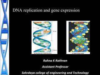
  • 1. DNA replication and gene expression C T A A T CG GC A C G AT AT A T TA C TA 0.34 nm 3.4 nm (a) Key features of DNA structure G 1 nm G (c) Space-filling model T Rahna K Rathnan Assistant Professor Sahrdaya college of engineering and Technology
  • 2. DNA replication • The parent molecule unwinds, and two new daughter strands are built based on base-pairing rules (a) The parent molecule has two complementary strands of DNA. Each base is paired by hydrogen bonding with its specific partner, A with T and G with C. (b) The first step in replication is separation of the two DNA strands. (c) Each parental strand now serves as a template that determines the order of nucleotides along a new, complementary strand. (d) The nucleotides are connected to form the sugar-phosphate backbones of the new strands. Each “daughter” DNA molecule consists of one parentalstrand and one new strand. A C T A G A C T A G A C T A G A C T A G T G A T C T G A T C A C T A G A C T A G T G A T C T G A T C T G A T C T G A T C
  • 3. DNA Replication is “Semi-conservative” • Newly synthesised duplex DNA conserve one strand of parent DNA and other strand will be newly synthesised • One original strand was used as a template to make the new strand
  • 4. DNA Replication • Require – Template DNA – More than dozen enzymes and proteins • DNA synthesis enzyme : DNA polymerase • DNA synthesis direction ---- 5’ to 3’ • The replication of a DNA molecule begins at special sites called origins of replication • the two strands are separated
  • 5. Origins of Replication• A eukaryotic chromosome may have hundreds or even thousands of replication origins Replication begins at specific sites where the two parental strands separate and form replication bubbles. The bubbles expand laterally, as DNA replication proceeds in both directions. the replication bubbles fuse, and synthesis of the daughter strands is complete. 1 2 3 Origin of replication Bubble Parental (template) strand Daughter (new) strand Replication fork Two daughter DNA molecules In eukaryotes, DNA replication begins at many sites along the giant DNA molecule of each chromosome. In this micrograph, three replication bubbles are visible along the DNA of (b)(a) 0.25 µm
  • 6. Mechanism of DNA Replication • DNA replication is catalyzed by DNA polymerase – needs an RNA primer • RNA primase synthesizes primer on DNA strand • DNA polymerase adds nucleotides to the 3’ end of the growing strand
  • 7. Mechanism of DNA Replication • Nucleotides are added by complementary base pairing with the template strand • The substrates, deoxyribonucleoside triphosphates, are hydrolyzed and releasing energy for DNA synthesis.
  • 8. The Mechanism of DNA Replication • DNA synthesis on the leading strand is continuous • The lagging strand grows the same general direction as the leading strand (in the same direction as the Replication Fork). However, DNA is made in the 5’-to-3’ direction • DNA synthesis on the lagging strand is discontinuous • DNA is added as short fragments (Okasaki fragments) – subsequently ligated together
  • 9.
  • 10. DNA polymerase I degrades the RNA primer and replaces it with DNA
  • 11. The Mechanism of DNA Replication • DNA helicases unwind the double helix • Single-stranded DNA binding proteins stabilize the SS DNA • RNA primase catalyzes the synthesis of short RNA primers, to which nucleotides are added. • DNA polymerase III extends the strand in the 5’-to- 3’ direction • DNA polymerase I degrades the RNA primer and replaces it with DNA • DNA ligase joins the DNA fragments into a continuous daughter strand
  • 12. Enzymes in DNA replication Helicase unwinds parental double helix Binding proteins stabilize separate strands DNA polymerase III binds nucleotides to form new strands Ligase joins Okazaki fragments and seals other nicks in sugar- phosphate backbone Primase adds short primer to template strand DNA polymerase I (Exonuclease) removes RNA primer and inserts the correct bases
  • 13. • Helicase protein binds to DNA sequences called origins and unwinds DNA strands. • Single strand Binding proteins prevent single strands from rewinding. • Primase protein makes a short segment of RNA complementary to the DNA, a primer. 5’ 3’ 5’ 3’ 3’5’ 5’3’ Replication
  • 14. Overall direction of replication 5’3’ 5’ 3’ 5’ 3’ 3’5’ DNA polymerase enzyme adds DNA nucleotides to the RNA primer. Replication
  • 15. • DNA polymerase enzyme adds DNA nucleotides to the RNA primer. • DNA polymerase proofreads bases added and replaces incorrect nucleotides. 5’ 5’ Overall direction of replication 5’ 3’ 5’ 3’ 3’ 3’ Replication
  • 16. 5’ 5’3’ 5’ 3’ 3’ 5’ 3’ Overall direction of replication • Leading strand synthesis continues in a 5’ to 3’ direction. Replication
  • 17. 3’5’ 5’ 5’3’ 5’ 3’ 3’ 5’ 3’ Overall direction of replication Okazaki fragment • Discontinuous synthesis produces 5’ to 3’ DNA segments called Okazaki fragments. Replication
  • 18. 5’ 5’ 3’ 5’ 3’ 3’ 5’ 3’ 3’ 5’ 5’3’ Leading strand synthesis continues in a 5’ to 3’ direction. Discontinuous synthesis produces 5’ to 3’ DNA segments called Okazaki fragments. Replication
  • 19. 3’ 5’ 3’ 5’ 5’ 3’ 5’ 3’ 3’ 5’ 5’3’ Leading strand synthesis continues in a 5’ to 3’ direction. Discontinuous synthesis produces 5’ to 3’ DNA segments called Okazaki fragments. Replication
  • 20. 5’ 5’ 3’ 3’ 5’ 3’ 5’ 3’ 5’ 3’ 3’ 5’ • Exonuclease activity of DNA polymerase I removes RNA primers. Replication
  • 21. Polymerase activity of DNA polymerase I fills the gaps. Ligase forms bonds between sugar-phosphate backbone. 3’ 5’ 3’ 5’ 3’ 5’ 3’ 3’ 5’ Replication
  • 23. Gene Expression • Synthesis of protein from DNA through RNA • The process by which DNA directs protein synthesis, gene expression includes two stages – transcription – translation
  • 24. Transcription and Translation • Cells are governed by a cellular chain of command – DNA → RNA → protein • Transcription – Is the synthesis of RNA under the direction of DNA – Produces messenger RNA (mRNA) • Translation – Is the actual synthesis of a polypeptide – occurs under the direction of mRNA – Occurs on ribosomes
  • 25. Transcription and Translation • In a eukaryotic cell the nuclear envelope separates transcription from translation • Extensive RNA processing occurs in the nucleus Eukaryotic cell. The nucleus provides a separate compartment for transcription. The original RNA transcript, called pre-mRNA, is processed in various ways before leaving the nucleus as mRNA. (b) TRANSCRIPTION RNA PROCESSING TRANSLATION mRNA DNA Pre-mRNA Polypeptide Ribosome Nuclear envelope
  • 26. Transcription • Transcription is the DNA-directed synthesis of RNA • RNA synthesis – Is catalyzed by RNA polymerase – uracil substitutes for thymine
  • 27. RNA • RNA is single stranded, not double stranded like DNA • RNA is short, only 1 gene long, where DNA is very long and contains many genes • RNA uses the sugar ribose instead of deoxyribose in DNA • RNA uses the base uracil (U) instead of thymine (T) in DNA. Table 17.1
  • 28. Synthesis of an RNA Transcript • The stages of transcription are – Initiation – Elongation – Termination Promoter Transcription unit RNA polymerase Start point 5′ 3′ 3′ 5′ 3′ 5′ 5′ 3′ 5′ 3′ 3′ 5′ 5′ 3′ 3′ 5′ 5′ 5′ Rewound RNA R N A tr a ns cri pt 3′ 3′ Completed RNA transcript Unwound DNA RNA transcript Template strand of DNA DNA 1 Initiation. After RNA polymerase binds to the promoter, the DNA strands unwind, and the polymerase initiates RNA synthesis at the start point on the template strand. 2 Elongation. The polymerase moves downstream, unwinding the DNA and elongating the RNA transcript 5′ → 3 ′. In the wake of transcription, the DNA strands re-form a double helix. 3 Termination. Eventually, the RNA transcript is released, and the polymerase detaches from the DNA.
  • 29. • Promoters signal the initiation of RNA synthesis • Transcription factors help eukaryotic RNA polymerase recognize promoter sequences TRANSCRIPTION RNA PROCESSING TRANSLATION DNA Pre-mRNA mRNA Ribosome Polypeptide T A T AAA A ATAT T T T TATA box Start point Template DNA strand 5′ 3′ 3′ 5′ Transcription factors 5′ 3′ 3′ 5′ Promoter 5′ 3′ 3′ 5′5′ RNA polymerase II Transcription factors RNA transcript Transcription initiation complex Eukaryotic promoters1 Several transcription factors 2 Additional transcription factors 3 Synthesis of an RNA Transcript - Initiation
  • 30. Synthesis of an RNA Transcript - Elongation • RNA polymerase synthesizes a single strand of RNA against the DNA template strand (anti-sense strand), adding nucleotides to the 3’ end of the RNA chain • As RNA polymerase moves along the DNA it continues to untwist the double helix, exposing about 10 to 20 DNA bases at a time for pairing with RNA nucleotides Elongation RNA polymerase Non-template strand of DNA RNA nucleotides 3′ end C A E G C A A U T A G G T T A A C G U A T C A T C C A A T T G G 3′ 5′ 5′ Newly made RNA Direction of transcription (“downstream”) Template strand of DNA
  • 31. • Specific sequences in the DNA signal termination of transcription • When one of these is encountered by the polymerase, the RNA transcript is released from the DNA and the double helix can zip up again. Synthesis of an RNA Transcript - Termination
  • 33. • Most eukaryotic mRNAs aren’t ready to be translated into protein directly after being transcribed from DNA. • mRNA requires processing. • Transcription of RNA processing occur in the nucleus. – the messenger RNA moves to the cytoplasm for translation. • The cell adds a protective cap to one end, and a tail of A’s to the other end. • These both function to protect the RNA from enzymes that would degrade Post Termination RNA Processing
  • 34.
  • 35. • Most of the genome consists of non-coding regions called introns – Non-coding regions may have specific chromosomal functions or have regulatory purposes – Introns also allow for alternative RNA splicing • Thus, an RNA copy of a gene is converted into messenger RNA by doing 2 things: – Add protective bases to the ends – Cut out the introns Post Termination RNA Processing
  • 36. Alteration of mRNA Ends • Each end of a pre-mRNA molecule is modified in a particular way – The 5′ end receives a modified nucleotide cap – The 3′ end gets a poly-A tail A modified guanine nucleotide added to the 5′ end 50 to 250 adenine nucleotides added to the 3′ end Protein-coding segment Polyadenylation signal Poly-A tail3′ UTR Stop codonStart codon 5′ Cap 5′ UTR AAUAAA AAA…AAA TRANSCRIPTION RNA PROCESSING DNA Pre-mRNA mRNA TRANSLATION Ribosome Polypeptide G P P P 5′ 3′
  • 37. RNA Processing - Splicing • The original transcript from the DNA is called pre-mRNA. • It contains transcripts of both introns and exons. • The introns are removed by a process called splicing to produce messenger RNA (mRNA)
  • 38. RNA Processing • Proteins often have a modular architecture consisting of discrete structural and functional regions called domains • In many cases different exons code for the different domains in a protein Figure 17.12 Gene DNA Exon 1 Intron Exon 2 Intron Exon 3 Transcription RNA processing Translation Domain 3 Domain 1 Domain 2 Polypeptide
  • 39. Translation • Translation is the RNA-directed synthesis of a polypeptide • Translation involves – mRNA – Ribosomes - Ribosomal RNA – Transfer RNA – Genetic coding - codons TRANSCRIPTION TRANSLATION DNA mRNA Ribosome Polypeptide Polypeptide Amino acids tRNA with amino acid attachedRibosome tRNA Anticodon mRNA Trp Phe Gly A G C A A A C C G U G G U U U G G C Codons5′ 3′
  • 40. The Genetic Code • Genetic information is encoded as a sequence of nonoverlapping base triplets, or codons DNA molecule Gene 1 Gene 2 Gene 3 DNA strand (template) TRANSCRIPTION mRNA Protein TRANSLATION Amino acid A C C A A A C C G A G T U G G U U U G G C U C A Trp Phe Gly Ser Codon 3′ 5′ 3′5′
  • 41. The Genetic Code • Codons: 3 base code for the production of a specific amino acid, • Present on mRNA • there are 64 possible codons • 64 codons but only 20 amino acids, therefore most have more than 1 codon • 3 of the 64 codons are used as STOP signals – they are found at the end of every gene and mark the end of the protein • One codon is used as a START signal: it is at the start of every protein • Universal: in all living organisms
  • 42. The Genetic Code • A codon in messenger RNA is either translated into an amino acid or serves as a translational start/stop signal Second mRNA base U C A G U C A G UUU UUC UUA UUG CUU CUC CUA CUG AUU AUC AUA AUG GUU GUC GUA GUG Met or start Phe Leu Leu lle Val UCU UCC UCA UCG CCU CCC CCA CCG ACU ACC ACA ACG GCU GCC GCA GCG Ser Pro Thr Ala UAU UAC UGU UGC Tyr Cys CAU CAC CAA CAG CGU CGC CGA CGG AAU AAC AAA AAG AGU AGC AGA AGG GAU GAC GAA GAG GGU GGC GGA GGG UGG UAA UAG Stop Stop UGA Stop Trp His Gln Asn Lys Asp Arg Ser Arg Gly U C A G U C A G U C A G U C A G FirstmRNAbase(5′end) ThirdmRNAbase(3′end) Glu
  • 43. Transfer RNA • Consists of a single RNA strand that is only about 80 nucleotides long • Each carries a specific amino acid on one end and has an anticodon on the other end • A special group of enzymes pairs up the proper tRNA molecules with their corresponding amino acids. • tRNA brings the amino acids to the ribosomes Two-dimensional structure. The four base-paired regions and three loops are characteristic of all tRNAs, as is the base sequence of the amino acid attachment site at the 3′ end. The anticodon triplet is unique to each tRNA type. (The asterisks mark bases that have been chemically modified, a characteristic of tRNA.) (a) 3′ C C A C G C U U A A GACACCU * G C * * G U G U *CU * G AG G U * *A * A A G U C A G A C C * C G A G A G G G * * GA CUC*AU U U A G G C G 5′ Amino acid attachment site Hydrogen bonds Anticodon A The “anticodon” is the 3 RNA bases that matches the 3 bases of the codon on the mRNA molecule
  • 44. Ribosomes • Ribosomes facilitate the specific coupling of tRNA anticodons with mRNA codons during protein synthesis • The 2 ribosomal subunits are constructed of proteins and RNA molecules named ribosomal RNA or rRNA
  • 45. Composition of whole ribosomes and of ribosomal subunits in mammalian cells
  • 46. Building a Polypeptide • divide translation into three stages – Initiation – Elongation – Termination
  • 47. Building a Polypeptide • The AUG start codon is recognized by methionyl-tRNA or Met • Once the start codon has been identified, the ribosome incorporates amino acids into a polypeptide chain • RNA is decoded by tRNA (transfer RNA) molecules, which each transport specific amino acids to the growing chain • Translation ends when a stop codon (UAA, UAG, UGA) is reached
  • 48. • Translation starts (primarily) at AUG • AUG codes for methionine • 16S rRNA of the small 30S ribosomal subunit recognizes the ribosomal binding site on the mRNA -Shine-Dalgarno sequence or SD • Shine-Dalgarno sequence is complementary to sequence on 16S rRNA. • SD helps to correctly position the ribosome onto the mRNA so that the P site is directly on the AUG initiation codon INITIATION
  • 49.
  • 50.
  • 51.
  • 52.
  • 53. Initiation of Translation • The initiation stage of translation brings together mRNA, tRNA bearing the first amino acid of the polypeptide, and two subunits of a ribosome Large ribosomal subunit The arrival of a large ribosomal subunit completes the initiation complex. Proteins called initiation factors (not shown) are required to bring all the translation components together. GTP provides the energy for the assembly. The initiator tRNA is in the P site; the A site is available to the tRNA bearing the next amino acid. 2 Initiator tRNA mRNA mRNA binding site Small ribosomal subunit Translation initiation complex P site GDPGTP Start codon A small ribosomal subunit binds to a molecule of mRNA. In a prokaryotic cell, the mRNA binding site on this subunit recognizes a specific nucleotide sequence on the mRNA just upstream of the start codon. An initiator tRNA, with the anticodon UAC, base-pairs with the start codon, AUG. This tRNA carries the amino acid methionine (Met). 1 Met Met U A C A U G E A 3′ 5′ 5′ 3′ 3′5′ 3′5′
  • 54. Amino end Growing polypeptide Next amino acid to be added to polypeptide chain tRNA mRNA Codons 3′ 5′ Schematic model with mRNA and tRNA. A tRNA fits into a binding site when its anticodon base- pairs with an mRNA codon. The P site holds the tRNA attached to the growing polypeptide. The A site holds the tRNA carrying the next amino acid to be added to the polypeptide chain. Discharged tRNA leaves via the E site. (c) Building a Polypeptide
  • 55. Elongation of the Polypeptide Chain • In the elongation stage, amino acids are added one by one to the preceding amino acid Amino end of polypeptide mRNA Ribosome ready for next aminoacyl tRNA E P A E P A E P A E P A GDP GTP GTP GDP 2 2 site site5′ 3′ TRANSCRIPTION TRANSLATION DNA mRNA Ribosome Polypeptide Codon recognition. The anticodon of an incoming aminoacyl tRNA base-pairs with the complementary mRNA codon in the A site. Hydrolysis of GTP increases the accuracy and efficiency of this step. 1 Peptide bond formation. An rRNA molecule of the large subunit catalyzes the formation of a peptide bond between the new amino acid in the A site and the carboxyl end of the growing polypeptide in the P site. This step attaches the polypeptide to the tRNA in the A site. 2 Translocation. The ribosome translocates the tRNA in the A site to the P site. The empty tRNA in the P site is moved to the E site, where it is released. The mRNA moves along with its bound tRNAs, bringing the next codon to be translated into the A site. 3
  • 56. Termination of Translation • The final stage is termination when the ribosome reaches a stop codon in the mRNA Release factor Free polypeptide Stop codon (UAG, UAA, or UGA) 5′ 3′ 3′ 5′ 3′ 5′ When a ribosome reaches a stop codon on mRNA, the A site of the ribosome accepts a protein called a release factor instead of tRNA. 1 The release factor hydrolyzes the bond between the tRNA in the P site and the last amino acid of the polypeptide chain. The polypeptide is thus freed from the ribosome. 2 3 The two ribosomal subunits and the other components of the assembly dissociate.
  • 57. Translation • The final step in translation is termination. When the ribosome reaches a STOP codon, there is no corresponding transfer RNA. • Instead, a small protein called a “release factor” attaches to the stop codon. • The release factor causes the whole complex to fall apart: messenger RNA, the two ribosome subunits, the new polypeptide. • The messenger RNA can be translated many times, to
  • 58. A summary of transcription and translation in a eukaryotic cell Figure 17.26 TRANSCRIPTION RNA is transcribed from a DNA template. DNA RNA polymerase RNA transcript RNA PROCESSING In eukaryotes, the RNA transcript (pre- mRNA) is spliced and modified to produce mRNA, which moves from the nucleus to the cytoplasm. Exon Poly-A RNA transcript (pre-mRNA) Intron NUCLEUS Cap FORMATION OF INITIATION COMPLEX After leaving the nucleus, mRNA attaches to the ribosome. CYTOPLASM mRNA Poly-A Growing polypeptide Ribosomal subunits Cap Aminoacyl-tRNA synthetase Amino acid tRNA AMINO ACID ACTIVATION Each amino acid attaches to its proper tRNA with the help of a specific enzyme and ATP. Activated amino acid TRANSLATION A succession of tRNAs add their amino acids to the polypeptide chain as the mRNA is moved through the ribosome one codon at a time. (When completed, the polypeptide is released from the ribosome.) Anticodon A C C A A A U G G U U U A U G U A CE A Ribosome 1 Poly-A 5′ 5′ 3′ Codon 2 3 4 5
  • 59. Post-translation • The new polypeptide is now floating loose in the cytoplasm if translated by a free ribosme. • It might also be inserted into a membrane, if translated by a ribosome bound to the endoplasmic reticulum. • Polypeptides fold spontaneously into their active configuration, and they spontaneously join with other polypeptides to form the final proteins. • Sometimes other molecules are also attached to the polypeptides: sugars, lipids, phosphates, etc. All of these have special purposes for protein function.