2. pleural effusion and chest drain tube vvi
•
8 recomendaciones•6,063 vistas

chest tube drainage

Recomendados
Más contenido relacionado
La actualidad más candente
La actualidad más candente (20)
Destacado
Similar a 2. pleural effusion and chest drain tube vvi
Similar a 2. pleural effusion and chest drain tube vvi (20)
Más de BP KOIRALA INSTITUTE OF HELATH SCIENCS,, NEPAL
Más de BP KOIRALA INSTITUTE OF HELATH SCIENCS,, NEPAL (20)
2. pleural effusion and chest drain tube vvi
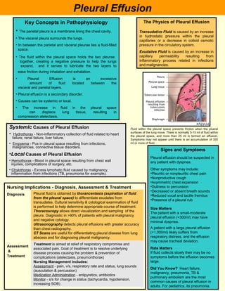
- 1. Pleural Effusion Key Concepts in Pathophysiology The Physics of Pleural Effusion • The parietal pleura is a membrane lining the chest cavity. Transudative Fluid is caused by an increase in hydrostatic pressure within the pleural • The visceral pleura surrounds the lungs. capillaries or a decrease in colloid osmotic • In between the parietal and visceral pleurae lies a fluid-filled pressure in the circulatory system. space. Exudative Fluid is caused by an increase in • The fluid within the pleural space holds the two pleurae capillary permeability resulting from together, creating a negative pressure to help the lungs inflammatory process related in infections expand, and it serves to lubricate the two layers to and malignancies. ease friction during inhalation and exhalation. • Pleural Effusion is an excessive amount of fluid located between the visceral and parietal layers. • Pleural effusion is a secondary disorder. • Causes can be systemic or local. • The increase in fluid in the pleural space can displace lung tissue, resulting in compression atelectasis. Systemic Causes of Pleural Effusion Fluid within the pleural space prevents friction when the plueral surfaces of the lung move. There is normally 5-15 ml of fluid within • Hydrothorax - Non-inflammatory collection of fluid related to heart the pleural space, and more than 25 ml is termed an effusion. failure, renal failure, liver failure. Symptoms may not appear until there is an accumulation of 300 • Empyema - Pus in pleural space resulting from infections, ml or more of fluid. malignancies, connective tissue disorders. Signs and Symptoms •Local Causes of Pleural Effusion Pleural effusion should be suspected in • Hemothorax - Blood in pleural space resulting from chest wall any patient with dyspnea. injuries, complications of surgery, etc. • Chylothorax - Excess lymphatic fluid caused by malignancy, Other symptoms may include: inflammation from infections (TB, pneumonia for example). •Pleuritic or nonpleuritic chest pain •Nonproductive cough •Asymmetric chest expansion Nursing Implications - Diagnosis, Assessment & Treatment •Dullness to percussion •Decreased or absent breath sounds Diagnosis Pleural fluid is obtained by thorancentesis (aspiration of fluid •Reduced vocal and tactile fremitus from the pleural space) to differentiate exudates from •Presence of a pleural rub transudates. Cultural sensitivity & cytological examination of fluid is performed to help determine appropriate course of treatment. Size Matters Thoracoscopy allows direct visualization and sampling of the The patient with a small-moderate pleura. Diagnostic in >90% of patients with pleural malignancy pleural effusion (<300ml) may have and negative cytology. minimal dyspnea. Ultrasonography detects pleural effusions with greater accuracy than chest radiography. A patient with a large pleural effusion CT Scans are useful for differentiating pleural disease from lung (>1,000ml) likely suffers from abscess and for diagnosing pleural malignancy. respiratory distress, and the effusion may cause tracheal deviation. Treatment is aimed at relief of respiratory compromise and Assessment associated pain. Goal of treatment is to resolve underlying Rate Matters & If fluid collects slowly their may be no disease process causing the problem & prevention of Treatment complications (atelectasis, pneumonthorax) symptoms before the effusion becomes Nursing Management includes: large. Assessment - pain, v/s, respiratory rate and status, lung sounds Did You Know? Heart failure, (ausculation & percussion) malignancy, pneumonia, TB & Medication Administration - antipyretics, antibiotics pulmonary embolism are the most Monitor - s/s for change in status (tachycardia, hypotension, common causes of pleural effusion in increasing SOB) adults. For pediatrics, its pneumonia.
- 2. Nursing Considerations Interventions • Manage patient’s anxiety • Manage pain • Provide support during thoracentesis • Monitor for s/s of complications after procedure (e.g., reexpansion pulmonary edema, effusion) • Monitor chest tube drainage system & record drainage • Position on unaffected side to relieve pressure Pain Management R/T Procedures • Tell him what to expect • Educate him on pain management options • Assess his understanding of the pain management regimen • Find out what has worked for him in the past • Don’t assume - sedation isn’t analgesia. Pain medications are still needed • Establish signals that he can use during procedure to indicate needs for more pain meds Management of Patient With Chest Tubing • Monitor v/s q2hr - RR, pattern, depth, SpO2 highest priority • Assess for symmetry of breath sounds bilaterally • Assess insertion site for subcutaneous emphysema • Encourage deep breathing & coughing to promote drainage and lung expansion • Keep tubing free of kinks • Avoid clamping of tubes for extended period of time (prevents escape of air/fluid & increases risk of pneumothorax or cardiac tamponade • Check to ensure connections are secure to chest wall • Keep collection apparatus below patient’s chest level • Ensure water seal fluctuates with respiratory effort (tidaling). If not, check for tube kinks • Record drainage amount & characteristics per protocol - mark chamber levels with date/time • Include drainage in fluid I/O records • Report >70 ml/hr of bright red blood or free-flowing drainage • Assist with ambulation as tolerate Handout References - Coughlin, A.M. & Parchinsky, C. (2006). Go with the flow of chest tube therapy. Nursing, 36(3), 36-42. - D’arcy, Y. (2004). Managing procedural pain. Nursing, 34(12), 76. - Dev, S.P. & Nascimiento B. (2007). Chest-tube insertion. New England Journal of Medicine, 357(15), 17. - Hogan, M.A. & Hill, K. (2004). Pathophysiology Reviews and Rationales. Upper Saddle River, New Jersey: Prentice Hall. - Pendharkar, S.R. (2007). Guidance on how to identify the cause - a diagnostic approach to pleural effusion. Journal of Respiratory Diseases, 28(12), 565. - Porcel, J.M. & Light, R.W. (2006). Diagnostic approach to pleural effusion in adults. American Family Physician, 73(7), 1211-1220. - Smeltzer, S. & Bare, B. (2003). Bunner & Suddarth’s Textbook of Medical-Surgical Nursing (10th ed.). Philadelphia, PA: Lippinott Williams & Wilkins. Abstract obtiained from Nursing: Understanding Pleural Effusion,, 2004, 34(8), 64.