SlideShare una empresa de Scribd logo
1 de 11
Descargar para leer sin conexión
Glucocorticoids Modulate MicroRNA Expression and
Processing during Lymphocyte Apoptosis*□S
Received for publication,July 7, 2010, and in revised form, September 3, 2010 Published, JBC Papers in Press,September 16, 2010, DOI 10.1074/jbc.M110.162123
Lindsay K. Smith‡§
, Ruchir R. Shah¶
, and John A. Cidlowski‡1
From the ‡
Molecular Endocrinology Group, Laboratory of Signal Transduction, NIEHS, National Institutes of Health, Department of
Health and Human Services, Research Triangle Park, North Carolina 27709, the §
Curriculum in Toxicology, University of North
Carolina Chapel Hill, Chapel Hill, North Carolina 27599, and ¶
SRA International, Durham, North Carolina 27713
Glucocorticoids modulate immune development and func-
tion through the induction of lymphocyte apoptosis via mecha-
nisms requiring alterations in gene expression. Recently, short,
noncoding, microRNAs have been identified as key regulators of
lymphocyte function; however, it is unknown whether glucocor-
ticoids regulate noncoding microRNAs and whether this regu-
lation contributes to lymphocyte apoptosis. We now show by
both microarray and deep sequencing analysis that microRNAs
are substantially repressed during glucocorticoid-induced apo-
ptosis of primary rat thymocytes. Mechanistic studies revealed
that primary microRNA transcripts were not repressed, whereas
the expression of the key microRNA processing enzymes: Dicer,
Drosha, and DGCR8/Pasha, were significantly reduced at both
the mRNA and protein levels during glucocorticoid-induced
apoptosis. To delineate the role of Dicer depletion and
microRNA repression in apoptosis, we silenced Dicer expres-
sion in two human leukemic cell lines and demonstrated that
Dicer depletion significantly enhanced glucocorticoid-induced
apoptosis in both model systems. Finally, in vitro and in vivo
overexpression of the conserved miR-17–92 polycistron, which
was repressed significantly by dexamethasone treatment in both
our microarray and deep sequencing studies, blunted glucocor-
ticoid-induced apoptosis. These studies provide evidence of
altered post-transcriptional microRNA expression and the
repression of the microRNA bioprocessing pathway during glu-
cocorticoid-induced apoptosis of lymphocytes, suggesting a role
for microRNA processors and specific microRNAs in cell life/
death decisions.
Recently, noncoding microRNAs have emerged as important
gene expression regulatory elements. Transcribed by RNA
polymerase II, microRNA precursors undergo extensive post-
transcriptional processing by the nuclear RNase III enzyme
Drosha and its co-factor DGCR8 (DiGeorge syndrome critical
region 8)/Pasha followed by Ran-GTP/Exportin-5-dependent
nuclear export. In the cytoplasm, primary microRNAs are fur-
ther cleaved by the RNase III enzyme Dicer to yield a
microRNA duplex. The biologically active, mature, single-
stranded microRNA is then incorporated into the microRNA-
induced silencing complex. The microRNA-bearing microRNA-
induced silencing complex proceeds to affect gene expression
through the inhibitory engagement of complimentary “seed
sequences” within the 3Ј-UTRs of target mRNAs, resulting in
translational inhibition (1, 2).
Through the modulation of gene expression, microRNAs are
key effectors of fundamental biological processes including
development, differentiation, and apoptosis (3). Apoptosis of
lymphocytes is a highly coordinated process governing the
development of the T-cell repertoire during thymocyte selec-
tion as well as the emergence of lymphoproliferative disease
caused by the escape of malignant cells from apoptotic con-
straint. Recently, microRNAs have been implicated in both the
regulation of lymphocyte apoptosis and the development of
hematomalignancy. For example, hsa-miR-15 and -16 are
strongly down-regulated in chronic lymphocytic leukemia and
the expression of the anti-apoptotic oncogene Bcl-2 is inversely
correlated with the expression of these microRNAs (4, 5). Fur-
thermore, overexpression of hsa-miR-15 and -16 in a leukemic
cell line resulted in the reduced expression of Bcl-2 and the
induction of apoptosis, suggesting a pro-apoptotic tumor sup-
pressor function for microRNAs 15 and 16 in human lympho-
cytes (6). Conversely, members of the hsa-miR-17–92 polycis-
tron are up-regulated in B-cell lymphomas. Adoptive transfer
of hematopoietic stem cells bearing a truncated portion of the
mmu-miR-17–92 polycistron in c-Myc transgenic mice
resulted in the rapid onset of malignant lymphomas. These
lymphomas exhibited resistance to apoptosis and increased
proliferation (7). Transgenic overexpression of the entire hsa-
miR-17–92 polycistron in the murine hematopoietic compart-
ment led to the development of lymphoproliferative disease
and increased lethality. Lymphocytes derived from these ani-
mals displayed reduced sensitivity to Fas ligand-induced cell
death as well as increased proliferation (8). Finally, the expres-
sion of the pro-apoptotic proteins, Bim2
and phosphatase and
tensin homolog, was decreased in response to hsa-miR-17–92
overexpression, suggesting a conserved, anti-apoptotic func-
tion for the miR-17–92 polycistron in lymphocytes.
Glucocorticoids are key effectors of lymphocyte develop-
ment through the induction of apoptosis and the mutual antag-
onism of T-cell receptor activation-induced apoptosis during
thymocyte selection (9). Because of their ability to induce rapid
* Thisworkwassupported,inwholeorinpart,byNationalInstitutesofHealth
Grant Z01E5090057-12.
□S
The on-line version of this article (available at http://www.jbc.org) contains
supplemental text, Table S1, and Figs. S1 and S2.
1
To whom correspondence should be addressed: 111 TW Alexander Dr., Rm.
F349, Research Triangle Park, NC 27709. Tel.: 919-541-1564; Fax: 919-541-
1367; E-mail: cidlows1@niehs.nih.gov.
2
Theabbreviationsusedare:Bim,Bcl-2interactingmediatorofcelldeath;GR,
glucocorticoid receptor; miRNA, microRNA.
THE JOURNAL OF BIOLOGICAL CHEMISTRY VOL. 285, NO. 47, pp. 36698–36708, November 19, 2010
Printed in the U.S.A.
36698 JOURNAL OF BIOLOGICAL CHEMISTRY VOLUME 285•NUMBER 47•NOVEMBER 19, 2010
atUNIVOFSOUTHFLORIDA,onSeptember8,2011www.jbc.orgDownloadedfrom
http://www.jbc.org/content/suppl/2010/09/16/M110.162123.DC1.html
Supplemental Material can be found at:
apoptosis of lymphocytes, synthetic glucocorticoids are also
critical components of hematomalignant chemotherapy regi-
mens. Glucocorticoid-induced apoptosis of lymphocytes is a
multifaceted process, requiring signaling through the glucocor-
ticoid receptor (GR) and the subsequent transactivation of apo-
ptotic effector genes (10–13). The requirement for de novo
transcriptional events in the execution of glucocorticoid-in-
duced apoptosis is well established. However, it is not clear
whether the expression of microRNAs is altered during glu-
cocorticoid-induced apoptosis of lymphocytes and, if so,
whether such alterations contribute to the cell death process.
Here, we provide evidence of prevalent post-transcriptional
repression of microRNA expression during glucocorticoid-in-
duced apoptosis of primary rat thymocytes and further demon-
strate the repression of the microRNA bioprocessors, Dicer,
Drosha, and DGCR8/Pasha. Additionally, the stable depletion
of Dicer in glucocorticoid-sensitive leukemic cell lines signifi-
cantly enhanced glucocorticoid-induced apoptosis and overex-
pression of the glucocorticoid-repressed hsa-miR-17–92 poly-
cistron-blunted glucocorticoid-induced apoptosis, indicating
that the repression of microRNA expression and processing
contributes to the progression of glucocorticoid-induced lym-
phocyte apoptosis.
EXPERIMENTAL PROCEDURES
Rat Primary Thymocyte Isolation—Rat primary thymoyctes
were isolated from adrenalectomized male Sprague-Dawley
rats (60–75 g) ϳ1–2 weeks after surgery. Following decapita-
tion, the thymi of three animals were removed and pooled in
RPMI 1640 medium containing 10% heat-inactivated fetal
bovine serum, 4 mM glutamine, 75 units/ml streptomycin,
and 100 units/ml penicillin. Thymi were gently sheared with
surgical scissors at room temperature. Sheared cells were
filtered through 200-micron nylon mesh twice and centri-
fuged at 3,000 ϫ g for 5 min at room temperature. The cell
pellet was then resuspended in fresh media and filtered into
a sterile conical tube. The cells were cultured at a final con-
centration of 2 ϫ 106
cells/ml and incubated at 37 °C, 5%
CO2 atmosphere.
Mouse Primary Thymocyte Isolation—Primary thymocytes
were isolated from adrenalectomized male B57BL/6 wild-type
control or hsa-miR-17–92 transgenic mice at 20 weeks of age.
Following cervical dislocation, individual thymi were
removed and gently sheared to dissociate free thymocytes.
Sheared cells were filtered through 200-micron nylon mesh
twice and centrifuged at 3,000 ϫ g for 5 min at room tem-
perature. The cell pellet was then resuspended in fresh
medium and filtered into a sterile conical tube. The cells
were cultured at a final concentration of 2 ϫ 106
cells/ml and
incubated at 37 °C, 5% CO2 atmosphere.
Human Cell Culture—The Jurkat GR␣ cell line was kindly
generated by Dr. Nick Lu in our laboratory. Briefly, parental
Jurkat cells were transfected with pTRE hGR␣ vector (14) using
the Lonza Nucelofector System (Basel, Switzerland). Stably
overexpressing clones were established by hygromycin/G418
selective pressure (200 ␮g/ml). CEM-C7 leukemic cells were
obtained from Dr. Carl Bortner. For all experiments, Jurkat
GR␣ and CEM-C7 cells were cultured at a density of 2 ϫ 105
cells/ml in RPMI 1640 medium containing 10% heat-inacti-
vated fetal bovine serum, 4 mM glutamine, 75 units/ml strepto-
mycin, and 100 units/ml penicillin.
Dicer Depleted Cells—CEM-C7 and Jurkat GR␣ cells were
transduced with lentivirus encoding Dicer shRNA or nontar-
geting control at a multiplicity of infection of 5. Stable Dicer
depleted cells were established by puromycin selection (2
␮g/ml). Anti-Dicer and nontargeting control Mission shRNAs
were purchased from Sigma-Aldrich.
miR-17–92-overexpressing Cells—Jurkat GR␣ cells were
transduced with lentivirus encoding hsa-miR-17–92 pre-
miRNA or empty vector control at a multiplicity of infection of
10. Transduction at this multiplicity of infection resulted in a
transduction efficiency of Ͼ90% as determined by CopGFP
expression. Lenti-miR hsa-miR-17–92 precursor and empty
vector clones were purchased from System Biosciences (Moun-
tain View, CA).
Lentivirus Generation—All of the lentiviruses were packaged
in HEK293T cells according to established protocols (15).
HEK293T cells were transiently transfected with pMD2G,
psPAX2, and transfer vector containing the desired gene or
miRNA using Lipofectamine 2000. Supernatant was collected
48 h post-transfection and concentrated by centrifugation at
50,000 ϫ g for 2 h. The pellets were resuspended in PBS and
used for infection. All of the titers were determined by quanti-
tative PCR analysis of lentiviral integration into the host
genome. In addition, titration of viruses co-expressing fluores-
cent moieties was determined by flow cytometric analysis.
MicroRNA Microarray—Rat primary thymocytes were iso-
lated and cultured in the presence or absence of 100 nM dexa-
methasone for 6 h. Following treatment, total RNA was isolated
from untreated control and dexamethasone-treated samples
and subjected to microarray analysis using Agilent rat miRNA
V2 (design identification 019159) 8 ϫ 15 multiplex format
arrays (Agilent Technologies) following the Agilent one-color
miRNA complete labeling and hybridization kit protocol. Start-
ing with 70–100 ng of total RNA, the cytidine bisphosphate
reagent was used to selectively label mature miRNAs accord-
ing to manufacturer’s protocol. In combination with the
miRNA microarray probe design, the cytidine bisphosphate
reagent ensures detection of both low abundance and highly
homologous miRNAs. Each labeled sample was hybridized
for 20 h in a rotating hybridization oven. The slides were
washed and then scanned with an Agilent scanner. The data
were obtained using the Agilent feature extraction software
(v9.5), using the miRNA defaults for all parameters. The Agi-
lent feature extraction software performed error modeling,
adjusting for additive and multiplicative noise. The resulting
data were processed using the Rosetta Resolver௡ system
(version 7.2) (Rosetta Biosoftware, Kirkland, WA). Error-
weighted ratios and the associated p values were generated
by Resolver to identify differentially expressed probes. A p
value of Ͻ0.05 was used to generate gene lists of differen-
tially expressed microRNAs.
MicroRNA Deep Sequencing—Rat primary thymocytes were
isolated and cultured in the presence of absence of 100 nM dexa-
methasone for 6 h. Following treatment, total RNA was isolated
from untreated control and dexamethasone-treated samples
Glucocorticoids Modulate MicroRNA Expression and Processing
NOVEMBER 19, 2010•VOLUME 285•NUMBER 47 JOURNAL OF BIOLOGICAL CHEMISTRY 36699
atUNIVOFSOUTHFLORIDA,onSeptember8,2011www.jbc.orgDownloadedfrom
and subjected to microRNA deep sequencing. Small RNA
cDNA libraries were prepared according to the manufactur-
er’s protocol (small RNA sample prep kit, oligonucleotide
only, protocol 71003; Illumina, Inc., San Diego, CA). Small
RNA cDNA libraries were then sequenced according to the
manufacturer’s instructions on the Illumina Genome Ana-
lyzer II.
Bioinformatics—Reads were aligned to the reference rat
genome (2004 assembly) using Bowtie (16). Given that mature
microRNAs are usually ϳ18–20 nucleotides in length, we used
the first 20 nucleotides from the 5Ј end of the read sequence for
the alignment (to avoid the adapter sequence that may be pres-
ent in the read sequence). We allowed up to two mismatches
and up to three alignments of same quality per read sequence.
Reads that aligned at more than three unique genomic locations
were removed from further analysis to avoid repetitive ele-
ments. Fold changes were calculated as a ratio of total number
of reads in the control divided by the total number of reads in
FIGURE 1. Evaluation of glucocorticoid-induced apoptosis of primary thymocytes. Primary thymocytes were isolated and treated with 100 nM dexameth-
asone (Dex) for 0, 6, or 12 h. Apoptotic progression was monitored via flow cytometric analysis of phosphatidylserine exposure, plasma membrane integrity,
and caspase-3 activation. A, as cells undergo apoptosis, the internal phosphatidylserine component of the plasma membrane becomes externalized. This
increased externalization was detected via fluorescently labeled annexin V. Additionally, the cells undergoing apoptosis exhibit a decrease in plasma mem-
brane integrity. This loss of plasma membrane integrity was detected via the uptake of propidium iodide (PI) vital dye. The results are expressed as percentages
of positive mean values Ϯ S.E. for three independent experiments. *, p Ն 0.05). B, apoptosis culminates in the activation of the effector caspase, caspase-3.
Caspase-3 activation was monitored via the binding of fluorescently labeled CaspaTag to active caspase-3. The results are expressed as percentage positive
mean values Ϯ S.E. for three independent experiments. *, p Ն 0.05). Con, control.
Glucocorticoids Modulate MicroRNA Expression and Processing
36700 JOURNAL OF BIOLOGICAL CHEMISTRY VOLUME 285•NUMBER 47•NOVEMBER 19, 2010
atUNIVOFSOUTHFLORIDA,onSeptember8,2011www.jbc.orgDownloadedfrom
dexamethasone treatment, as implemented in Erange (17) for
sites that contained at least 20 aligned reads in either of the two
samples. Sites that were within 100 bp of a previously annotated
microRNA and displayed fold change of 2.0 or greater in either
direction were selected for detailed analyses.
Quantitative PCR Analysis—Total RNAs were isolated using
the Ambion mirVana miRNA isolation kit (Austin, TX). For
mature microRNA and primary microRNA analysis, total
RNAs were reverse transcribed
using the TaqMan microRNA
reverse transcription kit and ana-
lyzed using TaqMan mature
microRNA and primary microRNA
assays (Applied Biosystems). For
gene expression analysis, total
RNAs were reverse transcribed
using the TaqMan reverse tran-
scription kit and analyzed with Taq-
Man Gene Expression Assays
(Applied Biosystems). All of the
quantitative PCRs were performed
on a 7900HT quantitative PCR plat-
form (Applied Biosystems).
Western Blotting and Antibodies—
The cells were harvested and lysed
in 1ϫ Laemmli buffer containing
2.5% ␤-mercaptoethanol (Bio-Rad).
Whole cell lysates (20–30 ␮g) were
fractionated on 6% or 8% Tris-gly-
cine gels (Invitrogen) and trans-
ferred to nitrocellulose membranes.
The membranes were probed over-
night with the following antibodies:
Dicer rabbit polyclonal, 1:200
(Santa Cruz Biotechnology, Santa
Cruz, CA); Drosha rabbit poly-
clonal, 1:200 (Santa Cruz Biotech-
nology); DGCR8/Pasha goat poly-
clonal, 1:1000 (Abnova, Taipei City,
Taiwan); and ␤-actin, 1:10,000 (Mil-
lipore, Billerica, MA).
Apoptosis Assays—Apoptosis was
evaluated via annexin V/propidium
iodide staining or propidium iodide
staining alone. For annexin V analy-
sis, the cells were stained with
annexin V according to the manu-
facturer’s protocol and counter-
stained with propidium iodide
(Trevigen, Gaithersburg, MD).
Caspase-3 activation was moni-
tored via CaspaTag fluorescence,
and the cells were stained accord-
ing to manufacturer’s instructions
(Millipore). For propidium iodide
staining alone, propidium iodide
(Invitrogen) was added to achieve a
final concentration of 10 ␮g/ml.
Following staining, the cells were immediately analyzed on a
Becton Dickinson FACSort cytometer equipped with
CellQuest software.
Animals—Adrenalectomized hsa-miR-17–92 transgenic
mice or C57BL/6 wild-type controls were obtained from Jack-
son Labs (Bar Harbor, ME). Adrenalectomized rats were
obtained from Charles River Laboratories (Wilmington, MA).
All of the experiments were performed in accordance with the
FIGURE 2. Repression of microRNA expression during glucocorticoid-induced apoptosis of primary thy-
mocytes. Primary thymocytes were isolated and treated with 100 nM dexamethasone for 6 h. Total RNAs were
subjected to microRNA microarray analysis on the Agilent rat genome microRNA array. A, heat map represen-
tation of microRNAs differentially expressed in control (Con) and dexamethasone (Dex)-treated primary thy-
mocytes. Red indicates microRNAs induced by dexamethasone treatment, and green indicates microRNAs
repressed (three independent biological replicates, p Ͻ 0.05). B, percentage of repression of abundantly
expressed microRNAs (Micro, basal intensity valuesϾ200). The results are calculated as intensity dexametha-
sone-treated/intensityuntreated.C,quantitativePCRvalidationofmicroRNAmicroarray.MicroRNAmicroarray
results were validated by quantitative PCR analysis of three microRNAs significantly regulated on the array as
well as one microRNA (rno-miR-17) not significantly regulated on the array. The results are expressed as per-
centages of control mean values Ϯ S.E. for three independent experiments. *, p Ն 0.05. D, quantitative PCR
analysis of primary microRNA expression. rno-miR-17 and rno-miR-let-7a primary microRNA transcript expres-
sion was analyzed via quantitative PCR and normalized to the GusB housekeeping gene. The results are
expressed as percentages of control mean values Ϯ S.E. for three independent experiments.
Glucocorticoids Modulate MicroRNA Expression and Processing
NOVEMBER 19, 2010•VOLUME 285•NUMBER 47 JOURNAL OF BIOLOGICAL CHEMISTRY 36701
atUNIVOFSOUTHFLORIDA,onSeptember8,2011www.jbc.orgDownloadedfrom
NIEHS, National Institutes of Health Institutional Animal Care
and Use Committee.
RESULTS
Repression of MicroRNA Expression during Glucocorticoid-
induced Apoptosis—For these studies, primary rat thymocytes,
a classic model of glucocorticoid-induced apoptosis, were cul-
tured in vitro in the presence or absence of the synthetic glu-
cocorticoid dexamethasone for 6 h. Apoptotic progression was
monitored in control and glucocorticoid-treated cells via flow
cytometric analysis of phosphatidylserine exposure, plasma
membrane integrity, and caspase-3 activation. Following 6 h of
glucocorticoid treatment, we observed an apoptotic population
of cells. These cells exhibited phosphatidylserine externaliza-
tion as determined by a significant increase in annexin-FITC
fluorescence and decreased plasma membrane integrity dem-
onstrated by a modest increase in propidium iodide fluores-
cence (Fig. 1A). These cells also exhibited an increase in
caspase-3 activity determined by an increase in CaspaTag fluo-
rescence, indicating the clear induction of apoptosis following
6 h of glucocorticoid treatment (Fig. 1B). These apoptotic indi-
cators persisted following 12 h of treatment, resulting in signif-
icant cell death (Fig. 1). Therefore, we performed our
microRNA expression analysis early
in the apoptotic process (prior to
cell death) after 6 h of glucocorti-
coid treatment.
Following 6 h of treatment, total
RNAs from control and glucocorti-
coid-treated cells were prepared
and subjected to microRNA
microarray analysis. Of the 350
microRNAs represented on our
chip, 56 were differentially ex-
pressed in response to glucocorti-
coid-induced apoptosis (Fig. 2A).
Surprisingly, the majority of these
microRNAs (79% or 44 microRNAs)
were repressed, and only 12 low
abundance microRNAs were
weakly induced during this time
frame. Given the low basal ex-
pression levels of these induced
microRNAs, we were unable to con-
firm their up-regulation via quanti-
tative PCR. Because microRNA
expression levels can vary widely,
we focused our efforts on the abun-
dantly expressed and thus poten-
tially biologically active microRNAs
(basal intensity values Ͼ200). The
percent repression values (inten-
sity glucocorticoid/intensity con-
trol) were generated for these
microRNAs (Fig. 2B). Interest-
ingly, these abundantly expressed
microRNAs were repressed uni-
formly irrespective of their genomic
locations, suggesting that the repression of microRNA expres-
sion during glucocorticoid-induced apoptosis occurs post-
transcriptionally, perhaps at the level of bioprocessing. The
repression of a subset of microRNAs identified as differentially
expressed (rno-let-7a, rno-miR-15b, and rno-miR-25) as well as
the repression of a microRNA not significantly regulated in the
microarray (rno-miR-17) was validated via quantitative PCR
analysis (Fig. 2C). Interestingly, the expression of rno-miR-17
and rno-let-7a primary transcripts was increased during glu-
cocorticoid-induced apoptosis, providing important evidence
that the repression of the resultant mature microRNAs occurs
post-transcriptionally (Fig. 2D).
To evaluate the expression of the microRNAnome during
glucocorticoid-induced apoptosis by an alternative approach,
we performed next generation sequencing or deep sequencing
of microRNAs from the control and glucocorticoid-treated
samples. These samples were obtained from independent ani-
mals rather than those described in Fig. 2; however, these cells
exhibited apoptotic kinetics similar to those described in Fig. 1.
Deep sequencing of microRNAs is an open-ended and hybrid-
ization-independent approach that facilitates the detection of
both annotated and novel microRNAs. In our study, massively
parallel sequencing of control and glucocorticoid-treated RNA
FIGURE3.MicroRNAdeepsequencingconfirmstheprevalentrepressionofmicroRNAexpressionduring
glucocorticoid-induced apoptosis. Primary thymocytes were isolated and treated with 100 nM dexametha-
sone (Dex) for 6 h. Total RNAs were subjected to microRNA deep sequencing analysis. A, deep sequencing of
two biological replicates was performed using the Illumina Solexa platform for each treatment group, and the
resulting sequences from replicates were combined for further analyses. Sequences were aligned to the refer-
ence rat genome and sites that were enriched with reads were selected for further analysis. B, peak detection
and ratio calculation revealed prevalent down-regulation of microRNAs in response to dexamethasone treat-
ment.Theratioswerecalculatedascontrolversusdexamethasone(microRNAsrepressed)anddexamethasone
versus control (microRNAs induced). C, percentage of repression of abundantly expressed microRNAs corre-
lates with microRNA microarray results. The results are calculated as intensity dexamethasone-treated/inten-
sity untreated presented as percentages of control. ND denotes microRNAs detected by microarray analysis
but not deep sequencing analysis. MicroRNAs highlighted in gray indicate a subset of microRNAs repressed in
deep sequencing analysis but not microarray analysis.
Glucocorticoids Modulate MicroRNA Expression and Processing
36702 JOURNAL OF BIOLOGICAL CHEMISTRY VOLUME 285•NUMBER 47•NOVEMBER 19, 2010
atUNIVOFSOUTHFLORIDA,onSeptember8,2011www.jbc.orgDownloadedfrom
samples generated millions of short sequences (commonly
referred to as “reads”), which were aligned to the reference rat
genome (Fig. 3A). We identified a set of 1782 sites (genomic loci
that may be encoding microRNAs) that were down-regulated
and 238 sites that were up-regulated by glucocorticoid treat-
ment (see “Experimental Procedures” for more details). Of the
1782 sites, 132 sites were within 100 bp of an annotated
microRNA, which correspond to 129 individual microRNAs
(supplemental Table S1). It is also possible that the sites not
immediately adjacent to a known microRNA may house novel
microRNAs, which warrants fur-
ther evaluation. Similar to the afore-
mentioned microarray study, we
detected a number of sites that
were up-regulated by dexametha-
sone treatment; however, none of
the 238 up-regulated sites were in
close proximity to an annotated
microRNA (Fig. 3B). These results
obtained by deep sequencing mirror
the microarray findings of prevalent
microRNA repression and minimal
microRNA induction during glu-
cocorticoid-induced apoptosis of
primary thymocytes. In addition,
the percent repression values of
abundantly expressed microRNAs
generated using deep sequencing
data were comparable with those
generated using microarray data, fur-
ther suggesting the post-transcrip-
tional repression of microRNAs
during glucocorticoid-induced apo-
ptosis of lymphocytes (Fig. 3C).
Deep sequencing analysis con-
firmed the repression of 35 of the 44
microRNAs repressed in the
microarray analysis (79%). More-
over, microRNA deep sequencing
was also able to detect repressed
microRNAs not detected by mi-
croarray (representative values in
gray in Fig. 3C), suggesting that
deep sequencing of microRNAs is a
more sensitive technique that may
be used in conjunction with
microRNA microarray and quanti-
tative PCR as a novel “weight of evi-
dence” approach to comprehensive
profiling of the microRNAnome.
MicroRNA Processing Enzymes
Are Dysregulated during Gluco-
corticoid-induced Apoptosis—The
broad, genome-wide down-regula-
tion of microRNA expression
detected by both microarray and
deep sequencing analysis and the
absence of primary microRNA tran-
script repression suggest post-transcriptional repression of
mature microRNAs during glucocorticoid-induced apoptosis
of lymphocytes. To evaluate this hypothesis, we examined the
expression of the key microRNA processing enzymes Drosha,
DGCR8/Pasha, and Dicer during glucocorticoid-induced apo-
ptosis of primary thymocytes. Following 6 h of glucocorticoid
treatment, the mRNA expression of each processor is
repressed, with the repression of Dicer and DGCR8/Pasha
mRNA achieving statistical significance (Fig. 4A). After 12 h of
treatment, the mRNA levels of each processor are significantly
FIGURE 4. MicroRNA processing enzymes are repressed during glucocorticoid-induced apoptosis of pri-
mary thymocytes. Primary thymocytes were isolated and treated with 100 nM dexamethasone (Dex) for 6 and
12 h. A, the mRNA expression of the microRNA processing enzymes Drosha, DGCR8/Pasha, and Dicer was
evaluated by quantitative PCR and normalized to the GusB housekeeping gene. The results are expressed as
the percentages of control (Con) mean values Ϯ S.E. for three independent experiments. *, p Ն 0.05. B, whole
cell lysates were immunoblotted with Drosha, DGCR8/Pasha, or Dicer polyclonal antibodies. Actin immuno-
blotting served as a loading control, and the results are representative of at least three independent experi-
ments. C, induction of Bim and Gelsolin mRNA expression during glucocorticoid-induced apoptosis. Primary
thymocyteswereisolatedandtreatedwith100nM dexamethasonefor6and12h.ThemRNAexpressionofBim
and Gelsolin was evaluated by quantitative PCR and normalized to the GusB housekeeping gene. The results
are expressed as the percentages of control mean values Ϯ S.E. for three independent experiments. *, p Ն 0.05.
Glucocorticoids Modulate MicroRNA Expression and Processing
NOVEMBER 19, 2010•VOLUME 285•NUMBER 47 JOURNAL OF BIOLOGICAL CHEMISTRY 36703
atUNIVOFSOUTHFLORIDA,onSeptember8,2011www.jbc.orgDownloadedfrom
repressed in response to glucocorticoid-induced apoptosis (Fig.
4A). The reduced expression of microRNA processing enzymes
is not due to an apoptosis-related global increase in RNase
activity because the expression of the glucocorticoid-respon-
sive genes Gelsolin and Bim are strongly induced under these
conditions in our cells (Fig. 4C).
The repression of microRNA processing enzymes was also
evident at the level of protein expression (Fig. 4B). Drosha pro-
Glucocorticoids Modulate MicroRNA Expression and Processing
36704 JOURNAL OF BIOLOGICAL CHEMISTRY VOLUME 285•NUMBER 47•NOVEMBER 19, 2010
atUNIVOFSOUTHFLORIDA,onSeptember8,2011www.jbc.orgDownloadedfrom
tein expression was decreased following 6 h of glucocorticoid
treatment and was essentially undetectable at 12 h. The expres-
sion of Drosha’s co-factor DGCR8/Pasha was also reduced in
response to glucocorticoid treatment and upon overexposure, a
clear cleavage pattern emerged following 12 h of treatment.
Furthermore, the protein expression of the cytoplasmic pro-
cessor, Dicer, was also markedly reduced following 6 h of glu-
cocorticoid treatment, whereas at 12 h full-length Dicer was
barely detectable (Fig. 4C). This repression of the microRNA
bioprocessing pathway results in the nuclear accumulation of
unprocessed primary microRNAs (supplemental Fig. S1),
accounting for the increased expression of microRNA primary
transcripts observed during glucocorticoid-induced apoptosis
(Fig. 2D).
MicroRNA Repression Enhances Glucocorticoid-induced
Apoptosis of Lymphocytes—To assess whether the repression of
microRNA expression contributes to the cell death process, we
silenced the expression of Dicer, the enzyme responsible for
mature microRNA biosynthesis, in two human lymphoid cell
lines. The transition to cultured cell lines was necessary because
primary rat thymocytes are difficult to genetically manipulate
in vitro. Therefore, we performed our functional analyses in the
Jurkat GR␣ and the CEM-C7 leukemic cell lines. The Jurkat
GR␣ cell line was generated by the exogenous expression of the
full-length GR␣ isoform in the glucocorticoid-resistant Jurkat
ALL parental cell line (18), and stable expression of GR␣
restores glucocorticoid sensitivity to Jurkat cells (Fig. 5A). The
glucocorticoid-sensitive CEM-C7 cell line contains an endoge-
nous, hormone-responsive GR␣ and exhibits slower apoptotic
kinetics in response to glucocorticoid compared with the Jurkat
GR␣ cell line (Fig. 5A). Interestingly, similar to the results
obtained in primary cell culture, Dicer expression is repressed
at both the mRNA and protein levels in each leukemic cell line
during glucocorticoid-induced apoptosis (Fig. 5B). Stable Dicer
depletion in these cells was achieved via lentiviral transduction
of shRNA (Fig. 5C), and the depletion of Dicer resulted in the
reduced expression of a subset of microRNAs (Fig. 5D). Dicer
depletion and the subsequent repression of microRNA expres-
sion augmented glucocorticoid induced apoptosis in both cell
lines as determined by flow cytometric evaluation of phosphati-
dylserine exposure and plasma membrane integrity (Fig. 5E).
Importantly, this increased sensitivity to glucocorticoid-in-
duced apoptosis was not due to increased GR expression in the
absence of Dicer expression (data not shown). These data sug-
gest that Dicer depletion and subsequent microRNA repression
enhance the progression of glucocorticoid-induced apoptosis
in lymphocytes.
Overexpression of the Repressed miR-17–92 Polycistron
Blunts Glucocorticoid-induced Apoptosis—To delineate the
potential role of specific microRNAs down-regulated during
glucocorticoid-induced apoptosis, we sought to overexpress
the anti-apoptotic miR-17–92 polycistron. Members of this
polycistron were repressed by glucocorticoid treatment in pri-
mary thymocytes, as determined by both the microarray and
deep sequencing studies. Furthermore, mature members of this
polycistron are also down-regulated during glucocorticoid-in-
duced apoptosis of cultured leukemic cells (Fig. 6A). Overex-
pression of the hsa-miR-17–92 polycistron via lentiviral trans-
duction of hsa-miR-17–92 precursor microRNA resulted in
efficient overexpression of individual mature hsa-miR-17–92
polycistron members in the Jurkat GR␣ cell line (Fig. 6B). Over-
expression of hsa-miR-17–92 polycistron members in the Jur-
kat GR␣ background significantly diminished the ability of both
subsaturating and saturating doses of glucocorticoid to induce
glucocorticoid-induced apoptosis (Fig. 6C). This diminished
sensitivity was not due to decreased GR expression in response
to hsa-miR-17–92 overexpression (data not shown). In addi-
tion, primary thymocytes derived from mice transgenic for the
hsa-miR-17–92 polycistron (ϳ1.5–2-fold overexpression of
mature hsa-miR-17–92 polycistron members (8)) in the lym-
phocyte compartment exhibited decreased sensitivity to glu-
cocorticoid-induced apoptosis (Fig. 6D). This decreased sensi-
tivity to glucocorticoid-induced apoptosis may also contribute
to the hematomalignant phenotype of these mice. These data
suggest that the specific repression of miR-17–92 polycistron
contributes to glucocorticoid-induced apoptosis of lympho-
cytes in vitro and in vivo.
DISCUSSION
Glucocorticoid-induced apoptosis is a key component of
thymocyte development. Furthermore, because of their potent
apoptosis-inducing properties, synthetic glucocorticoids are
common components of hematomalignant chemotherapy reg-
imens. Here we provide evidence of broad microRNA repres-
sion during glucocorticoid-induced apoptosis of lymphocytes.
This repression was associated with the reduction of both
nuclear (Drosha and DGCR8/Pasha) and cytoplasmic (Dicer)
FIGURE 5. MicroRNA depletion contributes to glucocorticoid-induced apoptosis in human leukemic cell lines. A, flow cytometric analysis of glucocorti-
coid-induced apoptosis in Jurkat GR␣ and CEM-C7 ALL leukemic cell lines. The cells were treated with 100 nM dexamethasone (Dex), and apoptotic progression
was monitored at 24, 48, and 72 h via flow cytometric analysis of propidium iodide (PI) uptake. The results are represented as the mean percentages of
PI-positive values Ϯ S.E. for six independent experiments. *, p Ն 0.05. B, repression of Dicer expression during glucocorticoid-induced apoptosis of human
leukemic cell lines. The cells were treated with 100 nM dexamethasone for 24 and 48 h. The expression of Dicer mRNA was evaluated in total RNA via
quantitative PCR. The expression of GusB mRNA served as an endogenous control (Con). The results are represented as percentages of control mean values Ϯ
S.E. for three independent experiments. *, p Ն 0.05. Whole cell lysates were immunoblotted with Dicer polyclonal antibody. Actin immunoblotting served as
a loading control. The results are representative of three independent experiments. C, stable silencing of Dicer protein expression in Jurkat GR␣ and CEM-C7
lymphocyte cell lines via lentiviral transduction of shRNA. Whole cell lysates from cells stably transduced with nontargeting control or anti-Dicer shRNA (Dicer
knock down (KD)), were immunoblotted with Dicer polyclonal antibody. Actin immunoblotting served as a loading control, and the results are representative
ofthreeindependentexperiments.D,repressionofmaturemicroRNAexpressionintheabsenceofDicerexpression.TotalRNAsfromnontargetingcontroland
Dicer KD cells were evaluated for the expression of three independent mature microRNAs via quantitative PCR. The expression of Rnu43 small nuclear RNA
served as an endogenous control. The results are reported as percentages of nontargeting control values Ϯ S.E. for three independent experiments. *, p Ն 0.05.
E, enhancement of glucocorticoid-induced apoptosis in Dicer KD cells. Flow cytometric analysis of annexin-FITC and propidium iodide uptake in nontargeting
control and Dicer KD stable cell lines in response to 48 h of 100 nM dexamethasone treatment. The results were normalized to untreated control and are
representative of three independent experiments. *, p Ն 0.05.
Glucocorticoids Modulate MicroRNA Expression and Processing
NOVEMBER 19, 2010•VOLUME 285•NUMBER 47 JOURNAL OF BIOLOGICAL CHEMISTRY 36705
atUNIVOFSOUTHFLORIDA,onSeptember8,2011www.jbc.orgDownloadedfrom
microRNA processing enzymes and the increased expression of
primary microRNA transcripts.
Dicer repression and cleavage during apoptosis has recently
been reported in laboratory-derived human endothelial cells,
HL-60 leukemic cells, and HeLa cervical carcinoma cells in
response to apoptotic stimuli; however, none of these reports
evaluated the expression of the nuclear processors Drosha and
DGCR8/Pasha during apoptosis (19–22). This reduction of
Dicer protein expression has recently been attributed to
caspase-3 cleavage (20). The precise mechanism of Dicer
mRNA repression during apoptosis remains undetermined.
Interestingly, we show that Dicer repression in response to glu-
cocorticoid-induced apoptosis is conserved in both healthy and
transformed lymphocytes. Therefore, the results of this study
expand our current understanding of Dicer repression in
response to apoptotic stimulation while being the first to detail
the repression of all three key microRNA processing enzymes
during glucocorticoid-induced apoptosis of lymphocytes.
Additional studies in our laboratory have revealed that alterna-
tive apoptotic stimuli, specifically Fas ligand and UVC, also pro-
mote the repression of microRNA expression and processing
enzymes during primary thymocyte apoptosis, suggesting that
the dsyregulation of microRNA expression and bioprocessing
machinery is a common convergence point for multiple apo-
ptotic pathways during apoptosis of lymphocytes.3
Several recent reports have suggested that sex steroid hor-
mones also exert post-transcriptional control through the reg-
ulation of microRNA processing and expression. For example,
ligand-bound estrogen receptor ␣ regulates microRNA expres-
sion in cultured MCF-7 breast cancer cells, myometrial, and
leiomyoma smooth muscle cells, rat mammary tissue, and
mouse splenic lymphocytes (23–28). MicroRNAs can also neg-
atively regulate the estrogen receptor ␣ transcriptional
response via translational inhibition of estrogen-responsive
genes and the p160 transcriptional co-activator, AIB1 (24, 25).
Additionally, estrogen treatment of ovarectomized mice led to
the repression of a subset of microRNAs (29). This study found
that ligand-activated estrogen receptor ␣ inhibits Drosha pro-
cessing of precursor microRNAs by mediating the dissociation
of the Drosha microprocessor machinery from the precursor
microRNA. These results provide additional mechanisms for
hormonal impairment of microRNA processing. Alternatively,
androgen treatment of androgen-responsive prostate cancer
cell lines induced the expression of 16 mature microRNAs in
the absence of microRNA repression (30). Further analysis
identified direct transcriptional activation of miR-21 via andro-
gen receptor binding to the miR-21 promoter region, indicating
that nuclear hormone receptors can also act via classical ligand-
3
L. K. Smith and J. A. Cidlowski, unpublished observations.
FIGURE 6. Overexpression of hsa-miR-17–92 blunts glucocorticoid-in-
duced apoptosis. A, repression of hsa-miR-17–92 expression during glu-
cocorticoid-induced apoptosis of Jurkat GR␣ cells. Jurkat GR␣ cells were
treated with 100 nM dexamethasone (Dex) for 24 and 48 h. The expression of
individual mature hsa-miR-17–92 polycistron members was evaluated via
quantitative PCR. The expression of Rnu43 small nuclear RNA served as an
endogenous control (Con). The results are represented as mean percentages
of control values Ϯ S.E. for three independent experiments. *, p Ն 0.05. B, ef-
ficient overexpression of individual polycistron members in Jurkat GR␣ cells
via lentiviral transduction. Jurkat GR␣ cells were stably transduced with len-
tivirus encoding the hsa-miR-17–92 precursor microRNA or empty vector
control. The overexpression of individual mature hsa-miR-17–92 polycistron
members was evaluated via quantitative PCR. The expression of Rnu43 small
nuclear RNA served as an endogenous control. The results are represented as
mean percentages of vector control values Ϯ S.E. for three independent
experiments. *, p Ն 0.05. C, Hsa-miR-17–92 overexpression blunts glucocor-
ticoid-induced apoptosis. Flow cytometric analysis of propidium iodide (PI)
uptake in empty vector or hsa-miR-17–92 stable overexpressors in response
to 10 nM and 100 nM dexamethasone treatment. The results were normal-
ized to untreated control and are representative of three independent
experiments. *, p Ն 0.05. D, transgenic hsa-miR-17–92 overexpression hin-
ders glucocorticoid-induced apoptosis of primary thymocytes. Primary thy-
mocytes were derived from wild-type control and hsa-miR-17–92 transgenic
mice and treated with 10 and 100 nM dexamethasone for 12 h. Apoptosis was
monitored by flow cytometric analysis of annexin-FITC/propidium iodide
staining. The results were normalized to untreated control and are represen-
tative of four independent experiments. *, p Ն 0.05.
Glucocorticoids Modulate MicroRNA Expression and Processing
36706 JOURNAL OF BIOLOGICAL CHEMISTRY VOLUME 285•NUMBER 47•NOVEMBER 19, 2010
atUNIVOFSOUTHFLORIDA,onSeptember8,2011www.jbc.orgDownloadedfrom
activated transcription factor signaling to induce microRNA
expression.
Previously, a single study sought to examine the expression
of mature microRNAs during glucocorticoid-induced apopto-
sis of lymphocytes. In conflict with our results, this group did
not detect the prevalent repression of microRNAs in response
to dexamethasone treatment (31). Moreover, this study could
not validate the down-regulation of any mature microRNA
repressed on their array via quantitative PCR and in contrast
reported the induction of three microRNAs: hsa-miR-15b, -16,
and -223. Interestingly, in our model system we observe the
strong repression of rno-miR-15b in response to glucocorti-
coid-induced apoptosis (Fig. 2C). These discrepancies are likely
due to the use of alternative experimental models, different
methods of data analysis, and distinct microarray platforms.
The findings of a separate analysis indicate that the hormonal
regulation of microRNAs is likely specific to cell type and phys-
iological context. For example, glucocorticoid treatment of
lung epithelial cells, which do not undergo glucocorticoid-in-
duced apoptosis, failed to significantly alter microRNA expres-
sion (32). These data suggest that the dramatic down-regula-
tion of microRNA expression reported in our study is likely due
to the physiological activation of the apoptotic cascade in
response to glucocorticoid treatment.
We also report that the depletion of Dicer and Dicer-depen-
dent microRNA expression enhanced glucocorticoid-induced
apoptosis in two human leukemic cell lines. Dicer has an essen-
tial role in the development of the adaptive immune system.
Conditional deletion of Dicer expression in the T-cell compart-
ment results in impaired T-cell development and diminished
regulatory T-cell function (33–35), and ablation of Dicer
expression in the B-cell compartment attenuates B-cell devel-
opment and alters the antibody repertoire (36). Therefore,
Dicer depletion hinders lymphocyte development and in-
creases sensitivity to glucocorticoid-induced apoptosis.
In our studies, the stable (ϳ2 weeks) depletion of Dicer in
human leukemic cell lines resulted in the partial (40–60%)
reduction of evaluated microRNAs. This partial reduction may
be due to the robust stability of mature microRNAs or to the
activity of an alternative microRNA processing pathway. In
fact, a recent report describes a novel Dicer-independent,
Argonaute-2-dependent microRNA processing pathway (37).
Interestingly, unlike other microRNA bioprocessors, Argo-
naute-2 expression is not reduced during glucocorticoid-in-
duced apoptosis of primary thymocytes (supplemental Fig. S2),
suggesting that microRNAs refractory to Dicer depletion may
be processed in an Argonaute-2-dependent manner during glu-
cocorticoid-induced apoptosis.
Furthermore, we determined that the overexpression of spe-
cific microRNAs repressed during glucocorticoid-induced
apoptosis of lymphocytes, the miR-17–92 polycistron, blunts
glucocorticoid-induced apoptosis in both human and murine-
derived lymphocytes. Members of this highly conserved poly-
cistron exhibit complete sequence identity in the human, rat,
and mouse, suggesting that their anti-apoptotic functions are
conserved across vertebrate species (38). Interestingly, trans-
genic overexpression of the hsa-miR-17–92 polycistron sup-
presses expression of the pro-apoptotic Bcl-2 family member,
Bim (8). The induction of Bim expression is a critical compo-
nent of glucocorticoid-induced apoptosis signaling (39–42).
Therefore, decreased basal Bim expression or weakened Bim
induction upon glucocorticoid treatment may account for the
decreased sensitivity of hsa-miR-17–92-overexpressing lym-
phocytes to glucocorticoid-induced apoptosis. In summary,
these studies establish a “glucocorticoid-induced apoptotic sig-
nature” of prevalent repression of microRNAs and microRNA
bioprocessing machinery and further delineate the functional
significance of this repression.
Acknowledgments—We acknowledge the generous contributions of
the NIEHS Microarray, Viral Vector, and Flow Cytometry facilities
for expert assistance. MicroRNA deep sequencing was ably performed
by the University of North Carolina High Throughput Sequencing
Facility. We also thank Dr. Nick Lu for the generation of the Jurkat
GR␣ stable cell line. We thank Dhiral Phadke of SRA International for
support in the analysis of deep sequencing data. Finally, we are grate-
ful to Lois Wyrick for expert assistance in graphics preparation.
REFERENCES
1. Kim, V. N. (2005) Nat. Rev. Mol. Cell Biol. 6, 376–385
2. Cullen, B. R. (2004) Mol. Cell 16, 861–865
3. Esquela-Kerscher, A., and Slack, F. J. (2006) Nat. Rev. Cancer 6, 259–269
4. Calin, G. A., Liu, C. G., Sevignani, C., Ferracin, M., Felli, N., Dumitru, C. D.,
Shimizu, M., Cimmino, A., Zupo, S., Dono, M., Dell’Aquila, M. L., Alder,
H., Rassenti, L., Kipps, T. J., Bullrich, F., Negrini, M., and Croce, C. M.
(2004) Proc. Natl. Acad. Sci. U.S.A. 101, 11755–11760
5. Calin, G. A., Ferracin, M., Cimmino, A., Di Leva, G., Shimizu, M., Wojcik,
S. E., Iorio, M. V., Visone, R., Sever, N. I., Fabbri, M., Iuliano, R., Palumbo,
T., Pichiorri, F., Roldo, C., Garzon, R., Sevignani, C., Rassenti, L., Alder, H.,
Volinia, S., Liu, C. G., Kipps, T. J., Negrini, M., and Croce, C. M. (2005) N.
Engl. J. Med. 353, 1793–1801
6. Cimmino, A., Calin, G. A., Fabbri, M., Iorio, M. V., Ferracin, M., Shimizu,
M., Wojcik, S. E., Aqeilan, R. I., Zupo, S., Dono, M., Rassenti, L., Alder, H.,
Volinia, S., Liu, C. G., Kipps, T. J., Negrini, M., and Croce, C. M. (2005)
Proc. Natl. Acad. Sci. U.S.A. 102, 13944–13949
7. He, L., Thomson, J. M., Hemann, M. T., Hernando-Monge, E., Mu, D.,
Goodson, S., Powers, S., Cordon-Cardo, C., Lowe, S. W., Hannon, G. J.,
and Hammond, S. M. (2005) Nature 435, 828–833
8. Xiao, C., Srinivasan, L., Calado, D. P., Patterson, H. C., Zhang, B., Wang, J.,
Henderson, J. M., Kutok, J. L., and Rajewsky, K. (2008) Nat. Immunol. 9,
405–414
9. Ashwell, J. D., Lu, F. W., and Vacchio, M. S. (2000) Annu. Rev. Immunol.
18, 309–345
10. Cifone, M. G., Migliorati, G., Parroni, R., Marchetti, C., Millimaggi, D.,
Santoni, A., and Riccardi, C. (1999) Blood 93, 2282–2296
11. Mann, C. L., Hughes, F. M., Jr., and Cidlowski, J. A. (2000) Endocrinology
141, 528–538
12. McConkey, D. J., Nicotera, P., Hartzell, P., Bellomo, G., Wyllie, A. H., and
Orrenius, S. (1989) Arch. Biochem. Biophys. 269, 365–370
13. Wang, D., Mu¨ller, N., McPherson, K. G., and Reichardt, H. M. (2006) J.
Immunol. 176, 1695–1702
14. Lu, N. Z., and Cidlowski, J. A. (2005) Mol. Cell 18, 331–342
15. Salmon, P., and Trono, D. (2007) Current Protocol in Human Genetics,
Chapter 12, Unit 12.10, John Wiley and Sons, Hoboken, NJ
16. Langmead, B., Trapnell, C., Pop, M., and Salzberg, S. L. (2009) Genome
Biol. 10, R25–R34
17. Mortazavi, A., Williams, B. A., McCue, K., Schaeffer, L., and Wold, B.
(2008) Nat. Methods 5, 621–628
18. Riml, S., Schmidt, S., Ausserlechner, M. J., Geley, S., and Kofler, R. (2004)
Cell Death Differ. 11, (Suppl. 1) S65–S72
19. Asada, S., Takahashi, T., Isodono, K., Adachi, A., Imoto, H., Ogata, T.,
Ueyama, T., Matsubara, H., and Oh, H. (2008) Am. J. Physiol. Heart Circ.
Glucocorticoids Modulate MicroRNA Expression and Processing
NOVEMBER 19, 2010•VOLUME 285•NUMBER 47 JOURNAL OF BIOLOGICAL CHEMISTRY 36707
atUNIVOFSOUTHFLORIDA,onSeptember8,2011www.jbc.orgDownloadedfrom
Physiol. 295, H2512–H2521
20. Ghodgaonkar, M. M., Shah, R. G., Kandan-Kulangara, F., Affar, E. B., Qi,
H. H., Wiemer, E., and Shah, G. M. (2009) Cell Death Differ. 16, 858–868
21. Matskevich, A. A., and Moelling, K. (2008) Biochem. J. 412, 527–534
22. Wiesen, J. L., and Tomasi, T. B. (2009) Mol. Immunol. 46, 1222–1228
23. Klinge, C. M. (2009) Curr. Genomics 10, 169–183
24. Bhat-Nakshatri, P., Wang, G., Collins, N. R., Thomson, M. J., Geistlinger,
T. R., Carroll, J. S., Brown, M., Hammond, S., Srour, E. F., Liu, Y., and
Nakshatri, H. (2009) Nucleic Acids Res. 37, 4850–4861
25. Castellano, L., Giamas, G., Jacob, J., Coombes, R. C., Lucchesi, W.,
Thiruchelvam, P., Barton, G., Jiao, L. R., Wait, R., Waxman, J., Hannon,
G. J., and Stebbing, J. (2009) Proc. Natl. Acad. Sci. U.S.A. 106,
15732–15737
26. Pan, Q., Luo, X., and Chegini, N. (2008) J. Cell Mol. Med. 12, 227–240
27. Kovalchuk, O., Tryndyak, V. P., Montgomery, B., Boyko, A., Kutanzi, K.,
Zemp, F., Warbritton, A. R., Latendresse, J. R., Kovalchuk, I., Beland, F. A.,
and Pogribny, I. P. (2007) Cell Cycle 6, 2010–2018
28. Dai, R., Phillips, R. A., Zhang, Y., Khan, D., Crasta, O., and Ahmed, S. A.
(2008) Blood 112, 4591–4597
29. Yamagata, K., Fujiyama, S., Ito, S., Ueda, T., Murata, T., Naitou, M.,
Takeyama, K., Minami, Y., O’Malley, B. W., and Kato, S. (2009) Mol. Cell
36, 340–347
30. Ribas, J., Ni, X., Haffner, M., Wentzel, E. A., Salmasi, A. H., Chowdhury,
W. H., Kudrolli, T. A., Yegnasubramanian, S., Luo, J., Rodriguez, R., Men-
dell, J. T., and Lupold, S. E. (2009) Cancer Res. 69, 7165–7169
31. Rainer, J., Ploner, C., Jesacher, S., Ploner, A., Eduardoff, M., Mansha, M.,
Wasim, M., Panzer-Gru¨mayer, R., Trajanoski, Z., Niederegger, H., and
Kofler, R. (2009) Leukemia 23, 746–752
32. Moschos, S. A., Williams, A. E., Perry, M. M., Birrell, M. A., Belvisi, M. G.,
and Lindsay, M. A. (2007) BMC Genomics 8, 240–245
33. Muljo, S. A., Ansel, K. M., Kanellopoulou, C., Livingston, D. M., Rao, A.,
and Rajewsky, K. (2005) J. Exp. Med. 202, 261–269
34. Cobb, B. S., Hertweck, A., Smith, J., O’Connor, E., Graf, D., Cook, T.,
Smale, S. T., Sakaguchi, S., Livesey, F. J., Fisher, A. G., and Merkenschlager,
M. (2006) J. Exp. Med. 203, 2519–2527
35. Liston, A., Lu, L. F., O’Carroll, D., Tarakhovsky, A., and Rudensky, A. Y.
(2008) J. Exp. Med. 205, 1993–2004
36. Koralov, S. B., Muljo, S. A., Galler, G. R., Krek, A., Chakraborty, T., Kanel-
lopoulou, C., Jensen, K., Cobb, B. S., Merkenschlager, M., Rajewsky, N.,
and Rajewsky, K. (2008) Cell 132, 860–874
37. Cifuentes, D., Xue, H., Taylor, D. W., Patnode, H., Mishima, Y., Cheloufi,
S., Ma, E., Mane, S., Hannon, G. J., Lawson, N. D., Wolfe, S. A., and Giral-
dez, A. J. (2010) Science 328, 1694–1698
38. Mendell, J. T. (2008) Cell 133, 217–222
39. Bouillet, P., Metcalf, D., Huang, D. C., Tarlinton, D. M., Kay, T. W., Ko¨nt-
gen, F., Adams, J. M., and Strasser, A. (1999) Science 286, 1735–1738
40. Abrams, M. T., Robertson, N. M., Yoon, K., and Wickstrom, E. (2004) J.
Biol. Chem. 279, 55809–55817
41. Lu, J., Quearry, B., and Harada, H. (2006) FEBS Lett. 580, 3539–3544
42. Ploner, C., Rainer, J., Niederegger, H., Eduardoff, M., Villunger, A., Geley,
S., and Kofler, R. (2008) Leukemia 22, 370–377
Glucocorticoids Modulate MicroRNA Expression and Processing
36708 JOURNAL OF BIOLOGICAL CHEMISTRY VOLUME 285•NUMBER 47•NOVEMBER 19, 2010
atUNIVOFSOUTHFLORIDA,onSeptember8,2011www.jbc.orgDownloadedfrom

Más contenido relacionado

La actualidad más candente

Organization and Regulation of Mitochondrial Protein Synthesis
Organization and Regulation of Mitochondrial Protein SynthesisOrganization and Regulation of Mitochondrial Protein Synthesis
Organization and Regulation of Mitochondrial Protein SynthesisHeena36363
 
Gene expression in prokaryotes
Gene expression in prokaryotesGene expression in prokaryotes
Gene expression in prokaryotesNamrata Chhabra
 
Mitochondrial gene expression
Mitochondrial gene expression Mitochondrial gene expression
Mitochondrial gene expression Ibad khan
 
Nezih Cereb - HLA Typing in the 21st Century High Resolution Typing for Everyone
Nezih Cereb - HLA Typing in the 21st Century High Resolution Typing for EveryoneNezih Cereb - HLA Typing in the 21st Century High Resolution Typing for Everyone
Nezih Cereb - HLA Typing in the 21st Century High Resolution Typing for EveryoneSingapore Society for Haematology
 
Epigenetic regulation of rice flowering and reproduction
Epigenetic regulation of rice flowering and reproductionEpigenetic regulation of rice flowering and reproduction
Epigenetic regulation of rice flowering and reproductionRoshan Parihar
 
2014 - cIAP1 regulates TNF-mediated cdc42 activation and filopodia formation
2014 - cIAP1 regulates TNF-mediated cdc42 activation and filopodia formation2014 - cIAP1 regulates TNF-mediated cdc42 activation and filopodia formation
2014 - cIAP1 regulates TNF-mediated cdc42 activation and filopodia formationSimon Gemble
 
PROGRAMMED CELL DEATH (APOPTOSIS )
PROGRAMMED CELL DEATH (APOPTOSIS ) PROGRAMMED CELL DEATH (APOPTOSIS )
PROGRAMMED CELL DEATH (APOPTOSIS ) Amany Elsayed
 
journal.pone.0126088.PDF
journal.pone.0126088.PDFjournal.pone.0126088.PDF
journal.pone.0126088.PDFStuart Teal
 
The Thiazide-sensitive NaCl
The Thiazide-sensitive NaClThe Thiazide-sensitive NaCl
The Thiazide-sensitive NaClAvin Snyder
 
Research Paper Presentation- Systematic protein location mapping reveals five...
Research Paper Presentation- Systematic protein location mapping reveals five...Research Paper Presentation- Systematic protein location mapping reveals five...
Research Paper Presentation- Systematic protein location mapping reveals five...Sneha Verghese
 
Seminar- recent advances in apoptosis
Seminar- recent advances in apoptosisSeminar- recent advances in apoptosis
Seminar- recent advances in apoptosisEkta Jajodia
 
Dissertation final complete1
Dissertation final complete1Dissertation final complete1
Dissertation final complete1Patrick Newton
 

La actualidad más candente (20)

Gpcr genetic variation
Gpcr genetic variationGpcr genetic variation
Gpcr genetic variation
 
Organization and Regulation of Mitochondrial Protein Synthesis
Organization and Regulation of Mitochondrial Protein SynthesisOrganization and Regulation of Mitochondrial Protein Synthesis
Organization and Regulation of Mitochondrial Protein Synthesis
 
PNS 0502002625
PNS 0502002625PNS 0502002625
PNS 0502002625
 
Gene expression in prokaryotes
Gene expression in prokaryotesGene expression in prokaryotes
Gene expression in prokaryotes
 
REGULATION OF GENE EXPRESSION IN PROKARYOTES & EUKARYOTES
REGULATION OF GENE EXPRESSION IN PROKARYOTES & EUKARYOTESREGULATION OF GENE EXPRESSION IN PROKARYOTES & EUKARYOTES
REGULATION OF GENE EXPRESSION IN PROKARYOTES & EUKARYOTES
 
Mitochondrial gene expression
Mitochondrial gene expression Mitochondrial gene expression
Mitochondrial gene expression
 
publication 1
publication 1publication 1
publication 1
 
Nezih Cereb - HLA Typing in the 21st Century High Resolution Typing for Everyone
Nezih Cereb - HLA Typing in the 21st Century High Resolution Typing for EveryoneNezih Cereb - HLA Typing in the 21st Century High Resolution Typing for Everyone
Nezih Cereb - HLA Typing in the 21st Century High Resolution Typing for Everyone
 
Epigenetic regulation of rice flowering and reproduction
Epigenetic regulation of rice flowering and reproductionEpigenetic regulation of rice flowering and reproduction
Epigenetic regulation of rice flowering and reproduction
 
2014 - cIAP1 regulates TNF-mediated cdc42 activation and filopodia formation
2014 - cIAP1 regulates TNF-mediated cdc42 activation and filopodia formation2014 - cIAP1 regulates TNF-mediated cdc42 activation and filopodia formation
2014 - cIAP1 regulates TNF-mediated cdc42 activation and filopodia formation
 
PROGRAMMED CELL DEATH (APOPTOSIS )
PROGRAMMED CELL DEATH (APOPTOSIS ) PROGRAMMED CELL DEATH (APOPTOSIS )
PROGRAMMED CELL DEATH (APOPTOSIS )
 
Mitocondria
MitocondriaMitocondria
Mitocondria
 
journal.pone.0126088.PDF
journal.pone.0126088.PDFjournal.pone.0126088.PDF
journal.pone.0126088.PDF
 
The Thiazide-sensitive NaCl
The Thiazide-sensitive NaClThe Thiazide-sensitive NaCl
The Thiazide-sensitive NaCl
 
micro RNA
micro RNAmicro RNA
micro RNA
 
Research Paper Presentation- Systematic protein location mapping reveals five...
Research Paper Presentation- Systematic protein location mapping reveals five...Research Paper Presentation- Systematic protein location mapping reveals five...
Research Paper Presentation- Systematic protein location mapping reveals five...
 
Seminar- recent advances in apoptosis
Seminar- recent advances in apoptosisSeminar- recent advances in apoptosis
Seminar- recent advances in apoptosis
 
Crispr trap
Crispr trapCrispr trap
Crispr trap
 
miRNA and Diabetes
miRNA and DiabetesmiRNA and Diabetes
miRNA and Diabetes
 
Dissertation final complete1
Dissertation final complete1Dissertation final complete1
Dissertation final complete1
 

Destacado

Destacado (18)

Cómo se comenta el caudal de un río
Cómo se comenta el caudal de un ríoCómo se comenta el caudal de un río
Cómo se comenta el caudal de un río
 
TIC Integrated Event Marketing
TIC Integrated Event MarketingTIC Integrated Event Marketing
TIC Integrated Event Marketing
 
Impacta2 001
Impacta2 001Impacta2 001
Impacta2 001
 
CIENCIA EX AEQUIO
CIENCIA EX AEQUIOCIENCIA EX AEQUIO
CIENCIA EX AEQUIO
 
Rolling with the Times: Using wheels, pbr, and Twine for Distributing and Ins...
Rolling with the Times: Using wheels, pbr, and Twine for Distributing and Ins...Rolling with the Times: Using wheels, pbr, and Twine for Distributing and Ins...
Rolling with the Times: Using wheels, pbr, and Twine for Distributing and Ins...
 
Unas sonrisas
Unas sonrisasUnas sonrisas
Unas sonrisas
 
Reporte de-inflacion-diciembre-2010
Reporte de-inflacion-diciembre-2010Reporte de-inflacion-diciembre-2010
Reporte de-inflacion-diciembre-2010
 
Solución de ecuaciones por matrices
Solución de ecuaciones por matricesSolución de ecuaciones por matrices
Solución de ecuaciones por matrices
 
La Definicion Tarskiana De La Verdad
La Definicion Tarskiana De La VerdadLa Definicion Tarskiana De La Verdad
La Definicion Tarskiana De La Verdad
 
Ingeniería en Economía Agrícola (INGEA)
Ingeniería en Economía Agrícola (INGEA)Ingeniería en Economía Agrícola (INGEA)
Ingeniería en Economía Agrícola (INGEA)
 
Movimento estidantil
Movimento estidantilMovimento estidantil
Movimento estidantil
 
Ensalada de frutas
Ensalada de frutasEnsalada de frutas
Ensalada de frutas
 
Target Marketing Presentation1
Target Marketing Presentation1Target Marketing Presentation1
Target Marketing Presentation1
 
SLIDESHARE
SLIDESHARESLIDESHARE
SLIDESHARE
 
Tema 1
Tema 1Tema 1
Tema 1
 
Launching the Heritage Reference List Strategic Initiative
Launching the Heritage Reference List Strategic InitiativeLaunching the Heritage Reference List Strategic Initiative
Launching the Heritage Reference List Strategic Initiative
 
Curso "Herramientas de comunicación: slide share"
Curso "Herramientas de comunicación: slide share"Curso "Herramientas de comunicación: slide share"
Curso "Herramientas de comunicación: slide share"
 
Very Pinteresting
Very PinterestingVery Pinteresting
Very Pinteresting
 

Similar a Glucocorticoids modulate micro rna expression and processing during lymphocyte apoptosis

N fk b signaling in cancer
N fk b signaling in cancerN fk b signaling in cancer
N fk b signaling in cancerSrilaxmiMenon
 
SEMINARIO BIOLOGÍA MOLECULAR. 3′-Hydroxy-3,4′-dimethoxyflavone-induced cell d...
SEMINARIO BIOLOGÍA MOLECULAR. 3′-Hydroxy-3,4′-dimethoxyflavone-induced cell d...SEMINARIO BIOLOGÍA MOLECULAR. 3′-Hydroxy-3,4′-dimethoxyflavone-induced cell d...
SEMINARIO BIOLOGÍA MOLECULAR. 3′-Hydroxy-3,4′-dimethoxyflavone-induced cell d...Luz Katherine Angel
 
Rho Kinase Promotes Alloimmune Responses by Regulating the Proliferation and ...
Rho Kinase Promotes Alloimmune Responses by Regulating the Proliferation and ...Rho Kinase Promotes Alloimmune Responses by Regulating the Proliferation and ...
Rho Kinase Promotes Alloimmune Responses by Regulating the Proliferation and ...Federal University of Bahia
 
Direct Lineage Reprogramming: Novel Factors involved in Lineage Reprogramming
Direct Lineage Reprogramming: Novel Factors involved in Lineage ReprogrammingDirect Lineage Reprogramming: Novel Factors involved in Lineage Reprogramming
Direct Lineage Reprogramming: Novel Factors involved in Lineage ReprogrammingAhmed Madni
 
Mitochondrial dynamics and cancer (2)
Mitochondrial dynamics and cancer (2)Mitochondrial dynamics and cancer (2)
Mitochondrial dynamics and cancer (2)soumik1997
 
Oncogenesis in Neurosurgery.pptx
Oncogenesis in Neurosurgery.pptxOncogenesis in Neurosurgery.pptx
Oncogenesis in Neurosurgery.pptxGypsyDanger6
 
mitogen activated protein kinase.pptx
mitogen activated protein kinase.pptxmitogen activated protein kinase.pptx
mitogen activated protein kinase.pptxmrithyunjeyan
 
Medullary thyroid carcinoma
Medullary thyroid carcinomaMedullary thyroid carcinoma
Medullary thyroid carcinomaSoM
 
CELLULAR REPROGRAMMING: Current Technology, Perspectives and Generation of iP...
CELLULAR REPROGRAMMING: Current Technology, Perspectives and Generation of iP...CELLULAR REPROGRAMMING: Current Technology, Perspectives and Generation of iP...
CELLULAR REPROGRAMMING: Current Technology, Perspectives and Generation of iP...Munna Yadav
 
Na f activates map ks and induces apoptosis in odontoblast-like
Na f activates map ks and induces apoptosis in odontoblast-likeNa f activates map ks and induces apoptosis in odontoblast-like
Na f activates map ks and induces apoptosis in odontoblast-likeGanesh Murthi
 
cancer nice paper.pdf
cancer nice paper.pdfcancer nice paper.pdf
cancer nice paper.pdfasthabisht789
 

Similar a Glucocorticoids modulate micro rna expression and processing during lymphocyte apoptosis (20)

N fk b signaling in cancer
N fk b signaling in cancerN fk b signaling in cancer
N fk b signaling in cancer
 
SEMINARIO BIOLOGÍA MOLECULAR. 3′-Hydroxy-3,4′-dimethoxyflavone-induced cell d...
SEMINARIO BIOLOGÍA MOLECULAR. 3′-Hydroxy-3,4′-dimethoxyflavone-induced cell d...SEMINARIO BIOLOGÍA MOLECULAR. 3′-Hydroxy-3,4′-dimethoxyflavone-induced cell d...
SEMINARIO BIOLOGÍA MOLECULAR. 3′-Hydroxy-3,4′-dimethoxyflavone-induced cell d...
 
Polymophism in the promoter
Polymophism in the promoterPolymophism in the promoter
Polymophism in the promoter
 
Rho Kinase Promotes Alloimmune Responses by Regulating the Proliferation and ...
Rho Kinase Promotes Alloimmune Responses by Regulating the Proliferation and ...Rho Kinase Promotes Alloimmune Responses by Regulating the Proliferation and ...
Rho Kinase Promotes Alloimmune Responses by Regulating the Proliferation and ...
 
Direct Lineage Reprogramming: Novel Factors involved in Lineage Reprogramming
Direct Lineage Reprogramming: Novel Factors involved in Lineage ReprogrammingDirect Lineage Reprogramming: Novel Factors involved in Lineage Reprogramming
Direct Lineage Reprogramming: Novel Factors involved in Lineage Reprogramming
 
Mitochondrial dynamics and cancer (2)
Mitochondrial dynamics and cancer (2)Mitochondrial dynamics and cancer (2)
Mitochondrial dynamics and cancer (2)
 
Oncogenesis in Neurosurgery.pptx
Oncogenesis in Neurosurgery.pptxOncogenesis in Neurosurgery.pptx
Oncogenesis in Neurosurgery.pptx
 
mitogen activated protein kinase.pptx
mitogen activated protein kinase.pptxmitogen activated protein kinase.pptx
mitogen activated protein kinase.pptx
 
ICC Final Paper
ICC Final PaperICC Final Paper
ICC Final Paper
 
Inflammation
InflammationInflammation
Inflammation
 
Medullary thyroid carcinoma
Medullary thyroid carcinomaMedullary thyroid carcinoma
Medullary thyroid carcinoma
 
Micro rna
Micro rnaMicro rna
Micro rna
 
CELLULAR REPROGRAMMING: Current Technology, Perspectives and Generation of iP...
CELLULAR REPROGRAMMING: Current Technology, Perspectives and Generation of iP...CELLULAR REPROGRAMMING: Current Technology, Perspectives and Generation of iP...
CELLULAR REPROGRAMMING: Current Technology, Perspectives and Generation of iP...
 
ijms-17-00518
ijms-17-00518ijms-17-00518
ijms-17-00518
 
Apoptosis
ApoptosisApoptosis
Apoptosis
 
plospathogen
plospathogenplospathogen
plospathogen
 
Ppat.1005652
Ppat.1005652Ppat.1005652
Ppat.1005652
 
Na f activates map ks and induces apoptosis in odontoblast-like
Na f activates map ks and induces apoptosis in odontoblast-likeNa f activates map ks and induces apoptosis in odontoblast-like
Na f activates map ks and induces apoptosis in odontoblast-like
 
cancer nice paper.pdf
cancer nice paper.pdfcancer nice paper.pdf
cancer nice paper.pdf
 
Seminario Biologia Molecular
Seminario Biologia Molecular Seminario Biologia Molecular
Seminario Biologia Molecular
 

Último

STS-UNIT 4 CLIMATE CHANGE POWERPOINT PRESENTATION
STS-UNIT 4 CLIMATE CHANGE POWERPOINT PRESENTATIONSTS-UNIT 4 CLIMATE CHANGE POWERPOINT PRESENTATION
STS-UNIT 4 CLIMATE CHANGE POWERPOINT PRESENTATIONrouseeyyy
 
module for grade 9 for distance learning
module for grade 9 for distance learningmodule for grade 9 for distance learning
module for grade 9 for distance learninglevieagacer
 
Call Girls Alandi Call Me 7737669865 Budget Friendly No Advance Booking
Call Girls Alandi Call Me 7737669865 Budget Friendly No Advance BookingCall Girls Alandi Call Me 7737669865 Budget Friendly No Advance Booking
Call Girls Alandi Call Me 7737669865 Budget Friendly No Advance Bookingroncy bisnoi
 
Pests of cotton_Sucking_Pests_Dr.UPR.pdf
Pests of cotton_Sucking_Pests_Dr.UPR.pdfPests of cotton_Sucking_Pests_Dr.UPR.pdf
Pests of cotton_Sucking_Pests_Dr.UPR.pdfPirithiRaju
 
9999266834 Call Girls In Noida Sector 22 (Delhi) Call Girl Service
9999266834 Call Girls In Noida Sector 22 (Delhi) Call Girl Service9999266834 Call Girls In Noida Sector 22 (Delhi) Call Girl Service
9999266834 Call Girls In Noida Sector 22 (Delhi) Call Girl Servicenishacall1
 
biology HL practice questions IB BIOLOGY
biology HL practice questions IB BIOLOGYbiology HL practice questions IB BIOLOGY
biology HL practice questions IB BIOLOGY1301aanya
 
Chemical Tests; flame test, positive and negative ions test Edexcel Internati...
Chemical Tests; flame test, positive and negative ions test Edexcel Internati...Chemical Tests; flame test, positive and negative ions test Edexcel Internati...
Chemical Tests; flame test, positive and negative ions test Edexcel Internati...ssuser79fe74
 
Conjugation, transduction and transformation
Conjugation, transduction and transformationConjugation, transduction and transformation
Conjugation, transduction and transformationAreesha Ahmad
 
High Profile 🔝 8250077686 📞 Call Girls Service in GTB Nagar🍑
High Profile 🔝 8250077686 📞 Call Girls Service in GTB Nagar🍑High Profile 🔝 8250077686 📞 Call Girls Service in GTB Nagar🍑
High Profile 🔝 8250077686 📞 Call Girls Service in GTB Nagar🍑Damini Dixit
 
pumpkin fruit fly, water melon fruit fly, cucumber fruit fly
pumpkin fruit fly, water melon fruit fly, cucumber fruit flypumpkin fruit fly, water melon fruit fly, cucumber fruit fly
pumpkin fruit fly, water melon fruit fly, cucumber fruit flyPRADYUMMAURYA1
 
Connaught Place, Delhi Call girls :8448380779 Model Escorts | 100% verified
Connaught Place, Delhi Call girls :8448380779 Model Escorts | 100% verifiedConnaught Place, Delhi Call girls :8448380779 Model Escorts | 100% verified
Connaught Place, Delhi Call girls :8448380779 Model Escorts | 100% verifiedDelhi Call girls
 
Locating and isolating a gene, FISH, GISH, Chromosome walking and jumping, te...
Locating and isolating a gene, FISH, GISH, Chromosome walking and jumping, te...Locating and isolating a gene, FISH, GISH, Chromosome walking and jumping, te...
Locating and isolating a gene, FISH, GISH, Chromosome walking and jumping, te...Silpa
 
Seismic Method Estimate velocity from seismic data.pptx
Seismic Method Estimate velocity from seismic  data.pptxSeismic Method Estimate velocity from seismic  data.pptx
Seismic Method Estimate velocity from seismic data.pptxAlMamun560346
 
PSYCHOSOCIAL NEEDS. in nursing II sem pptx
PSYCHOSOCIAL NEEDS. in nursing II sem pptxPSYCHOSOCIAL NEEDS. in nursing II sem pptx
PSYCHOSOCIAL NEEDS. in nursing II sem pptxSuji236384
 
FAIRSpectra - Enabling the FAIRification of Spectroscopy and Spectrometry
FAIRSpectra - Enabling the FAIRification of Spectroscopy and SpectrometryFAIRSpectra - Enabling the FAIRification of Spectroscopy and Spectrometry
FAIRSpectra - Enabling the FAIRification of Spectroscopy and SpectrometryAlex Henderson
 
Introduction,importance and scope of horticulture.pptx
Introduction,importance and scope of horticulture.pptxIntroduction,importance and scope of horticulture.pptx
Introduction,importance and scope of horticulture.pptxBhagirath Gogikar
 
Proteomics: types, protein profiling steps etc.
Proteomics: types, protein profiling steps etc.Proteomics: types, protein profiling steps etc.
Proteomics: types, protein profiling steps etc.Silpa
 
Feature-aligned N-BEATS with Sinkhorn divergence (ICLR '24)
Feature-aligned N-BEATS with Sinkhorn divergence (ICLR '24)Feature-aligned N-BEATS with Sinkhorn divergence (ICLR '24)
Feature-aligned N-BEATS with Sinkhorn divergence (ICLR '24)Joonhun Lee
 
Asymmetry in the atmosphere of the ultra-hot Jupiter WASP-76 b
Asymmetry in the atmosphere of the ultra-hot Jupiter WASP-76 bAsymmetry in the atmosphere of the ultra-hot Jupiter WASP-76 b
Asymmetry in the atmosphere of the ultra-hot Jupiter WASP-76 bSérgio Sacani
 

Último (20)

STS-UNIT 4 CLIMATE CHANGE POWERPOINT PRESENTATION
STS-UNIT 4 CLIMATE CHANGE POWERPOINT PRESENTATIONSTS-UNIT 4 CLIMATE CHANGE POWERPOINT PRESENTATION
STS-UNIT 4 CLIMATE CHANGE POWERPOINT PRESENTATION
 
module for grade 9 for distance learning
module for grade 9 for distance learningmodule for grade 9 for distance learning
module for grade 9 for distance learning
 
Call Girls Alandi Call Me 7737669865 Budget Friendly No Advance Booking
Call Girls Alandi Call Me 7737669865 Budget Friendly No Advance BookingCall Girls Alandi Call Me 7737669865 Budget Friendly No Advance Booking
Call Girls Alandi Call Me 7737669865 Budget Friendly No Advance Booking
 
Pests of cotton_Sucking_Pests_Dr.UPR.pdf
Pests of cotton_Sucking_Pests_Dr.UPR.pdfPests of cotton_Sucking_Pests_Dr.UPR.pdf
Pests of cotton_Sucking_Pests_Dr.UPR.pdf
 
9999266834 Call Girls In Noida Sector 22 (Delhi) Call Girl Service
9999266834 Call Girls In Noida Sector 22 (Delhi) Call Girl Service9999266834 Call Girls In Noida Sector 22 (Delhi) Call Girl Service
9999266834 Call Girls In Noida Sector 22 (Delhi) Call Girl Service
 
biology HL practice questions IB BIOLOGY
biology HL practice questions IB BIOLOGYbiology HL practice questions IB BIOLOGY
biology HL practice questions IB BIOLOGY
 
Chemical Tests; flame test, positive and negative ions test Edexcel Internati...
Chemical Tests; flame test, positive and negative ions test Edexcel Internati...Chemical Tests; flame test, positive and negative ions test Edexcel Internati...
Chemical Tests; flame test, positive and negative ions test Edexcel Internati...
 
Conjugation, transduction and transformation
Conjugation, transduction and transformationConjugation, transduction and transformation
Conjugation, transduction and transformation
 
High Profile 🔝 8250077686 📞 Call Girls Service in GTB Nagar🍑
High Profile 🔝 8250077686 📞 Call Girls Service in GTB Nagar🍑High Profile 🔝 8250077686 📞 Call Girls Service in GTB Nagar🍑
High Profile 🔝 8250077686 📞 Call Girls Service in GTB Nagar🍑
 
pumpkin fruit fly, water melon fruit fly, cucumber fruit fly
pumpkin fruit fly, water melon fruit fly, cucumber fruit flypumpkin fruit fly, water melon fruit fly, cucumber fruit fly
pumpkin fruit fly, water melon fruit fly, cucumber fruit fly
 
Connaught Place, Delhi Call girls :8448380779 Model Escorts | 100% verified
Connaught Place, Delhi Call girls :8448380779 Model Escorts | 100% verifiedConnaught Place, Delhi Call girls :8448380779 Model Escorts | 100% verified
Connaught Place, Delhi Call girls :8448380779 Model Escorts | 100% verified
 
Locating and isolating a gene, FISH, GISH, Chromosome walking and jumping, te...
Locating and isolating a gene, FISH, GISH, Chromosome walking and jumping, te...Locating and isolating a gene, FISH, GISH, Chromosome walking and jumping, te...
Locating and isolating a gene, FISH, GISH, Chromosome walking and jumping, te...
 
Seismic Method Estimate velocity from seismic data.pptx
Seismic Method Estimate velocity from seismic  data.pptxSeismic Method Estimate velocity from seismic  data.pptx
Seismic Method Estimate velocity from seismic data.pptx
 
PSYCHOSOCIAL NEEDS. in nursing II sem pptx
PSYCHOSOCIAL NEEDS. in nursing II sem pptxPSYCHOSOCIAL NEEDS. in nursing II sem pptx
PSYCHOSOCIAL NEEDS. in nursing II sem pptx
 
FAIRSpectra - Enabling the FAIRification of Spectroscopy and Spectrometry
FAIRSpectra - Enabling the FAIRification of Spectroscopy and SpectrometryFAIRSpectra - Enabling the FAIRification of Spectroscopy and Spectrometry
FAIRSpectra - Enabling the FAIRification of Spectroscopy and Spectrometry
 
Introduction,importance and scope of horticulture.pptx
Introduction,importance and scope of horticulture.pptxIntroduction,importance and scope of horticulture.pptx
Introduction,importance and scope of horticulture.pptx
 
Proteomics: types, protein profiling steps etc.
Proteomics: types, protein profiling steps etc.Proteomics: types, protein profiling steps etc.
Proteomics: types, protein profiling steps etc.
 
Feature-aligned N-BEATS with Sinkhorn divergence (ICLR '24)
Feature-aligned N-BEATS with Sinkhorn divergence (ICLR '24)Feature-aligned N-BEATS with Sinkhorn divergence (ICLR '24)
Feature-aligned N-BEATS with Sinkhorn divergence (ICLR '24)
 
Asymmetry in the atmosphere of the ultra-hot Jupiter WASP-76 b
Asymmetry in the atmosphere of the ultra-hot Jupiter WASP-76 bAsymmetry in the atmosphere of the ultra-hot Jupiter WASP-76 b
Asymmetry in the atmosphere of the ultra-hot Jupiter WASP-76 b
 
Site Acceptance Test .
Site Acceptance Test                    .Site Acceptance Test                    .
Site Acceptance Test .
 

Glucocorticoids modulate micro rna expression and processing during lymphocyte apoptosis

  • 1. Glucocorticoids Modulate MicroRNA Expression and Processing during Lymphocyte Apoptosis*□S Received for publication,July 7, 2010, and in revised form, September 3, 2010 Published, JBC Papers in Press,September 16, 2010, DOI 10.1074/jbc.M110.162123 Lindsay K. Smith‡§ , Ruchir R. Shah¶ , and John A. Cidlowski‡1 From the ‡ Molecular Endocrinology Group, Laboratory of Signal Transduction, NIEHS, National Institutes of Health, Department of Health and Human Services, Research Triangle Park, North Carolina 27709, the § Curriculum in Toxicology, University of North Carolina Chapel Hill, Chapel Hill, North Carolina 27599, and ¶ SRA International, Durham, North Carolina 27713 Glucocorticoids modulate immune development and func- tion through the induction of lymphocyte apoptosis via mecha- nisms requiring alterations in gene expression. Recently, short, noncoding, microRNAs have been identified as key regulators of lymphocyte function; however, it is unknown whether glucocor- ticoids regulate noncoding microRNAs and whether this regu- lation contributes to lymphocyte apoptosis. We now show by both microarray and deep sequencing analysis that microRNAs are substantially repressed during glucocorticoid-induced apo- ptosis of primary rat thymocytes. Mechanistic studies revealed that primary microRNA transcripts were not repressed, whereas the expression of the key microRNA processing enzymes: Dicer, Drosha, and DGCR8/Pasha, were significantly reduced at both the mRNA and protein levels during glucocorticoid-induced apoptosis. To delineate the role of Dicer depletion and microRNA repression in apoptosis, we silenced Dicer expres- sion in two human leukemic cell lines and demonstrated that Dicer depletion significantly enhanced glucocorticoid-induced apoptosis in both model systems. Finally, in vitro and in vivo overexpression of the conserved miR-17–92 polycistron, which was repressed significantly by dexamethasone treatment in both our microarray and deep sequencing studies, blunted glucocor- ticoid-induced apoptosis. These studies provide evidence of altered post-transcriptional microRNA expression and the repression of the microRNA bioprocessing pathway during glu- cocorticoid-induced apoptosis of lymphocytes, suggesting a role for microRNA processors and specific microRNAs in cell life/ death decisions. Recently, noncoding microRNAs have emerged as important gene expression regulatory elements. Transcribed by RNA polymerase II, microRNA precursors undergo extensive post- transcriptional processing by the nuclear RNase III enzyme Drosha and its co-factor DGCR8 (DiGeorge syndrome critical region 8)/Pasha followed by Ran-GTP/Exportin-5-dependent nuclear export. In the cytoplasm, primary microRNAs are fur- ther cleaved by the RNase III enzyme Dicer to yield a microRNA duplex. The biologically active, mature, single- stranded microRNA is then incorporated into the microRNA- induced silencing complex. The microRNA-bearing microRNA- induced silencing complex proceeds to affect gene expression through the inhibitory engagement of complimentary “seed sequences” within the 3Ј-UTRs of target mRNAs, resulting in translational inhibition (1, 2). Through the modulation of gene expression, microRNAs are key effectors of fundamental biological processes including development, differentiation, and apoptosis (3). Apoptosis of lymphocytes is a highly coordinated process governing the development of the T-cell repertoire during thymocyte selec- tion as well as the emergence of lymphoproliferative disease caused by the escape of malignant cells from apoptotic con- straint. Recently, microRNAs have been implicated in both the regulation of lymphocyte apoptosis and the development of hematomalignancy. For example, hsa-miR-15 and -16 are strongly down-regulated in chronic lymphocytic leukemia and the expression of the anti-apoptotic oncogene Bcl-2 is inversely correlated with the expression of these microRNAs (4, 5). Fur- thermore, overexpression of hsa-miR-15 and -16 in a leukemic cell line resulted in the reduced expression of Bcl-2 and the induction of apoptosis, suggesting a pro-apoptotic tumor sup- pressor function for microRNAs 15 and 16 in human lympho- cytes (6). Conversely, members of the hsa-miR-17–92 polycis- tron are up-regulated in B-cell lymphomas. Adoptive transfer of hematopoietic stem cells bearing a truncated portion of the mmu-miR-17–92 polycistron in c-Myc transgenic mice resulted in the rapid onset of malignant lymphomas. These lymphomas exhibited resistance to apoptosis and increased proliferation (7). Transgenic overexpression of the entire hsa- miR-17–92 polycistron in the murine hematopoietic compart- ment led to the development of lymphoproliferative disease and increased lethality. Lymphocytes derived from these ani- mals displayed reduced sensitivity to Fas ligand-induced cell death as well as increased proliferation (8). Finally, the expres- sion of the pro-apoptotic proteins, Bim2 and phosphatase and tensin homolog, was decreased in response to hsa-miR-17–92 overexpression, suggesting a conserved, anti-apoptotic func- tion for the miR-17–92 polycistron in lymphocytes. Glucocorticoids are key effectors of lymphocyte develop- ment through the induction of apoptosis and the mutual antag- onism of T-cell receptor activation-induced apoptosis during thymocyte selection (9). Because of their ability to induce rapid * Thisworkwassupported,inwholeorinpart,byNationalInstitutesofHealth Grant Z01E5090057-12. □S The on-line version of this article (available at http://www.jbc.org) contains supplemental text, Table S1, and Figs. S1 and S2. 1 To whom correspondence should be addressed: 111 TW Alexander Dr., Rm. F349, Research Triangle Park, NC 27709. Tel.: 919-541-1564; Fax: 919-541- 1367; E-mail: cidlows1@niehs.nih.gov. 2 Theabbreviationsusedare:Bim,Bcl-2interactingmediatorofcelldeath;GR, glucocorticoid receptor; miRNA, microRNA. THE JOURNAL OF BIOLOGICAL CHEMISTRY VOL. 285, NO. 47, pp. 36698–36708, November 19, 2010 Printed in the U.S.A. 36698 JOURNAL OF BIOLOGICAL CHEMISTRY VOLUME 285•NUMBER 47•NOVEMBER 19, 2010 atUNIVOFSOUTHFLORIDA,onSeptember8,2011www.jbc.orgDownloadedfrom http://www.jbc.org/content/suppl/2010/09/16/M110.162123.DC1.html Supplemental Material can be found at:
  • 2. apoptosis of lymphocytes, synthetic glucocorticoids are also critical components of hematomalignant chemotherapy regi- mens. Glucocorticoid-induced apoptosis of lymphocytes is a multifaceted process, requiring signaling through the glucocor- ticoid receptor (GR) and the subsequent transactivation of apo- ptotic effector genes (10–13). The requirement for de novo transcriptional events in the execution of glucocorticoid-in- duced apoptosis is well established. However, it is not clear whether the expression of microRNAs is altered during glu- cocorticoid-induced apoptosis of lymphocytes and, if so, whether such alterations contribute to the cell death process. Here, we provide evidence of prevalent post-transcriptional repression of microRNA expression during glucocorticoid-in- duced apoptosis of primary rat thymocytes and further demon- strate the repression of the microRNA bioprocessors, Dicer, Drosha, and DGCR8/Pasha. Additionally, the stable depletion of Dicer in glucocorticoid-sensitive leukemic cell lines signifi- cantly enhanced glucocorticoid-induced apoptosis and overex- pression of the glucocorticoid-repressed hsa-miR-17–92 poly- cistron-blunted glucocorticoid-induced apoptosis, indicating that the repression of microRNA expression and processing contributes to the progression of glucocorticoid-induced lym- phocyte apoptosis. EXPERIMENTAL PROCEDURES Rat Primary Thymocyte Isolation—Rat primary thymoyctes were isolated from adrenalectomized male Sprague-Dawley rats (60–75 g) ϳ1–2 weeks after surgery. Following decapita- tion, the thymi of three animals were removed and pooled in RPMI 1640 medium containing 10% heat-inactivated fetal bovine serum, 4 mM glutamine, 75 units/ml streptomycin, and 100 units/ml penicillin. Thymi were gently sheared with surgical scissors at room temperature. Sheared cells were filtered through 200-micron nylon mesh twice and centri- fuged at 3,000 ϫ g for 5 min at room temperature. The cell pellet was then resuspended in fresh media and filtered into a sterile conical tube. The cells were cultured at a final con- centration of 2 ϫ 106 cells/ml and incubated at 37 °C, 5% CO2 atmosphere. Mouse Primary Thymocyte Isolation—Primary thymocytes were isolated from adrenalectomized male B57BL/6 wild-type control or hsa-miR-17–92 transgenic mice at 20 weeks of age. Following cervical dislocation, individual thymi were removed and gently sheared to dissociate free thymocytes. Sheared cells were filtered through 200-micron nylon mesh twice and centrifuged at 3,000 ϫ g for 5 min at room tem- perature. The cell pellet was then resuspended in fresh medium and filtered into a sterile conical tube. The cells were cultured at a final concentration of 2 ϫ 106 cells/ml and incubated at 37 °C, 5% CO2 atmosphere. Human Cell Culture—The Jurkat GR␣ cell line was kindly generated by Dr. Nick Lu in our laboratory. Briefly, parental Jurkat cells were transfected with pTRE hGR␣ vector (14) using the Lonza Nucelofector System (Basel, Switzerland). Stably overexpressing clones were established by hygromycin/G418 selective pressure (200 ␮g/ml). CEM-C7 leukemic cells were obtained from Dr. Carl Bortner. For all experiments, Jurkat GR␣ and CEM-C7 cells were cultured at a density of 2 ϫ 105 cells/ml in RPMI 1640 medium containing 10% heat-inacti- vated fetal bovine serum, 4 mM glutamine, 75 units/ml strepto- mycin, and 100 units/ml penicillin. Dicer Depleted Cells—CEM-C7 and Jurkat GR␣ cells were transduced with lentivirus encoding Dicer shRNA or nontar- geting control at a multiplicity of infection of 5. Stable Dicer depleted cells were established by puromycin selection (2 ␮g/ml). Anti-Dicer and nontargeting control Mission shRNAs were purchased from Sigma-Aldrich. miR-17–92-overexpressing Cells—Jurkat GR␣ cells were transduced with lentivirus encoding hsa-miR-17–92 pre- miRNA or empty vector control at a multiplicity of infection of 10. Transduction at this multiplicity of infection resulted in a transduction efficiency of Ͼ90% as determined by CopGFP expression. Lenti-miR hsa-miR-17–92 precursor and empty vector clones were purchased from System Biosciences (Moun- tain View, CA). Lentivirus Generation—All of the lentiviruses were packaged in HEK293T cells according to established protocols (15). HEK293T cells were transiently transfected with pMD2G, psPAX2, and transfer vector containing the desired gene or miRNA using Lipofectamine 2000. Supernatant was collected 48 h post-transfection and concentrated by centrifugation at 50,000 ϫ g for 2 h. The pellets were resuspended in PBS and used for infection. All of the titers were determined by quanti- tative PCR analysis of lentiviral integration into the host genome. In addition, titration of viruses co-expressing fluores- cent moieties was determined by flow cytometric analysis. MicroRNA Microarray—Rat primary thymocytes were iso- lated and cultured in the presence or absence of 100 nM dexa- methasone for 6 h. Following treatment, total RNA was isolated from untreated control and dexamethasone-treated samples and subjected to microarray analysis using Agilent rat miRNA V2 (design identification 019159) 8 ϫ 15 multiplex format arrays (Agilent Technologies) following the Agilent one-color miRNA complete labeling and hybridization kit protocol. Start- ing with 70–100 ng of total RNA, the cytidine bisphosphate reagent was used to selectively label mature miRNAs accord- ing to manufacturer’s protocol. In combination with the miRNA microarray probe design, the cytidine bisphosphate reagent ensures detection of both low abundance and highly homologous miRNAs. Each labeled sample was hybridized for 20 h in a rotating hybridization oven. The slides were washed and then scanned with an Agilent scanner. The data were obtained using the Agilent feature extraction software (v9.5), using the miRNA defaults for all parameters. The Agi- lent feature extraction software performed error modeling, adjusting for additive and multiplicative noise. The resulting data were processed using the Rosetta Resolver௡ system (version 7.2) (Rosetta Biosoftware, Kirkland, WA). Error- weighted ratios and the associated p values were generated by Resolver to identify differentially expressed probes. A p value of Ͻ0.05 was used to generate gene lists of differen- tially expressed microRNAs. MicroRNA Deep Sequencing—Rat primary thymocytes were isolated and cultured in the presence of absence of 100 nM dexa- methasone for 6 h. Following treatment, total RNA was isolated from untreated control and dexamethasone-treated samples Glucocorticoids Modulate MicroRNA Expression and Processing NOVEMBER 19, 2010•VOLUME 285•NUMBER 47 JOURNAL OF BIOLOGICAL CHEMISTRY 36699 atUNIVOFSOUTHFLORIDA,onSeptember8,2011www.jbc.orgDownloadedfrom
  • 3. and subjected to microRNA deep sequencing. Small RNA cDNA libraries were prepared according to the manufactur- er’s protocol (small RNA sample prep kit, oligonucleotide only, protocol 71003; Illumina, Inc., San Diego, CA). Small RNA cDNA libraries were then sequenced according to the manufacturer’s instructions on the Illumina Genome Ana- lyzer II. Bioinformatics—Reads were aligned to the reference rat genome (2004 assembly) using Bowtie (16). Given that mature microRNAs are usually ϳ18–20 nucleotides in length, we used the first 20 nucleotides from the 5Ј end of the read sequence for the alignment (to avoid the adapter sequence that may be pres- ent in the read sequence). We allowed up to two mismatches and up to three alignments of same quality per read sequence. Reads that aligned at more than three unique genomic locations were removed from further analysis to avoid repetitive ele- ments. Fold changes were calculated as a ratio of total number of reads in the control divided by the total number of reads in FIGURE 1. Evaluation of glucocorticoid-induced apoptosis of primary thymocytes. Primary thymocytes were isolated and treated with 100 nM dexameth- asone (Dex) for 0, 6, or 12 h. Apoptotic progression was monitored via flow cytometric analysis of phosphatidylserine exposure, plasma membrane integrity, and caspase-3 activation. A, as cells undergo apoptosis, the internal phosphatidylserine component of the plasma membrane becomes externalized. This increased externalization was detected via fluorescently labeled annexin V. Additionally, the cells undergoing apoptosis exhibit a decrease in plasma mem- brane integrity. This loss of plasma membrane integrity was detected via the uptake of propidium iodide (PI) vital dye. The results are expressed as percentages of positive mean values Ϯ S.E. for three independent experiments. *, p Ն 0.05). B, apoptosis culminates in the activation of the effector caspase, caspase-3. Caspase-3 activation was monitored via the binding of fluorescently labeled CaspaTag to active caspase-3. The results are expressed as percentage positive mean values Ϯ S.E. for three independent experiments. *, p Ն 0.05). Con, control. Glucocorticoids Modulate MicroRNA Expression and Processing 36700 JOURNAL OF BIOLOGICAL CHEMISTRY VOLUME 285•NUMBER 47•NOVEMBER 19, 2010 atUNIVOFSOUTHFLORIDA,onSeptember8,2011www.jbc.orgDownloadedfrom
  • 4. dexamethasone treatment, as implemented in Erange (17) for sites that contained at least 20 aligned reads in either of the two samples. Sites that were within 100 bp of a previously annotated microRNA and displayed fold change of 2.0 or greater in either direction were selected for detailed analyses. Quantitative PCR Analysis—Total RNAs were isolated using the Ambion mirVana miRNA isolation kit (Austin, TX). For mature microRNA and primary microRNA analysis, total RNAs were reverse transcribed using the TaqMan microRNA reverse transcription kit and ana- lyzed using TaqMan mature microRNA and primary microRNA assays (Applied Biosystems). For gene expression analysis, total RNAs were reverse transcribed using the TaqMan reverse tran- scription kit and analyzed with Taq- Man Gene Expression Assays (Applied Biosystems). All of the quantitative PCRs were performed on a 7900HT quantitative PCR plat- form (Applied Biosystems). Western Blotting and Antibodies— The cells were harvested and lysed in 1ϫ Laemmli buffer containing 2.5% ␤-mercaptoethanol (Bio-Rad). Whole cell lysates (20–30 ␮g) were fractionated on 6% or 8% Tris-gly- cine gels (Invitrogen) and trans- ferred to nitrocellulose membranes. The membranes were probed over- night with the following antibodies: Dicer rabbit polyclonal, 1:200 (Santa Cruz Biotechnology, Santa Cruz, CA); Drosha rabbit poly- clonal, 1:200 (Santa Cruz Biotech- nology); DGCR8/Pasha goat poly- clonal, 1:1000 (Abnova, Taipei City, Taiwan); and ␤-actin, 1:10,000 (Mil- lipore, Billerica, MA). Apoptosis Assays—Apoptosis was evaluated via annexin V/propidium iodide staining or propidium iodide staining alone. For annexin V analy- sis, the cells were stained with annexin V according to the manu- facturer’s protocol and counter- stained with propidium iodide (Trevigen, Gaithersburg, MD). Caspase-3 activation was moni- tored via CaspaTag fluorescence, and the cells were stained accord- ing to manufacturer’s instructions (Millipore). For propidium iodide staining alone, propidium iodide (Invitrogen) was added to achieve a final concentration of 10 ␮g/ml. Following staining, the cells were immediately analyzed on a Becton Dickinson FACSort cytometer equipped with CellQuest software. Animals—Adrenalectomized hsa-miR-17–92 transgenic mice or C57BL/6 wild-type controls were obtained from Jack- son Labs (Bar Harbor, ME). Adrenalectomized rats were obtained from Charles River Laboratories (Wilmington, MA). All of the experiments were performed in accordance with the FIGURE 2. Repression of microRNA expression during glucocorticoid-induced apoptosis of primary thy- mocytes. Primary thymocytes were isolated and treated with 100 nM dexamethasone for 6 h. Total RNAs were subjected to microRNA microarray analysis on the Agilent rat genome microRNA array. A, heat map represen- tation of microRNAs differentially expressed in control (Con) and dexamethasone (Dex)-treated primary thy- mocytes. Red indicates microRNAs induced by dexamethasone treatment, and green indicates microRNAs repressed (three independent biological replicates, p Ͻ 0.05). B, percentage of repression of abundantly expressed microRNAs (Micro, basal intensity valuesϾ200). The results are calculated as intensity dexametha- sone-treated/intensityuntreated.C,quantitativePCRvalidationofmicroRNAmicroarray.MicroRNAmicroarray results were validated by quantitative PCR analysis of three microRNAs significantly regulated on the array as well as one microRNA (rno-miR-17) not significantly regulated on the array. The results are expressed as per- centages of control mean values Ϯ S.E. for three independent experiments. *, p Ն 0.05. D, quantitative PCR analysis of primary microRNA expression. rno-miR-17 and rno-miR-let-7a primary microRNA transcript expres- sion was analyzed via quantitative PCR and normalized to the GusB housekeeping gene. The results are expressed as percentages of control mean values Ϯ S.E. for three independent experiments. Glucocorticoids Modulate MicroRNA Expression and Processing NOVEMBER 19, 2010•VOLUME 285•NUMBER 47 JOURNAL OF BIOLOGICAL CHEMISTRY 36701 atUNIVOFSOUTHFLORIDA,onSeptember8,2011www.jbc.orgDownloadedfrom
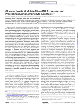
  • 5. NIEHS, National Institutes of Health Institutional Animal Care and Use Committee. RESULTS Repression of MicroRNA Expression during Glucocorticoid- induced Apoptosis—For these studies, primary rat thymocytes, a classic model of glucocorticoid-induced apoptosis, were cul- tured in vitro in the presence or absence of the synthetic glu- cocorticoid dexamethasone for 6 h. Apoptotic progression was monitored in control and glucocorticoid-treated cells via flow cytometric analysis of phosphatidylserine exposure, plasma membrane integrity, and caspase-3 activation. Following 6 h of glucocorticoid treatment, we observed an apoptotic population of cells. These cells exhibited phosphatidylserine externaliza- tion as determined by a significant increase in annexin-FITC fluorescence and decreased plasma membrane integrity dem- onstrated by a modest increase in propidium iodide fluores- cence (Fig. 1A). These cells also exhibited an increase in caspase-3 activity determined by an increase in CaspaTag fluo- rescence, indicating the clear induction of apoptosis following 6 h of glucocorticoid treatment (Fig. 1B). These apoptotic indi- cators persisted following 12 h of treatment, resulting in signif- icant cell death (Fig. 1). Therefore, we performed our microRNA expression analysis early in the apoptotic process (prior to cell death) after 6 h of glucocorti- coid treatment. Following 6 h of treatment, total RNAs from control and glucocorti- coid-treated cells were prepared and subjected to microRNA microarray analysis. Of the 350 microRNAs represented on our chip, 56 were differentially ex- pressed in response to glucocorti- coid-induced apoptosis (Fig. 2A). Surprisingly, the majority of these microRNAs (79% or 44 microRNAs) were repressed, and only 12 low abundance microRNAs were weakly induced during this time frame. Given the low basal ex- pression levels of these induced microRNAs, we were unable to con- firm their up-regulation via quanti- tative PCR. Because microRNA expression levels can vary widely, we focused our efforts on the abun- dantly expressed and thus poten- tially biologically active microRNAs (basal intensity values Ͼ200). The percent repression values (inten- sity glucocorticoid/intensity con- trol) were generated for these microRNAs (Fig. 2B). Interest- ingly, these abundantly expressed microRNAs were repressed uni- formly irrespective of their genomic locations, suggesting that the repression of microRNA expres- sion during glucocorticoid-induced apoptosis occurs post- transcriptionally, perhaps at the level of bioprocessing. The repression of a subset of microRNAs identified as differentially expressed (rno-let-7a, rno-miR-15b, and rno-miR-25) as well as the repression of a microRNA not significantly regulated in the microarray (rno-miR-17) was validated via quantitative PCR analysis (Fig. 2C). Interestingly, the expression of rno-miR-17 and rno-let-7a primary transcripts was increased during glu- cocorticoid-induced apoptosis, providing important evidence that the repression of the resultant mature microRNAs occurs post-transcriptionally (Fig. 2D). To evaluate the expression of the microRNAnome during glucocorticoid-induced apoptosis by an alternative approach, we performed next generation sequencing or deep sequencing of microRNAs from the control and glucocorticoid-treated samples. These samples were obtained from independent ani- mals rather than those described in Fig. 2; however, these cells exhibited apoptotic kinetics similar to those described in Fig. 1. Deep sequencing of microRNAs is an open-ended and hybrid- ization-independent approach that facilitates the detection of both annotated and novel microRNAs. In our study, massively parallel sequencing of control and glucocorticoid-treated RNA FIGURE3.MicroRNAdeepsequencingconfirmstheprevalentrepressionofmicroRNAexpressionduring glucocorticoid-induced apoptosis. Primary thymocytes were isolated and treated with 100 nM dexametha- sone (Dex) for 6 h. Total RNAs were subjected to microRNA deep sequencing analysis. A, deep sequencing of two biological replicates was performed using the Illumina Solexa platform for each treatment group, and the resulting sequences from replicates were combined for further analyses. Sequences were aligned to the refer- ence rat genome and sites that were enriched with reads were selected for further analysis. B, peak detection and ratio calculation revealed prevalent down-regulation of microRNAs in response to dexamethasone treat- ment.Theratioswerecalculatedascontrolversusdexamethasone(microRNAsrepressed)anddexamethasone versus control (microRNAs induced). C, percentage of repression of abundantly expressed microRNAs corre- lates with microRNA microarray results. The results are calculated as intensity dexamethasone-treated/inten- sity untreated presented as percentages of control. ND denotes microRNAs detected by microarray analysis but not deep sequencing analysis. MicroRNAs highlighted in gray indicate a subset of microRNAs repressed in deep sequencing analysis but not microarray analysis. Glucocorticoids Modulate MicroRNA Expression and Processing 36702 JOURNAL OF BIOLOGICAL CHEMISTRY VOLUME 285•NUMBER 47•NOVEMBER 19, 2010 atUNIVOFSOUTHFLORIDA,onSeptember8,2011www.jbc.orgDownloadedfrom
  • 6. samples generated millions of short sequences (commonly referred to as “reads”), which were aligned to the reference rat genome (Fig. 3A). We identified a set of 1782 sites (genomic loci that may be encoding microRNAs) that were down-regulated and 238 sites that were up-regulated by glucocorticoid treat- ment (see “Experimental Procedures” for more details). Of the 1782 sites, 132 sites were within 100 bp of an annotated microRNA, which correspond to 129 individual microRNAs (supplemental Table S1). It is also possible that the sites not immediately adjacent to a known microRNA may house novel microRNAs, which warrants fur- ther evaluation. Similar to the afore- mentioned microarray study, we detected a number of sites that were up-regulated by dexametha- sone treatment; however, none of the 238 up-regulated sites were in close proximity to an annotated microRNA (Fig. 3B). These results obtained by deep sequencing mirror the microarray findings of prevalent microRNA repression and minimal microRNA induction during glu- cocorticoid-induced apoptosis of primary thymocytes. In addition, the percent repression values of abundantly expressed microRNAs generated using deep sequencing data were comparable with those generated using microarray data, fur- ther suggesting the post-transcrip- tional repression of microRNAs during glucocorticoid-induced apo- ptosis of lymphocytes (Fig. 3C). Deep sequencing analysis con- firmed the repression of 35 of the 44 microRNAs repressed in the microarray analysis (79%). More- over, microRNA deep sequencing was also able to detect repressed microRNAs not detected by mi- croarray (representative values in gray in Fig. 3C), suggesting that deep sequencing of microRNAs is a more sensitive technique that may be used in conjunction with microRNA microarray and quanti- tative PCR as a novel “weight of evi- dence” approach to comprehensive profiling of the microRNAnome. MicroRNA Processing Enzymes Are Dysregulated during Gluco- corticoid-induced Apoptosis—The broad, genome-wide down-regula- tion of microRNA expression detected by both microarray and deep sequencing analysis and the absence of primary microRNA tran- script repression suggest post-transcriptional repression of mature microRNAs during glucocorticoid-induced apoptosis of lymphocytes. To evaluate this hypothesis, we examined the expression of the key microRNA processing enzymes Drosha, DGCR8/Pasha, and Dicer during glucocorticoid-induced apo- ptosis of primary thymocytes. Following 6 h of glucocorticoid treatment, the mRNA expression of each processor is repressed, with the repression of Dicer and DGCR8/Pasha mRNA achieving statistical significance (Fig. 4A). After 12 h of treatment, the mRNA levels of each processor are significantly FIGURE 4. MicroRNA processing enzymes are repressed during glucocorticoid-induced apoptosis of pri- mary thymocytes. Primary thymocytes were isolated and treated with 100 nM dexamethasone (Dex) for 6 and 12 h. A, the mRNA expression of the microRNA processing enzymes Drosha, DGCR8/Pasha, and Dicer was evaluated by quantitative PCR and normalized to the GusB housekeeping gene. The results are expressed as the percentages of control (Con) mean values Ϯ S.E. for three independent experiments. *, p Ն 0.05. B, whole cell lysates were immunoblotted with Drosha, DGCR8/Pasha, or Dicer polyclonal antibodies. Actin immuno- blotting served as a loading control, and the results are representative of at least three independent experi- ments. C, induction of Bim and Gelsolin mRNA expression during glucocorticoid-induced apoptosis. Primary thymocyteswereisolatedandtreatedwith100nM dexamethasonefor6and12h.ThemRNAexpressionofBim and Gelsolin was evaluated by quantitative PCR and normalized to the GusB housekeeping gene. The results are expressed as the percentages of control mean values Ϯ S.E. for three independent experiments. *, p Ն 0.05. Glucocorticoids Modulate MicroRNA Expression and Processing NOVEMBER 19, 2010•VOLUME 285•NUMBER 47 JOURNAL OF BIOLOGICAL CHEMISTRY 36703 atUNIVOFSOUTHFLORIDA,onSeptember8,2011www.jbc.orgDownloadedfrom
  • 7. repressed in response to glucocorticoid-induced apoptosis (Fig. 4A). The reduced expression of microRNA processing enzymes is not due to an apoptosis-related global increase in RNase activity because the expression of the glucocorticoid-respon- sive genes Gelsolin and Bim are strongly induced under these conditions in our cells (Fig. 4C). The repression of microRNA processing enzymes was also evident at the level of protein expression (Fig. 4B). Drosha pro- Glucocorticoids Modulate MicroRNA Expression and Processing 36704 JOURNAL OF BIOLOGICAL CHEMISTRY VOLUME 285•NUMBER 47•NOVEMBER 19, 2010 atUNIVOFSOUTHFLORIDA,onSeptember8,2011www.jbc.orgDownloadedfrom
  • 8. tein expression was decreased following 6 h of glucocorticoid treatment and was essentially undetectable at 12 h. The expres- sion of Drosha’s co-factor DGCR8/Pasha was also reduced in response to glucocorticoid treatment and upon overexposure, a clear cleavage pattern emerged following 12 h of treatment. Furthermore, the protein expression of the cytoplasmic pro- cessor, Dicer, was also markedly reduced following 6 h of glu- cocorticoid treatment, whereas at 12 h full-length Dicer was barely detectable (Fig. 4C). This repression of the microRNA bioprocessing pathway results in the nuclear accumulation of unprocessed primary microRNAs (supplemental Fig. S1), accounting for the increased expression of microRNA primary transcripts observed during glucocorticoid-induced apoptosis (Fig. 2D). MicroRNA Repression Enhances Glucocorticoid-induced Apoptosis of Lymphocytes—To assess whether the repression of microRNA expression contributes to the cell death process, we silenced the expression of Dicer, the enzyme responsible for mature microRNA biosynthesis, in two human lymphoid cell lines. The transition to cultured cell lines was necessary because primary rat thymocytes are difficult to genetically manipulate in vitro. Therefore, we performed our functional analyses in the Jurkat GR␣ and the CEM-C7 leukemic cell lines. The Jurkat GR␣ cell line was generated by the exogenous expression of the full-length GR␣ isoform in the glucocorticoid-resistant Jurkat ALL parental cell line (18), and stable expression of GR␣ restores glucocorticoid sensitivity to Jurkat cells (Fig. 5A). The glucocorticoid-sensitive CEM-C7 cell line contains an endoge- nous, hormone-responsive GR␣ and exhibits slower apoptotic kinetics in response to glucocorticoid compared with the Jurkat GR␣ cell line (Fig. 5A). Interestingly, similar to the results obtained in primary cell culture, Dicer expression is repressed at both the mRNA and protein levels in each leukemic cell line during glucocorticoid-induced apoptosis (Fig. 5B). Stable Dicer depletion in these cells was achieved via lentiviral transduction of shRNA (Fig. 5C), and the depletion of Dicer resulted in the reduced expression of a subset of microRNAs (Fig. 5D). Dicer depletion and the subsequent repression of microRNA expres- sion augmented glucocorticoid induced apoptosis in both cell lines as determined by flow cytometric evaluation of phosphati- dylserine exposure and plasma membrane integrity (Fig. 5E). Importantly, this increased sensitivity to glucocorticoid-in- duced apoptosis was not due to increased GR expression in the absence of Dicer expression (data not shown). These data sug- gest that Dicer depletion and subsequent microRNA repression enhance the progression of glucocorticoid-induced apoptosis in lymphocytes. Overexpression of the Repressed miR-17–92 Polycistron Blunts Glucocorticoid-induced Apoptosis—To delineate the potential role of specific microRNAs down-regulated during glucocorticoid-induced apoptosis, we sought to overexpress the anti-apoptotic miR-17–92 polycistron. Members of this polycistron were repressed by glucocorticoid treatment in pri- mary thymocytes, as determined by both the microarray and deep sequencing studies. Furthermore, mature members of this polycistron are also down-regulated during glucocorticoid-in- duced apoptosis of cultured leukemic cells (Fig. 6A). Overex- pression of the hsa-miR-17–92 polycistron via lentiviral trans- duction of hsa-miR-17–92 precursor microRNA resulted in efficient overexpression of individual mature hsa-miR-17–92 polycistron members in the Jurkat GR␣ cell line (Fig. 6B). Over- expression of hsa-miR-17–92 polycistron members in the Jur- kat GR␣ background significantly diminished the ability of both subsaturating and saturating doses of glucocorticoid to induce glucocorticoid-induced apoptosis (Fig. 6C). This diminished sensitivity was not due to decreased GR expression in response to hsa-miR-17–92 overexpression (data not shown). In addi- tion, primary thymocytes derived from mice transgenic for the hsa-miR-17–92 polycistron (ϳ1.5–2-fold overexpression of mature hsa-miR-17–92 polycistron members (8)) in the lym- phocyte compartment exhibited decreased sensitivity to glu- cocorticoid-induced apoptosis (Fig. 6D). This decreased sensi- tivity to glucocorticoid-induced apoptosis may also contribute to the hematomalignant phenotype of these mice. These data suggest that the specific repression of miR-17–92 polycistron contributes to glucocorticoid-induced apoptosis of lympho- cytes in vitro and in vivo. DISCUSSION Glucocorticoid-induced apoptosis is a key component of thymocyte development. Furthermore, because of their potent apoptosis-inducing properties, synthetic glucocorticoids are common components of hematomalignant chemotherapy reg- imens. Here we provide evidence of broad microRNA repres- sion during glucocorticoid-induced apoptosis of lymphocytes. This repression was associated with the reduction of both nuclear (Drosha and DGCR8/Pasha) and cytoplasmic (Dicer) FIGURE 5. MicroRNA depletion contributes to glucocorticoid-induced apoptosis in human leukemic cell lines. A, flow cytometric analysis of glucocorti- coid-induced apoptosis in Jurkat GR␣ and CEM-C7 ALL leukemic cell lines. The cells were treated with 100 nM dexamethasone (Dex), and apoptotic progression was monitored at 24, 48, and 72 h via flow cytometric analysis of propidium iodide (PI) uptake. The results are represented as the mean percentages of PI-positive values Ϯ S.E. for six independent experiments. *, p Ն 0.05. B, repression of Dicer expression during glucocorticoid-induced apoptosis of human leukemic cell lines. The cells were treated with 100 nM dexamethasone for 24 and 48 h. The expression of Dicer mRNA was evaluated in total RNA via quantitative PCR. The expression of GusB mRNA served as an endogenous control (Con). The results are represented as percentages of control mean values Ϯ S.E. for three independent experiments. *, p Ն 0.05. Whole cell lysates were immunoblotted with Dicer polyclonal antibody. Actin immunoblotting served as a loading control. The results are representative of three independent experiments. C, stable silencing of Dicer protein expression in Jurkat GR␣ and CEM-C7 lymphocyte cell lines via lentiviral transduction of shRNA. Whole cell lysates from cells stably transduced with nontargeting control or anti-Dicer shRNA (Dicer knock down (KD)), were immunoblotted with Dicer polyclonal antibody. Actin immunoblotting served as a loading control, and the results are representative ofthreeindependentexperiments.D,repressionofmaturemicroRNAexpressionintheabsenceofDicerexpression.TotalRNAsfromnontargetingcontroland Dicer KD cells were evaluated for the expression of three independent mature microRNAs via quantitative PCR. The expression of Rnu43 small nuclear RNA served as an endogenous control. The results are reported as percentages of nontargeting control values Ϯ S.E. for three independent experiments. *, p Ն 0.05. E, enhancement of glucocorticoid-induced apoptosis in Dicer KD cells. Flow cytometric analysis of annexin-FITC and propidium iodide uptake in nontargeting control and Dicer KD stable cell lines in response to 48 h of 100 nM dexamethasone treatment. The results were normalized to untreated control and are representative of three independent experiments. *, p Ն 0.05. Glucocorticoids Modulate MicroRNA Expression and Processing NOVEMBER 19, 2010•VOLUME 285•NUMBER 47 JOURNAL OF BIOLOGICAL CHEMISTRY 36705 atUNIVOFSOUTHFLORIDA,onSeptember8,2011www.jbc.orgDownloadedfrom
  • 9. microRNA processing enzymes and the increased expression of primary microRNA transcripts. Dicer repression and cleavage during apoptosis has recently been reported in laboratory-derived human endothelial cells, HL-60 leukemic cells, and HeLa cervical carcinoma cells in response to apoptotic stimuli; however, none of these reports evaluated the expression of the nuclear processors Drosha and DGCR8/Pasha during apoptosis (19–22). This reduction of Dicer protein expression has recently been attributed to caspase-3 cleavage (20). The precise mechanism of Dicer mRNA repression during apoptosis remains undetermined. Interestingly, we show that Dicer repression in response to glu- cocorticoid-induced apoptosis is conserved in both healthy and transformed lymphocytes. Therefore, the results of this study expand our current understanding of Dicer repression in response to apoptotic stimulation while being the first to detail the repression of all three key microRNA processing enzymes during glucocorticoid-induced apoptosis of lymphocytes. Additional studies in our laboratory have revealed that alterna- tive apoptotic stimuli, specifically Fas ligand and UVC, also pro- mote the repression of microRNA expression and processing enzymes during primary thymocyte apoptosis, suggesting that the dsyregulation of microRNA expression and bioprocessing machinery is a common convergence point for multiple apo- ptotic pathways during apoptosis of lymphocytes.3 Several recent reports have suggested that sex steroid hor- mones also exert post-transcriptional control through the reg- ulation of microRNA processing and expression. For example, ligand-bound estrogen receptor ␣ regulates microRNA expres- sion in cultured MCF-7 breast cancer cells, myometrial, and leiomyoma smooth muscle cells, rat mammary tissue, and mouse splenic lymphocytes (23–28). MicroRNAs can also neg- atively regulate the estrogen receptor ␣ transcriptional response via translational inhibition of estrogen-responsive genes and the p160 transcriptional co-activator, AIB1 (24, 25). Additionally, estrogen treatment of ovarectomized mice led to the repression of a subset of microRNAs (29). This study found that ligand-activated estrogen receptor ␣ inhibits Drosha pro- cessing of precursor microRNAs by mediating the dissociation of the Drosha microprocessor machinery from the precursor microRNA. These results provide additional mechanisms for hormonal impairment of microRNA processing. Alternatively, androgen treatment of androgen-responsive prostate cancer cell lines induced the expression of 16 mature microRNAs in the absence of microRNA repression (30). Further analysis identified direct transcriptional activation of miR-21 via andro- gen receptor binding to the miR-21 promoter region, indicating that nuclear hormone receptors can also act via classical ligand- 3 L. K. Smith and J. A. Cidlowski, unpublished observations. FIGURE 6. Overexpression of hsa-miR-17–92 blunts glucocorticoid-in- duced apoptosis. A, repression of hsa-miR-17–92 expression during glu- cocorticoid-induced apoptosis of Jurkat GR␣ cells. Jurkat GR␣ cells were treated with 100 nM dexamethasone (Dex) for 24 and 48 h. The expression of individual mature hsa-miR-17–92 polycistron members was evaluated via quantitative PCR. The expression of Rnu43 small nuclear RNA served as an endogenous control (Con). The results are represented as mean percentages of control values Ϯ S.E. for three independent experiments. *, p Ն 0.05. B, ef- ficient overexpression of individual polycistron members in Jurkat GR␣ cells via lentiviral transduction. Jurkat GR␣ cells were stably transduced with len- tivirus encoding the hsa-miR-17–92 precursor microRNA or empty vector control. The overexpression of individual mature hsa-miR-17–92 polycistron members was evaluated via quantitative PCR. The expression of Rnu43 small nuclear RNA served as an endogenous control. The results are represented as mean percentages of vector control values Ϯ S.E. for three independent experiments. *, p Ն 0.05. C, Hsa-miR-17–92 overexpression blunts glucocor- ticoid-induced apoptosis. Flow cytometric analysis of propidium iodide (PI) uptake in empty vector or hsa-miR-17–92 stable overexpressors in response to 10 nM and 100 nM dexamethasone treatment. The results were normal- ized to untreated control and are representative of three independent experiments. *, p Ն 0.05. D, transgenic hsa-miR-17–92 overexpression hin- ders glucocorticoid-induced apoptosis of primary thymocytes. Primary thy- mocytes were derived from wild-type control and hsa-miR-17–92 transgenic mice and treated with 10 and 100 nM dexamethasone for 12 h. Apoptosis was monitored by flow cytometric analysis of annexin-FITC/propidium iodide staining. The results were normalized to untreated control and are represen- tative of four independent experiments. *, p Ն 0.05. Glucocorticoids Modulate MicroRNA Expression and Processing 36706 JOURNAL OF BIOLOGICAL CHEMISTRY VOLUME 285•NUMBER 47•NOVEMBER 19, 2010 atUNIVOFSOUTHFLORIDA,onSeptember8,2011www.jbc.orgDownloadedfrom
  • 10. activated transcription factor signaling to induce microRNA expression. Previously, a single study sought to examine the expression of mature microRNAs during glucocorticoid-induced apopto- sis of lymphocytes. In conflict with our results, this group did not detect the prevalent repression of microRNAs in response to dexamethasone treatment (31). Moreover, this study could not validate the down-regulation of any mature microRNA repressed on their array via quantitative PCR and in contrast reported the induction of three microRNAs: hsa-miR-15b, -16, and -223. Interestingly, in our model system we observe the strong repression of rno-miR-15b in response to glucocorti- coid-induced apoptosis (Fig. 2C). These discrepancies are likely due to the use of alternative experimental models, different methods of data analysis, and distinct microarray platforms. The findings of a separate analysis indicate that the hormonal regulation of microRNAs is likely specific to cell type and phys- iological context. For example, glucocorticoid treatment of lung epithelial cells, which do not undergo glucocorticoid-in- duced apoptosis, failed to significantly alter microRNA expres- sion (32). These data suggest that the dramatic down-regula- tion of microRNA expression reported in our study is likely due to the physiological activation of the apoptotic cascade in response to glucocorticoid treatment. We also report that the depletion of Dicer and Dicer-depen- dent microRNA expression enhanced glucocorticoid-induced apoptosis in two human leukemic cell lines. Dicer has an essen- tial role in the development of the adaptive immune system. Conditional deletion of Dicer expression in the T-cell compart- ment results in impaired T-cell development and diminished regulatory T-cell function (33–35), and ablation of Dicer expression in the B-cell compartment attenuates B-cell devel- opment and alters the antibody repertoire (36). Therefore, Dicer depletion hinders lymphocyte development and in- creases sensitivity to glucocorticoid-induced apoptosis. In our studies, the stable (ϳ2 weeks) depletion of Dicer in human leukemic cell lines resulted in the partial (40–60%) reduction of evaluated microRNAs. This partial reduction may be due to the robust stability of mature microRNAs or to the activity of an alternative microRNA processing pathway. In fact, a recent report describes a novel Dicer-independent, Argonaute-2-dependent microRNA processing pathway (37). Interestingly, unlike other microRNA bioprocessors, Argo- naute-2 expression is not reduced during glucocorticoid-in- duced apoptosis of primary thymocytes (supplemental Fig. S2), suggesting that microRNAs refractory to Dicer depletion may be processed in an Argonaute-2-dependent manner during glu- cocorticoid-induced apoptosis. Furthermore, we determined that the overexpression of spe- cific microRNAs repressed during glucocorticoid-induced apoptosis of lymphocytes, the miR-17–92 polycistron, blunts glucocorticoid-induced apoptosis in both human and murine- derived lymphocytes. Members of this highly conserved poly- cistron exhibit complete sequence identity in the human, rat, and mouse, suggesting that their anti-apoptotic functions are conserved across vertebrate species (38). Interestingly, trans- genic overexpression of the hsa-miR-17–92 polycistron sup- presses expression of the pro-apoptotic Bcl-2 family member, Bim (8). The induction of Bim expression is a critical compo- nent of glucocorticoid-induced apoptosis signaling (39–42). Therefore, decreased basal Bim expression or weakened Bim induction upon glucocorticoid treatment may account for the decreased sensitivity of hsa-miR-17–92-overexpressing lym- phocytes to glucocorticoid-induced apoptosis. In summary, these studies establish a “glucocorticoid-induced apoptotic sig- nature” of prevalent repression of microRNAs and microRNA bioprocessing machinery and further delineate the functional significance of this repression. Acknowledgments—We acknowledge the generous contributions of the NIEHS Microarray, Viral Vector, and Flow Cytometry facilities for expert assistance. MicroRNA deep sequencing was ably performed by the University of North Carolina High Throughput Sequencing Facility. We also thank Dr. Nick Lu for the generation of the Jurkat GR␣ stable cell line. We thank Dhiral Phadke of SRA International for support in the analysis of deep sequencing data. Finally, we are grate- ful to Lois Wyrick for expert assistance in graphics preparation. REFERENCES 1. Kim, V. N. (2005) Nat. Rev. Mol. Cell Biol. 6, 376–385 2. Cullen, B. R. (2004) Mol. Cell 16, 861–865 3. Esquela-Kerscher, A., and Slack, F. J. (2006) Nat. Rev. Cancer 6, 259–269 4. Calin, G. A., Liu, C. G., Sevignani, C., Ferracin, M., Felli, N., Dumitru, C. D., Shimizu, M., Cimmino, A., Zupo, S., Dono, M., Dell’Aquila, M. L., Alder, H., Rassenti, L., Kipps, T. J., Bullrich, F., Negrini, M., and Croce, C. M. (2004) Proc. Natl. Acad. Sci. U.S.A. 101, 11755–11760 5. Calin, G. A., Ferracin, M., Cimmino, A., Di Leva, G., Shimizu, M., Wojcik, S. E., Iorio, M. V., Visone, R., Sever, N. I., Fabbri, M., Iuliano, R., Palumbo, T., Pichiorri, F., Roldo, C., Garzon, R., Sevignani, C., Rassenti, L., Alder, H., Volinia, S., Liu, C. G., Kipps, T. J., Negrini, M., and Croce, C. M. (2005) N. Engl. J. Med. 353, 1793–1801 6. Cimmino, A., Calin, G. A., Fabbri, M., Iorio, M. V., Ferracin, M., Shimizu, M., Wojcik, S. E., Aqeilan, R. I., Zupo, S., Dono, M., Rassenti, L., Alder, H., Volinia, S., Liu, C. G., Kipps, T. J., Negrini, M., and Croce, C. M. (2005) Proc. Natl. Acad. Sci. U.S.A. 102, 13944–13949 7. He, L., Thomson, J. M., Hemann, M. T., Hernando-Monge, E., Mu, D., Goodson, S., Powers, S., Cordon-Cardo, C., Lowe, S. W., Hannon, G. J., and Hammond, S. M. (2005) Nature 435, 828–833 8. Xiao, C., Srinivasan, L., Calado, D. P., Patterson, H. C., Zhang, B., Wang, J., Henderson, J. M., Kutok, J. L., and Rajewsky, K. (2008) Nat. Immunol. 9, 405–414 9. Ashwell, J. D., Lu, F. W., and Vacchio, M. S. (2000) Annu. Rev. Immunol. 18, 309–345 10. Cifone, M. G., Migliorati, G., Parroni, R., Marchetti, C., Millimaggi, D., Santoni, A., and Riccardi, C. (1999) Blood 93, 2282–2296 11. Mann, C. L., Hughes, F. M., Jr., and Cidlowski, J. A. (2000) Endocrinology 141, 528–538 12. McConkey, D. J., Nicotera, P., Hartzell, P., Bellomo, G., Wyllie, A. H., and Orrenius, S. (1989) Arch. Biochem. Biophys. 269, 365–370 13. Wang, D., Mu¨ller, N., McPherson, K. G., and Reichardt, H. M. (2006) J. Immunol. 176, 1695–1702 14. Lu, N. Z., and Cidlowski, J. A. (2005) Mol. Cell 18, 331–342 15. Salmon, P., and Trono, D. (2007) Current Protocol in Human Genetics, Chapter 12, Unit 12.10, John Wiley and Sons, Hoboken, NJ 16. Langmead, B., Trapnell, C., Pop, M., and Salzberg, S. L. (2009) Genome Biol. 10, R25–R34 17. Mortazavi, A., Williams, B. A., McCue, K., Schaeffer, L., and Wold, B. (2008) Nat. Methods 5, 621–628 18. Riml, S., Schmidt, S., Ausserlechner, M. J., Geley, S., and Kofler, R. (2004) Cell Death Differ. 11, (Suppl. 1) S65–S72 19. Asada, S., Takahashi, T., Isodono, K., Adachi, A., Imoto, H., Ogata, T., Ueyama, T., Matsubara, H., and Oh, H. (2008) Am. J. Physiol. Heart Circ. Glucocorticoids Modulate MicroRNA Expression and Processing NOVEMBER 19, 2010•VOLUME 285•NUMBER 47 JOURNAL OF BIOLOGICAL CHEMISTRY 36707 atUNIVOFSOUTHFLORIDA,onSeptember8,2011www.jbc.orgDownloadedfrom
  • 11. Physiol. 295, H2512–H2521 20. Ghodgaonkar, M. M., Shah, R. G., Kandan-Kulangara, F., Affar, E. B., Qi, H. H., Wiemer, E., and Shah, G. M. (2009) Cell Death Differ. 16, 858–868 21. Matskevich, A. A., and Moelling, K. (2008) Biochem. J. 412, 527–534 22. Wiesen, J. L., and Tomasi, T. B. (2009) Mol. Immunol. 46, 1222–1228 23. Klinge, C. M. (2009) Curr. Genomics 10, 169–183 24. Bhat-Nakshatri, P., Wang, G., Collins, N. R., Thomson, M. J., Geistlinger, T. R., Carroll, J. S., Brown, M., Hammond, S., Srour, E. F., Liu, Y., and Nakshatri, H. (2009) Nucleic Acids Res. 37, 4850–4861 25. Castellano, L., Giamas, G., Jacob, J., Coombes, R. C., Lucchesi, W., Thiruchelvam, P., Barton, G., Jiao, L. R., Wait, R., Waxman, J., Hannon, G. J., and Stebbing, J. (2009) Proc. Natl. Acad. Sci. U.S.A. 106, 15732–15737 26. Pan, Q., Luo, X., and Chegini, N. (2008) J. Cell Mol. Med. 12, 227–240 27. Kovalchuk, O., Tryndyak, V. P., Montgomery, B., Boyko, A., Kutanzi, K., Zemp, F., Warbritton, A. R., Latendresse, J. R., Kovalchuk, I., Beland, F. A., and Pogribny, I. P. (2007) Cell Cycle 6, 2010–2018 28. Dai, R., Phillips, R. A., Zhang, Y., Khan, D., Crasta, O., and Ahmed, S. A. (2008) Blood 112, 4591–4597 29. Yamagata, K., Fujiyama, S., Ito, S., Ueda, T., Murata, T., Naitou, M., Takeyama, K., Minami, Y., O’Malley, B. W., and Kato, S. (2009) Mol. Cell 36, 340–347 30. Ribas, J., Ni, X., Haffner, M., Wentzel, E. A., Salmasi, A. H., Chowdhury, W. H., Kudrolli, T. A., Yegnasubramanian, S., Luo, J., Rodriguez, R., Men- dell, J. T., and Lupold, S. E. (2009) Cancer Res. 69, 7165–7169 31. Rainer, J., Ploner, C., Jesacher, S., Ploner, A., Eduardoff, M., Mansha, M., Wasim, M., Panzer-Gru¨mayer, R., Trajanoski, Z., Niederegger, H., and Kofler, R. (2009) Leukemia 23, 746–752 32. Moschos, S. A., Williams, A. E., Perry, M. M., Birrell, M. A., Belvisi, M. G., and Lindsay, M. A. (2007) BMC Genomics 8, 240–245 33. Muljo, S. A., Ansel, K. M., Kanellopoulou, C., Livingston, D. M., Rao, A., and Rajewsky, K. (2005) J. Exp. Med. 202, 261–269 34. Cobb, B. S., Hertweck, A., Smith, J., O’Connor, E., Graf, D., Cook, T., Smale, S. T., Sakaguchi, S., Livesey, F. J., Fisher, A. G., and Merkenschlager, M. (2006) J. Exp. Med. 203, 2519–2527 35. Liston, A., Lu, L. F., O’Carroll, D., Tarakhovsky, A., and Rudensky, A. Y. (2008) J. Exp. Med. 205, 1993–2004 36. Koralov, S. B., Muljo, S. A., Galler, G. R., Krek, A., Chakraborty, T., Kanel- lopoulou, C., Jensen, K., Cobb, B. S., Merkenschlager, M., Rajewsky, N., and Rajewsky, K. (2008) Cell 132, 860–874 37. Cifuentes, D., Xue, H., Taylor, D. W., Patnode, H., Mishima, Y., Cheloufi, S., Ma, E., Mane, S., Hannon, G. J., Lawson, N. D., Wolfe, S. A., and Giral- dez, A. J. (2010) Science 328, 1694–1698 38. Mendell, J. T. (2008) Cell 133, 217–222 39. Bouillet, P., Metcalf, D., Huang, D. C., Tarlinton, D. M., Kay, T. W., Ko¨nt- gen, F., Adams, J. M., and Strasser, A. (1999) Science 286, 1735–1738 40. Abrams, M. T., Robertson, N. M., Yoon, K., and Wickstrom, E. (2004) J. Biol. Chem. 279, 55809–55817 41. Lu, J., Quearry, B., and Harada, H. (2006) FEBS Lett. 580, 3539–3544 42. Ploner, C., Rainer, J., Niederegger, H., Eduardoff, M., Villunger, A., Geley, S., and Kofler, R. (2008) Leukemia 22, 370–377 Glucocorticoids Modulate MicroRNA Expression and Processing 36708 JOURNAL OF BIOLOGICAL CHEMISTRY VOLUME 285•NUMBER 47•NOVEMBER 19, 2010 atUNIVOFSOUTHFLORIDA,onSeptember8,2011www.jbc.orgDownloadedfrom