Amalgam restoration
•
31 recomendaciones•2,828 vistas

Focused on essential elements in the cavity preparation for an amalgam restoration, also the possible causes for failure of an amalgam restoration.

Recomendados
Más contenido relacionado
La actualidad más candente
La actualidad más candente (20)
Destacado
Destacado (10)
Similar a Amalgam restoration
Similar a Amalgam restoration (20)
Último
Último (20)
Amalgam restoration
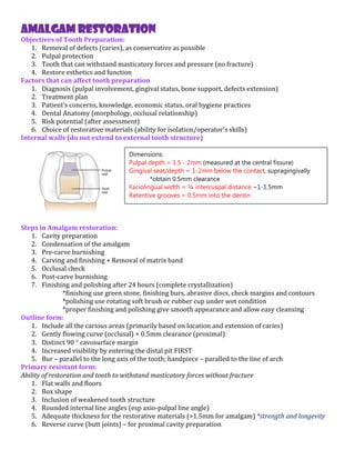
- 1. AMALGAM RESTORATION Objectives of Tooth Preparation: 1. Removal of defects (caries), as conservative as possible 2. Pulpal protection 3. Tooth that can withstand masticatory forces and pressure (no fracture) 4. Restore esthetics and function Factors that can affect tooth preparation 1. Diagnosis (pulpal involvement, gingival status, bone support, defects extension) 2. Treatment plan 3. Patient’s concerns, knowledge, economic status, oral hygiene practices 4. Dental Anatomy (morphology, occlusal relationship) 5. Risk potential (after assessment) 6. Choice of restorative materials (ability for isolation/operator’s skills) Internal walls (do not extend to external tooth structure) Steps in Amalgam restoration: 1. Cavity preparation 2. Condensation of the amalgam 3. Pre-carve burnishing 4. Carving and finishing + Removal of matrix band 5. Occlusal check 6. Post-carve burnishing 7. Finishing and polishing after 24 hours (complete crystallization) *finishing use green stone, finishing burs, abrasive discs, check margins and contours *polishing use rotating soft brush or rubber cup under wet condition *proper finishing and polishing give smooth appearance and allow easy cleansing Outline form: 1. Include all the carious areas (primarily based on location and extension of caries) 2. Gently flowing curve (occlusal) + 0.5mm clearance (proximal) 3. Distinct 90 cavosurface margin 4. Increased visibility by entering the distal pit FIRST 5. Bur – parallel to the long axis of the tooth; handpiece – paralled to the line of arch Primary resistant form: Ability of restoration and tooth to withstand masticatory forces without fracture 1. Flat walls and floors 2. Box shape 3. Inclusion of weakened tooth structure 4. Rounded internal line angles (esp axio-pulpal line angle) 5. Adequate thickness for the restorative materials (>1.5mm for amalgam) *strength and longevity 6. Reverse curve (butt joints) – for proximal cavity preparation Dimensions: Pulpal depth = 1.5 - 2mm (measured at the central fissure) Gingival seat/depth = 1-2mm below the contact, supragingivally *obtain 0.5mm clearance Faciolingual width = ¼ intercuspal distance ~1-1.5mm Retentive grooves = 0.5mm into the dentin
- 2. Primary retention form: Resistance to the displacement or removal of the materials from tipping/lifting forces 1. Convergence of external walls, occlusally 2. Occlusal dovetails (prevent tipping) Convenience form: To provide adequate observation, accessibility and ease of operation, but with minimal amount of reduction Secondary resistance and retention form: 1. Etching, priming and adhesive materials placement 2. Retentive undercuts - retentive grooves (proximal box) 3. Retention lock (0.2mm from DEJ) *can’t really be measured clinically Notes: For proximal lesions, unless there is a penetration into the dentin, treatment is usually not needed. For cases where two lesions are seen, whether to prepare TWO separate cavities or ONE joined cavity, depends on the degree of close proximity between the two carious lesions. Always remove any remaining pits and fissures, infected dentin and old restorative materials. Pulpal protection due to pulpal irritation (potential): 1. Ingredients in the restorative materials 2. Thermal conduction 3. Force of transmission to the dentin 4. Galvanic shock 5. Microleakage Matrix band application: 1. Retains amalgam in the cavity during condensation 2. Permits close adaptation of the amalgam *same goes to the use of cervical wedge 3. Helps to restore contact area and external contour of the crown Condensation of amalgam: 1. Pack in lateral and vertical direction 2. Allow buildup of excess amalgam at the cavity margins 3. Pre-carve burnishing enhance the benefits of condensation 4. Fracture of condensed amalgam during matrix band removal can be due to a. Poor cavity preparation b. Insufficient marginal ridge condensation (insufficient bulk of amalgam) c. Inadequate trimming of the ridge before band removal Signs of Failure of Amalgam restoration: 1. Fracture line 2. Marginal enamel ditching 3. Proximal overhangs 4. Poor anatomic contour 5. Marginal ridge incompatibility 6. Improper proximal contacts 7. Recurrent caries Indication for the removal of old restorative materials: 1. Poor estheric results 2. Compromise of new materials 3. Secondary caries 4. Symptomatic tooth pulp 5. Microleakage/non-intact periphery margin