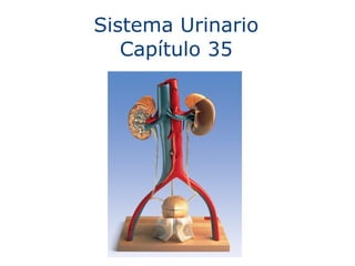
9. El Sistema Urinario Humano Riñón Ureter Vejiga Urinaria Uretra (en pene) Arteria Renal Vena Renal Vena Cava Aorta
10.
11.
12.
13.
14.
15. Juxta- medullary nephron Cortical nephron Collecting duct To renal pelvis Renal cortex Renal medulla 20 µm Afferent arteriole from renal artery Glomerulus Bowman’s capsule Proximal tubule Peritubular capillaries SEM Efferent arteriole from glomerulus Branch of renal vein Descending limb Ascending limb Loop of Henle Distal tubule Collecting duct (c) Nephron Vasa recta (d) Filtrate and blood flow
16.
17.
18.
19. Proximal tubule Filtrate H 2 O Salts (NaCl and others) HCO 3 – H + Urea Glucose; amino acids Some drugs Key Active transport Passive transport CORTEX OUTER MEDULLA INNER MEDULLA Descending limb of loop of Henle Thick segment of ascending limb Thin segment of ascending limb Collecting duct NaCl NaCl NaCl Distal tubule NaCl Nutrients Urea H 2 O NaCl H 2 O H 2 O HCO 3 K + H + NH 3 HCO 3 K + H + H 2 O 1 4 3 2 3 5
20. excretado en orina 3 En los riñones, urea + agua + otros desechos = orina Formación y Excreción de Urea 1 En las células, los aminoacidos son metabolizados, liberando amonia. Amonia NH 3 Se transporta en la sangre 2 En el hígado, amonia es combinada con CO 2 para producir urea Urea Se transporta en la sangre
21.
22. 2 En el hígado, amonia es combinada con CO 2 para producir urea Urea
23. 3 En los riñones, urea + agua + otros desechos = orina excretado en orina
FIGURE 35-2 (part 1) The formation and excretion of urea
Diagrammatic view of the human urinary system and its blood supply.
FIGURE 35-4 Cross section of a kidney A nephron is drawn considerably larger than normal (and further enlarged in the inset) to show its location in the kidney. Collecting ducts empty the urine into channels that lead to the renal pelvis, a chamber that funnels urine into the ureter and out of the kidney.
FIGURE 35-5 An individual nephron and its blood supply
FIGURE 35-6 Urine formation in the nephron
FIGURE 35-7 Dehydration stimulates ADH release and water retention

![Continuidad Dinámica ,[object Object],[object Object]](data:image/gif;base64,R0lGODlhAQABAIAAAAAAAP///yH5BAEAAAAALAAAAAABAAEAAAIBRAA7)