SlideShare una empresa de Scribd logo
1 de 40
Descargar para leer sin conexión
2012.10.30.


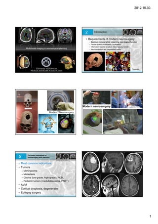
                                                          2           Introduction


                                                          • Requirements of modern neurosurgery
                                                              –   Maximum removal while preserving neurological function
                                                              –   Precise spatial visualization, localization
                                                              –   Information beyond structure: blood supply, function
        Multimodal imaging in neurosurgical planning          –   Neuronavigation and „augmented reality”
                                                                                                       reality


                                                                                                                  Trepanatio


                                                        Modern neurosurgery

                                 Modern? Neurosurgery


5        The main indications of
         neurosurgery and planning


• Most common indications
• Tumors
    – Meningeoma
    – Metastasis
    – Glioma (low-grade, high-grade), PCBL
    – Pediatric tumors (medulloblastoma, PNET)
• AVM
• Cortical dysplasia, degeneratio
• Epilepsy surgery


                                                                                                                               1
2012.10.30.


                                                                            Multimodal imaging for neurosurgery
    7          What clinicians should ask

                                                                 Anatomy
•    Location of craniotomy?                                                       Conventional MRI
                                                            Tumor localization          (Gd!)
•    Tumor localization, cortical eloquent areas
                                                                  Vessels
•    Vessels structure
                                                                                        DTI,
•    Eloquent areas                                          Eloquent t t
                                                             El     t tracts        tractography
      – White matter fibers
                                                            Function, laterality         fMRI
      – Functional domains
• Function laterality (speech, movement)                    Tumor metabolism
                                                                                        PET, MR
                                                                                      spectroscopy
• Tumor characterization                                       Stereotactic
                                                                                          CT
• Post operative imaging + controll                              planning

                                                               Localization        Neuronavigation systems


               Introduction
    9          Basics of multimodal imaging             10              MRI sequences


Multimodal                                              •     Conventional MRI sequences to display anatomy:
                                                        •     T1-weighted / post-Gd (vessels) or TOF time of flight
      – Parallel / fused display of different imaging
                                                        •     T2-weighted / flair
        modalities                                      •     Segmentation of cortical structure, vasculature
      – Their benefits multiply                         •     Segmentation of lesion / tumor
      – New information                                 •     Segmentation of markers
      – Image processing
         • Spatial alignment
         • Standard space
         • Normalization


               Information from                                         Information from
11             conventional MR images                   12              conventional MR images
    • Skin, muscles, skin markers for neurosurgery      • Vessels: (1) Contrast MRI (2) TOF


                                                                                                                               2
2012.10.30.


            Information from                                                        Diffusion information:
13          conventional MR images                                        14        DTI and fibertracking

• Cortex
• tumor


                                                                           •   Displaying white matter tracts
                                                                           •   Tumor adjacent tracts
                                                                           •   Diffusion mapping
                                                                           •   Characterizing pathologies


             Diffusion information:                                                 Diffusion information:
15           DTI and fibertracking                                        16        DTI and fibertracking


•   Térbeli illesztés, regisztráció                                       • Fibertracking: the method to display brain
•   Tenzorterek regisztrációja a strukturális felvételekhez (CT,MRI)
                                                                            tracts in vivo by MRI
                                                                          • Representation: line / tubes / probabilities


     –

•   Eredmény: kevesebb torzítás, pontosabb térbeli leképzés, koordináta
    rendszer.


             Diffusion information:                                                 Diffusion information:
17           DTI and fibertracking                                        18        DTI and fibertracking


• Segmenting and localizing main structures of the WM (pl. tr. cortico-
  spinalis, FLS, cc)


                                                                                                                               3
2012.10.30.


           Diffusion information:
19         DTI and fibertracking


• DTI acquisitions are distorted
• DTI has to be aligned with the T1-weighted brain image


• .


                                                                    4
2012.10.30.


         5
2012.10.30.


         6
2012.10.30.


                                              Bevezetés
                                         40   A multimodális képalkotás alapjai


     Bevezetés                                Bevezetés
41   A multimodális képalkotás alapjai   42   A multimodális képalkotás alapjai


                                                                                           7
2012.10.30.


     Bevezetés                                Bevezetés
43   A multimodális képalkotás alapjai   44   A multimodális képalkotás alapjai


     Bevezetés                                Bevezetés
45   A multimodális képalkotás alapjai   46   A multimodális képalkotás alapjai


     Bevezetés                                Bevezetés
47   A multimodális képalkotás alapjai   48   A multimodális képalkotás alapjai


                                                                                           8
2012.10.30.


             Bevezetés                                                                  Bevezetés
49           A multimodális képalkotás alapjai                           50             A multimodális képalkotás alapjai


                                                                            Second step: the actual fMRI acquisition
51           Functional MRI
                                                                         T2*-weighted images
                                                                             •   Image contrast relates to neuronal activity
                                                                             •   Low spatial resolution (3x3x5 mm)
                                                                             •   One volume of the brain is acquired in 2 seconds!
                                                                             •   We acquire many volumes in time (4D), ie. 150
                                                                             •   Repeated scanning


                                                                                                                                                     …

                                                                                                                             first volume
                                                                                                                             (2 sec to acquire)


                Paradigm and block design
Functional images
                           fMRI                   ROI
                                                                         54             fMRI
                  ~2 sec   signal                 Time
                           (% change              Course
                                                                           • fMRI also distorts, image alignment necessary
                                                                              – To patients anatomical (T1) images
                                                 Time            Tasks        – To standard neuroimaging spaces / atlases:
                                                                              – Talairach atlas /MNI atlas
                                                        Statistical
                                                        activation map
                                                        on T1 image


    Time


Region of interest                        ~ 5 minutes
kijelölés (ROI)


                                                                                                                                                           9
2012.10.30.


                                              Lesion in left precentral gyrus. Question:
                                                                 CST?
Right hand activation (finger-tapping test)


Forrás: Katona P., DEOEC                                                               Forrás: Katona P., Jakab A. DEOEC


      Summary – multimodality in
           neurosurgery
• Locate anatomy
• Locate vessels
• Locate eloquent (neighbouring) areas
•LLocate white matter t t
      t    hit    tt tracts                                              Case 1
                                                                              1.
• Locate functional domains

• Plan treatment


                                                                                                                           10
2012.10.30.


      Focal cortical dysplasias                                                  Dysplasia - Dysgenesis
•Cortical dysplasia is a congenital abnormality where the
neurons in an area of the brain failed to migrate in the                  • Greenfield’s Neuropathology – Dysplasia
proper formation in utero.                                                  of cerebral cortex
•Occasionally neuronswill develop that are larger than                      – Agyria
normal in certain areas.
•This causes the signals sent through the neurons in these                  – Pachygiria
areas to misfire, which sends an incorrect signal. It is                    – Polymicrogyria
commonly found near the cerebral cortex and is associated
with seizures.
                                                                            – Heterotopia
•Cortical dysplasia is estimated to be present in 1 in 2,500                – Focal cortical dysplasia (FCD)
newborns, making it one of the most common cortical
malformations.
                                                                          • Neuronal migration disorder


                                                                                      Palmini A, Najm I, Avanzini G, et al. Terminology and classification of the
            J Neurol Neurosurg Psychiatry. 1971 August; 34(4): 369–387.               cortical dysplasias. Neurology 2004;62(6 suppl 3):S2–S8.


         Abnormal proliferation
                                                                                               FCD Type I
         Focal cortical dysplasia
                                                                          • Abnormal cells anywhere from the ventricular
                                                                            wall to the cortex
                                                                          • Broadened gyrus, slightly irregular sulcus
                                                                          • Non enhancing
                                                                          • Adjacent cortex slightly thickened
                                                                          • Temporal localisation is common
                                                                          • Marked by slightly broadened sulcus
                                                                          • Hasznos a kontrollok során a fokozott figyelem,
                                                                            mert a korral egyértelmübbé válhat


                                                                                                                                                                    11
2012.10.30.


67         Case 1.                                                            68   Case 1.


     Infant, generalized epileptic seizures. CT negative. Background: focal
                                cortical dysplasia


                                                                                                     Case 2
                                                                                                          2.


                                                                                                              Case 2.

                   Pontine gliomas
•Brain stem tumors account for 10 percent of pediatric brain
tumors. The peak incidence is between ages 5 and 10.

Pontine Gliomas - The patients' symptoms often improve
dramatically during or after six weeks of irradiation.
Unfortunately, problems usually recur after six to nine
months, and progress rapidly. Survival past 12 to 14 months
is uncommon, and new approaches to treating these
tumors are urgently needed.
Midbrain/Medullary Gliomas - With the use of radiation
therapy, these patients often to well. Long-term survival
ranges from 65 to 90 percent for brain stem tumors that
arise from the midbrain or medulla.                                                3 yr, F, ICP signs, cerebellum – tonsillar herniation


                                                                                                                                                   12
2012.10.30.


        13
2012.10.30.


Jobb oldali nézet
                    Jobb oldali nézet


                                        14
2012.10.30.


                         Elülső nézet                felülnézet
                                              jobb                 Bal
jobb                                    Bal


       sin
             jug   ICA


                                                                          15
2012.10.30.


        16
2012.10.30.


Hátsó nézet a IV.
agykamra felől


                      17
2012.10.30.


        18
2012.10.30.


                  Glioblastoma multiforme
          •Glioblastoma multiforme (GBM) is the most common and
          most aggressive malignant primary brain tumor in humans,
          involving glial cells and accounting for 52% of all functional
          tissue brain tumor cases and 20% of all intracranial tumors.
          Despite being the most prevalent form of primary brain
Case 3.   tumor, GBM incidence is only 2–3 cases per 100,000
                ,                       y            p       ,
          people in Europe and North America. According to
          the WHO classification of the tumors of the central nervous
          system, the standard name for this brain tumor is
          "glioblastoma"; it presents two variants: giant cell
          glioblastoma and gliosarcoma.
          •Treatment can involve chemotherapy, radiation,
          radiosurgery, corticosteroids, antiangiogenic therapy,
          surgery[1] and experimental approaches such as gene
          transfer.[2]


                                                                           19
2012.10.30.


              ICE


          T


    ICE


T               ICE


                              20
2012.10.30.


T
                  T
              T       T


    Case 4
         4.


                                  21
2012.10.30.


        22
2012.10.30.


                          Low grade gliomas
          Gliomas are named according to the specific type of cell they share histological
          features with, but not necessarily originate from. The main types of gliomas are:
          Ependymomas — ependymal cells.
          Astrocytomas — astrocytes (glioblastoma multiforme is the most common
          astrocytoma).
          Oligodendrogliomas — oligodendrocytes.
Case 5
     5.   Mixed gliomas, such as oligoastrocytomas, contain cells from different types of
          glia.
                 g       ,           g        y       ,                            yp


          Gliomas are further categorized according to their grade, which is determined
          by pathologic evaluation of the tumor.

          Low-grade gliomas [WHO grade II] are well-differentiated (not anaplastic);
          these are not benign but still portend a better prognosis for the patient.
          High-grade [WHO grade III-IV] gliomas are undifferentiated or anaplastic; these
          are malignant and carry a worse prognosis.


                                                                                              23
2012.10.30.


        24
2012.10.30.


                                                                     VISUALIZATION OF STRUCTURE
                                                                     Recidive tumor, 2 foci, purple and magenta
                                                                     Markers on the skin


                                       removed temporal lobe parts


                                                                                                       Case 6
                                                                                                            6.
       OPTIC RADIATION


                 CORTICOSPINAL TRACT


VISUALIZATION OF FIBERS


                                                                                                                          25
2012.10.30.


                                       AVMs                                                       Diagnosis by angiography
•Arteriovenous malformation or AVM is an abnormal connection
between veins and arteries, usually congenital. This pathology is widely known because of its
occurrence in the central nervous system, but can appear in any location. An arteriovenous
malformation is a vascular anomaly. It is a RASopathy. The Spetzler-Martin grading system
developed at the Barrow Neurological Institute is utilized by neurosurgeons to determine
operative versus nonoperative management when approaching these lesions.


            Diagnosis by angiography                                                            Diagnosis by MRI (T1 and T2-w)


                                                                                                                                     26
2012.10.30.


        27
2012.10.30.


        28
2012.10.30.


        29
2012.10.30.


        30
2012.10.30.


Case 7. – large GBM
Treatment: d b lki
T           debulking


                                31
2012.10.30.


        32
2012.10.30.


C11 methionine – MRI – DTI
         fusions


                                     33
2012.10.30.


           PET imaging                 PET imaging
• fdsfsd                 • FDG: F-18 fluorodeoxyglucose positron
                           emission tomography (FDG-PET)
                           – Glucose metabolism, perfusion
                           – Necrosis: no FDG


                         • C11 – methionine
                           – Membrane turnover
                           – Cellular metabolism, tumor activity


                                                                           34
2012.10.30.


        35
2012.10.30.


                                         FDG – MRI – DTI fusions


215   Stereotaxic neurosurgery   216   Planning for Gamma Knife


                                               • Modalities:
                                                 – CT: 1x1x1 mm voxel size with FRAME
                                                 – MRI:
                                                    • 3DT1 (anatomy, vessels)
                                                    • T2 (pathology, oedema, tumor etc.)
                                                    • FIESTA: acoustic neurinomas
                                                 – DTI
                                                 – Multimodal image fusions for planning

                                                    DE OEC Gamma Radiosurgery Centre


                                                                                           36
2012.10.30.


      Műtéti tervezés                                              CT – 3DT1 MR regisztráció (gammakés)


         Jelszó: multimodalitás!
         Sztereotaxia / sugársebészet (gammakés)
            Anatómiai kép: 3DT1, de CT, mint koordináta rendszer
            és geometriai referencia
               g
            3DT1 MR – CT
            MRI – DTI
                                                                                                 - Automatic (maximalisation relative entropy)
            CT – DTI / PET / fMRI                                                                - Manual correction (with internal „landmarks”)
                                                                                                   To this time, manual correction was necessary in 60% of
                                                                                                 the cases
                                                                                                 - Optimalised automatic registration


    CT és T2-súlyozott MR fúziója                                  CT - TOF
                                                                     1,2x1,2x1,2 mm
       0,7x0,7x0,7 mm                                                190 slices
       40-60 slices                                                  6 mins
       4 mins                                                        Contrast agent


    Képregisztrációk (gammakés)                                    Képregisztrációk

                                                                   (3DT1 + DTI)


              T1-CT             Fiesta-CT             TOF-CT
2008: approx. 50 patients


                                                                                                                                                             37
2012.10.30.


Képregisztrációk (CT + DTI)


                                                                                                    Case 8
                                                                                                         8.


     CT + ADC map                                   CT + colorized FA map


               Acoustic neurinomas                                                          Acoustic neurinomas
•A vestibular schwannoma, often called an acoustic neuroma,[1] is
a benign primary intracranial tumor of the myelin-forming cells of the vestibulocochlear
nerve(CN VIII).[2] The term "vestibular schwannoma" involves the vestibular portion of
the 8th cranial nerve[3] and arises from Schwann cells, which are responsible for
themyelin sheath in the peripheral nervous system. Approximately 3,000 cases are
diagnosed each year in the United States with a prevalence of about 1 in 100,000
worldwide. It comprises 5-10% of all intracranial neoplasms in adults. Incidence peaks in
the fifth and sixth decades and both sexes are affected equally.


                                        28 éves nő                                                       28 éves nő
                                  Acousticus neurinoma                                              Acousticus neurinoma
                           Gamma Sugársebészeti Központ                                         Gamma Sugársebészeti Központ


                                                                                                                                       38
2012.10.30.


        39
2012.10.30.


Thank you for your attention!


                                   40

Más contenido relacionado

La actualidad más candente

Lec2: Digital Images and Medical Imaging Modalities
Lec2: Digital Images and Medical Imaging ModalitiesLec2: Digital Images and Medical Imaging Modalities
Lec2: Digital Images and Medical Imaging ModalitiesUlaş Bağcı
 
Segmentation of Diffusion Tensor Brain Tumor Images using Fuzzy C-Means Clust...
Segmentation of Diffusion Tensor Brain Tumor Images using Fuzzy C-Means Clust...Segmentation of Diffusion Tensor Brain Tumor Images using Fuzzy C-Means Clust...
Segmentation of Diffusion Tensor Brain Tumor Images using Fuzzy C-Means Clust...IJCSIS Research Publications
 
New Approach of MA Detection & Grading Using Different Classifiers
New Approach of MA Detection & Grading Using Different ClassifiersNew Approach of MA Detection & Grading Using Different Classifiers
New Approach of MA Detection & Grading Using Different ClassifiersIJSTA
 
Emerging MRI and metabolic neuroimaging techniques in mild traumatic brain in...
Emerging MRI and metabolic neuroimaging techniques in mild traumatic brain in...Emerging MRI and metabolic neuroimaging techniques in mild traumatic brain in...
Emerging MRI and metabolic neuroimaging techniques in mild traumatic brain in...IntesarAldweri
 
Tumor Detection Based On Symmetry Information
Tumor Detection Based On Symmetry InformationTumor Detection Based On Symmetry Information
Tumor Detection Based On Symmetry InformationIJERA Editor
 
Fuzzy Clustering Based Segmentation of Vertebrae in T1-Weighted Spinal MR Images
Fuzzy Clustering Based Segmentation of Vertebrae in T1-Weighted Spinal MR ImagesFuzzy Clustering Based Segmentation of Vertebrae in T1-Weighted Spinal MR Images
Fuzzy Clustering Based Segmentation of Vertebrae in T1-Weighted Spinal MR Imagesijfls
 
New methods to enhance diagnosis in dentistry
New methods to enhance diagnosis in dentistryNew methods to enhance diagnosis in dentistry
New methods to enhance diagnosis in dentistryYasmineHage
 
Automated Detection of Optic Disc in Retinal FundusImages Using PCA
Automated Detection of Optic Disc in Retinal FundusImages Using PCAAutomated Detection of Optic Disc in Retinal FundusImages Using PCA
Automated Detection of Optic Disc in Retinal FundusImages Using PCAiosrjce
 
3 d imaging in endodontics a new era in diagnosis and treatment
3 d imaging in endodontics a new era in diagnosis and treatment3 d imaging in endodontics a new era in diagnosis and treatment
3 d imaging in endodontics a new era in diagnosis and treatmentH. Myriam Ocampo Guabloche
 
Implementation of Medical Image Analysis using Image Processing Techniques
Implementation of Medical Image Analysis using Image Processing TechniquesImplementation of Medical Image Analysis using Image Processing Techniques
Implementation of Medical Image Analysis using Image Processing TechniquesYogeshIJTSRD
 
12 a survey on mri brain image segmentation technique
12 a survey on mri brain image segmentation technique12 a survey on mri brain image segmentation technique
12 a survey on mri brain image segmentation techniqueINFOGAIN PUBLICATION
 
Brain Tumor Detection System for MRI Image
Brain Tumor Detection System for MRI ImageBrain Tumor Detection System for MRI Image
Brain Tumor Detection System for MRI Imageijtsrd
 
Aneurysm Enhancement Poster AMIA 2009
Aneurysm Enhancement Poster AMIA 2009Aneurysm Enhancement Poster AMIA 2009
Aneurysm Enhancement Poster AMIA 2009ktdiedrich
 
+Lelandais belief
+Lelandais belief+Lelandais belief
+Lelandais belieffatmakarem
 

La actualidad más candente (19)

Gregoire H&N
Gregoire H&NGregoire H&N
Gregoire H&N
 
Lec2: Digital Images and Medical Imaging Modalities
Lec2: Digital Images and Medical Imaging ModalitiesLec2: Digital Images and Medical Imaging Modalities
Lec2: Digital Images and Medical Imaging Modalities
 
Segmentation of Diffusion Tensor Brain Tumor Images using Fuzzy C-Means Clust...
Segmentation of Diffusion Tensor Brain Tumor Images using Fuzzy C-Means Clust...Segmentation of Diffusion Tensor Brain Tumor Images using Fuzzy C-Means Clust...
Segmentation of Diffusion Tensor Brain Tumor Images using Fuzzy C-Means Clust...
 
M1803047782
M1803047782M1803047782
M1803047782
 
New Approach of MA Detection & Grading Using Different Classifiers
New Approach of MA Detection & Grading Using Different ClassifiersNew Approach of MA Detection & Grading Using Different Classifiers
New Approach of MA Detection & Grading Using Different Classifiers
 
Emerging MRI and metabolic neuroimaging techniques in mild traumatic brain in...
Emerging MRI and metabolic neuroimaging techniques in mild traumatic brain in...Emerging MRI and metabolic neuroimaging techniques in mild traumatic brain in...
Emerging MRI and metabolic neuroimaging techniques in mild traumatic brain in...
 
Tumor Detection Based On Symmetry Information
Tumor Detection Based On Symmetry InformationTumor Detection Based On Symmetry Information
Tumor Detection Based On Symmetry Information
 
Fuzzy Clustering Based Segmentation of Vertebrae in T1-Weighted Spinal MR Images
Fuzzy Clustering Based Segmentation of Vertebrae in T1-Weighted Spinal MR ImagesFuzzy Clustering Based Segmentation of Vertebrae in T1-Weighted Spinal MR Images
Fuzzy Clustering Based Segmentation of Vertebrae in T1-Weighted Spinal MR Images
 
New methods to enhance diagnosis in dentistry
New methods to enhance diagnosis in dentistryNew methods to enhance diagnosis in dentistry
New methods to enhance diagnosis in dentistry
 
Automated Detection of Optic Disc in Retinal FundusImages Using PCA
Automated Detection of Optic Disc in Retinal FundusImages Using PCAAutomated Detection of Optic Disc in Retinal FundusImages Using PCA
Automated Detection of Optic Disc in Retinal FundusImages Using PCA
 
Ijetcas14 313
Ijetcas14 313Ijetcas14 313
Ijetcas14 313
 
3 d imaging in endodontics a new era in diagnosis and treatment
3 d imaging in endodontics a new era in diagnosis and treatment3 d imaging in endodontics a new era in diagnosis and treatment
3 d imaging in endodontics a new era in diagnosis and treatment
 
Implementation of Medical Image Analysis using Image Processing Techniques
Implementation of Medical Image Analysis using Image Processing TechniquesImplementation of Medical Image Analysis using Image Processing Techniques
Implementation of Medical Image Analysis using Image Processing Techniques
 
643 648
643 648643 648
643 648
 
12 a survey on mri brain image segmentation technique
12 a survey on mri brain image segmentation technique12 a survey on mri brain image segmentation technique
12 a survey on mri brain image segmentation technique
 
Brain Tumor Detection System for MRI Image
Brain Tumor Detection System for MRI ImageBrain Tumor Detection System for MRI Image
Brain Tumor Detection System for MRI Image
 
Aneurysm Enhancement Poster AMIA 2009
Aneurysm Enhancement Poster AMIA 2009Aneurysm Enhancement Poster AMIA 2009
Aneurysm Enhancement Poster AMIA 2009
 
E010222124
E010222124E010222124
E010222124
 
+Lelandais belief
+Lelandais belief+Lelandais belief
+Lelandais belief
 

Destacado

Improvements in brain tumor surgery
Improvements in brain tumor surgeryImprovements in brain tumor surgery
Improvements in brain tumor surgeryMichel Triffaux
 
Simplicity in Neuronavigation
Simplicity in NeuronavigationSimplicity in Neuronavigation
Simplicity in NeuronavigationBrainlab
 
Advanced MRI Imaging Combined with Intraoperative MRI for Brain Tumors
Advanced MRI Imaging Combined with Intraoperative MRI for Brain TumorsAdvanced MRI Imaging Combined with Intraoperative MRI for Brain Tumors
Advanced MRI Imaging Combined with Intraoperative MRI for Brain TumorsAllina Health
 
Wilson: Neurosurgery for Everyone
Wilson: Neurosurgery for EveryoneWilson: Neurosurgery for Everyone
Wilson: Neurosurgery for EveryoneSMACC Conference
 
Tu Esmo Imaging Of Glioma Ppt
Tu Esmo Imaging Of Glioma PptTu Esmo Imaging Of Glioma Ppt
Tu Esmo Imaging Of Glioma Pptfondas vakalis
 
Radionucleide imaging of the brain
Radionucleide imaging of the brainRadionucleide imaging of the brain
Radionucleide imaging of the brainYassera Awan
 
PET/CT for Referring Physician
PET/CT for Referring PhysicianPET/CT for Referring Physician
PET/CT for Referring PhysicianELITE IMAGING
 
Advances in neuroimaging techniques
Advances in neuroimaging techniquesAdvances in neuroimaging techniques
Advances in neuroimaging techniquesSreenivasa Raju
 
Advances in oncological PET/CT Imaging
Advances in oncological PET/CT ImagingAdvances in oncological PET/CT Imaging
Advances in oncological PET/CT ImagingHussein Farghaly
 
Diagnostic Neuroradiology
Diagnostic NeuroradiologyDiagnostic Neuroradiology
Diagnostic NeuroradiologySumit Prajapati
 

Destacado (19)

Improvements in brain tumor surgery
Improvements in brain tumor surgeryImprovements in brain tumor surgery
Improvements in brain tumor surgery
 
Grid computing
Grid computingGrid computing
Grid computing
 
Image guidance in pit ad
Image guidance in pit adImage guidance in pit ad
Image guidance in pit ad
 
Simplicity in Neuronavigation
Simplicity in NeuronavigationSimplicity in Neuronavigation
Simplicity in Neuronavigation
 
Image guidance in neurosurgery
Image guidance in neurosurgeryImage guidance in neurosurgery
Image guidance in neurosurgery
 
Awake Craniotomy
Awake CraniotomyAwake Craniotomy
Awake Craniotomy
 
Advanced MRI Imaging Combined with Intraoperative MRI for Brain Tumors
Advanced MRI Imaging Combined with Intraoperative MRI for Brain TumorsAdvanced MRI Imaging Combined with Intraoperative MRI for Brain Tumors
Advanced MRI Imaging Combined with Intraoperative MRI for Brain Tumors
 
Sensory
SensorySensory
Sensory
 
Jy gauvrit imaging after traumatic brain injury jfim hanoi 2015
Jy gauvrit imaging after traumatic brain injury jfim hanoi 2015Jy gauvrit imaging after traumatic brain injury jfim hanoi 2015
Jy gauvrit imaging after traumatic brain injury jfim hanoi 2015
 
Wilson: Neurosurgery for Everyone
Wilson: Neurosurgery for EveryoneWilson: Neurosurgery for Everyone
Wilson: Neurosurgery for Everyone
 
Tu Esmo Imaging Of Glioma Ppt
Tu Esmo Imaging Of Glioma PptTu Esmo Imaging Of Glioma Ppt
Tu Esmo Imaging Of Glioma Ppt
 
Radionucleide imaging of the brain
Radionucleide imaging of the brainRadionucleide imaging of the brain
Radionucleide imaging of the brain
 
PET/CT for Referring Physician
PET/CT for Referring PhysicianPET/CT for Referring Physician
PET/CT for Referring Physician
 
DTI lecture 100710
DTI lecture 100710DTI lecture 100710
DTI lecture 100710
 
Advances in neuroimaging techniques
Advances in neuroimaging techniquesAdvances in neuroimaging techniques
Advances in neuroimaging techniques
 
Brain Tumors
Brain TumorsBrain Tumors
Brain Tumors
 
Advances in oncological PET/CT Imaging
Advances in oncological PET/CT ImagingAdvances in oncological PET/CT Imaging
Advances in oncological PET/CT Imaging
 
Neuroimaging
NeuroimagingNeuroimaging
Neuroimaging
 
Diagnostic Neuroradiology
Diagnostic NeuroradiologyDiagnostic Neuroradiology
Diagnostic Neuroradiology
 

Similar a Week 3. Neurosurgical planning with multimodal imaging

Application of Neuronavigation in Brain Surgery
Application of Neuronavigation in Brain SurgeryApplication of Neuronavigation in Brain Surgery
Application of Neuronavigation in Brain SurgeryDr Fakir Mohan Sahu
 
01 suh brain anatomy, planning and delivery hyderabad 2013 (cancer ci 2013) j...
01 suh brain anatomy, planning and delivery hyderabad 2013 (cancer ci 2013) j...01 suh brain anatomy, planning and delivery hyderabad 2013 (cancer ci 2013) j...
01 suh brain anatomy, planning and delivery hyderabad 2013 (cancer ci 2013) j...Dr. Vijay Anand P. Reddy
 
Week 2. Diffusion magnetic resonance imaging, tractography, mapping the brain...
Week 2. Diffusion magnetic resonance imaging, tractography, mapping the brain...Week 2. Diffusion magnetic resonance imaging, tractography, mapping the brain...
Week 2. Diffusion magnetic resonance imaging, tractography, mapping the brain...Dr. Jakab András
 
iMinds The Conference: Wim van Aarle
iMinds The Conference: Wim van AarleiMinds The Conference: Wim van Aarle
iMinds The Conference: Wim van Aarleimec
 
MSK Imaging Guidelines
MSK Imaging GuidelinesMSK Imaging Guidelines
MSK Imaging Guidelineskkallas
 
Igrt In Gynecologic Malignancies
Igrt In Gynecologic MalignanciesIgrt In Gynecologic Malignancies
Igrt In Gynecologic Malignanciesfondas vakalis
 
Imaging in orthopaedics
Imaging  in  orthopaedicsImaging  in  orthopaedics
Imaging in orthopaedicsBipulBorthakur
 
Presentation13 nanotech vela
Presentation13 nanotech velaPresentation13 nanotech vela
Presentation13 nanotech velabio-link
 
clinical application of CT/ MRI
clinical application of CT/ MRIclinical application of CT/ MRI
clinical application of CT/ MRIAvinash Chandra
 
Nyu wireless presentation
Nyu wireless presentationNyu wireless presentation
Nyu wireless presentation3GDR
 
MRI biomarkers for the spinal cord, webinar with Dr. Julien Cohen-Adad.
MRI biomarkers for the spinal cord, webinar with Dr. Julien Cohen-Adad.MRI biomarkers for the spinal cord, webinar with Dr. Julien Cohen-Adad.
MRI biomarkers for the spinal cord, webinar with Dr. Julien Cohen-Adad.jcohenadad
 
MEDICAL IMAGE PROCESSING
MEDICAL IMAGE PROCESSING MEDICAL IMAGE PROCESSING
MEDICAL IMAGE PROCESSING MOUMITA GHOSH
 
Radiological Evaluation of CNS Tumors
Radiological Evaluation of CNS TumorsRadiological Evaluation of CNS Tumors
Radiological Evaluation of CNS TumorsSubhash Thakur
 
Introduction to Magnetic resonance imaging (mri)
Introduction to Magnetic resonance imaging (mri)Introduction to Magnetic resonance imaging (mri)
Introduction to Magnetic resonance imaging (mri)CHINENYE OZOTA
 
Interpretability and informatics of deep learning in medical images3
Interpretability and informatics of deep learning in medical images3Interpretability and informatics of deep learning in medical images3
Interpretability and informatics of deep learning in medical images3Namkug Kim
 
Imaging in orthopaedics
Imaging  in  orthopaedicsImaging  in  orthopaedics
Imaging in orthopaedicsBipulBorthakur
 

Similar a Week 3. Neurosurgical planning with multimodal imaging (20)

Application of Neuronavigation in Brain Surgery
Application of Neuronavigation in Brain SurgeryApplication of Neuronavigation in Brain Surgery
Application of Neuronavigation in Brain Surgery
 
01 suh brain anatomy, planning and delivery hyderabad 2013 (cancer ci 2013) j...
01 suh brain anatomy, planning and delivery hyderabad 2013 (cancer ci 2013) j...01 suh brain anatomy, planning and delivery hyderabad 2013 (cancer ci 2013) j...
01 suh brain anatomy, planning and delivery hyderabad 2013 (cancer ci 2013) j...
 
Week 2. Diffusion magnetic resonance imaging, tractography, mapping the brain...
Week 2. Diffusion magnetic resonance imaging, tractography, mapping the brain...Week 2. Diffusion magnetic resonance imaging, tractography, mapping the brain...
Week 2. Diffusion magnetic resonance imaging, tractography, mapping the brain...
 
iMinds The Conference: Wim van Aarle
iMinds The Conference: Wim van AarleiMinds The Conference: Wim van Aarle
iMinds The Conference: Wim van Aarle
 
MSK Imaging Guidelines
MSK Imaging GuidelinesMSK Imaging Guidelines
MSK Imaging Guidelines
 
Igrt In Gynecologic Malignancies
Igrt In Gynecologic MalignanciesIgrt In Gynecologic Malignancies
Igrt In Gynecologic Malignancies
 
Imaging in orthopaedics
Imaging  in  orthopaedicsImaging  in  orthopaedics
Imaging in orthopaedics
 
Presentation13 nanotech vela
Presentation13 nanotech velaPresentation13 nanotech vela
Presentation13 nanotech vela
 
clinical application of CT/ MRI
clinical application of CT/ MRIclinical application of CT/ MRI
clinical application of CT/ MRI
 
Nyu wireless presentation
Nyu wireless presentationNyu wireless presentation
Nyu wireless presentation
 
MRI biomarkers for the spinal cord, webinar with Dr. Julien Cohen-Adad.
MRI biomarkers for the spinal cord, webinar with Dr. Julien Cohen-Adad.MRI biomarkers for the spinal cord, webinar with Dr. Julien Cohen-Adad.
MRI biomarkers for the spinal cord, webinar with Dr. Julien Cohen-Adad.
 
Dr. David Gutman: Development and Validation of Radiology Descriptors in Gliomas
Dr. David Gutman: Development and Validation of Radiology Descriptors in GliomasDr. David Gutman: Development and Validation of Radiology Descriptors in Gliomas
Dr. David Gutman: Development and Validation of Radiology Descriptors in Gliomas
 
MEDICAL IMAGE PROCESSING
MEDICAL IMAGE PROCESSING MEDICAL IMAGE PROCESSING
MEDICAL IMAGE PROCESSING
 
MRI.pptx
MRI.pptxMRI.pptx
MRI.pptx
 
Radiological Evaluation of CNS Tumors
Radiological Evaluation of CNS TumorsRadiological Evaluation of CNS Tumors
Radiological Evaluation of CNS Tumors
 
Introduction to Magnetic resonance imaging (mri)
Introduction to Magnetic resonance imaging (mri)Introduction to Magnetic resonance imaging (mri)
Introduction to Magnetic resonance imaging (mri)
 
Interpretability and informatics of deep learning in medical images3
Interpretability and informatics of deep learning in medical images3Interpretability and informatics of deep learning in medical images3
Interpretability and informatics of deep learning in medical images3
 
MRI Brain
MRI BrainMRI Brain
MRI Brain
 
Imaging in orthopaedics
Imaging  in  orthopaedicsImaging  in  orthopaedics
Imaging in orthopaedics
 
Normal CT BRAIN
Normal CT BRAINNormal CT BRAIN
Normal CT BRAIN
 

Más de Dr. Jakab András

Nemiszervek anatómiája - OLKD hallgatók részére
Nemiszervek anatómiája - OLKD hallgatók részéreNemiszervek anatómiája - OLKD hallgatók részére
Nemiszervek anatómiája - OLKD hallgatók részéreDr. Jakab András
 
Központi idegrendszer anatómiája OLKD-soknak, 2012
Központi idegrendszer anatómiája OLKD-soknak, 2012Központi idegrendszer anatómiája OLKD-soknak, 2012
Központi idegrendszer anatómiája OLKD-soknak, 2012Dr. Jakab András
 
Hasi zsigerek OLKDA anatómia konzultáció
Hasi zsigerek OLKDA anatómia konzultációHasi zsigerek OLKDA anatómia konzultáció
Hasi zsigerek OLKDA anatómia konzultációDr. Jakab András
 
Week 5. Basics and clinical uses of MR spectroscopy.
Week 5. Basics and clinical uses of MR spectroscopy.Week 5. Basics and clinical uses of MR spectroscopy.
Week 5. Basics and clinical uses of MR spectroscopy.Dr. Jakab András
 
05 - Légzőszervek anatómiája OLKDAsoknak
05 - Légzőszervek anatómiája OLKDAsoknak05 - Légzőszervek anatómiája OLKDAsoknak
05 - Légzőszervek anatómiája OLKDAsoknakDr. Jakab András
 
02 - Funkcionális Anatómia (OLKD, I. évf.): Izomtan, ízületek
02 - Funkcionális Anatómia (OLKD, I. évf.): Izomtan, ízületek02 - Funkcionális Anatómia (OLKD, I. évf.): Izomtan, ízületek
02 - Funkcionális Anatómia (OLKD, I. évf.): Izomtan, ízületekDr. Jakab András
 
01 - Funkcionális Anatómia (OLKD, I. évf.): Bevezetés
01 - Funkcionális Anatómia (OLKD, I. évf.): Bevezetés01 - Funkcionális Anatómia (OLKD, I. évf.): Bevezetés
01 - Funkcionális Anatómia (OLKD, I. évf.): BevezetésDr. Jakab András
 
Agykutatás hete 2012 - Dr. Jakab András - "Digitális agykutatás a III. évezre...
Agykutatás hete 2012 - Dr. Jakab András - "Digitális agykutatás a III. évezre...Agykutatás hete 2012 - Dr. Jakab András - "Digitális agykutatás a III. évezre...
Agykutatás hete 2012 - Dr. Jakab András - "Digitális agykutatás a III. évezre...Dr. Jakab András
 
Connectivity-augmented Surgical Targeting: Individualization of a 3D Atlas of...
Connectivity-augmented Surgical Targeting: Individualization of a 3D Atlas of...Connectivity-augmented Surgical Targeting: Individualization of a 3D Atlas of...
Connectivity-augmented Surgical Targeting: Individualization of a 3D Atlas of...Dr. Jakab András
 
A gerincvelő funkcionális anatómiája - OLKDA
A gerincvelő funkcionális anatómiája - OLKDAA gerincvelő funkcionális anatómiája - OLKDA
A gerincvelő funkcionális anatómiája - OLKDADr. Jakab András
 
A központi idegrendszer funkcionális anatómiája I
A központi idegrendszer funkcionális anatómiája IA központi idegrendszer funkcionális anatómiája I
A központi idegrendszer funkcionális anatómiája IDr. Jakab András
 
1 Diffúziós jelenségeken alapuló képalkotás
1 Diffúziós jelenségeken alapuló képalkotás1 Diffúziós jelenségeken alapuló képalkotás
1 Diffúziós jelenségeken alapuló képalkotásDr. Jakab András
 

Más de Dr. Jakab András (20)

Nemiszervek anatómiája - OLKD hallgatók részére
Nemiszervek anatómiája - OLKD hallgatók részéreNemiszervek anatómiája - OLKD hallgatók részére
Nemiszervek anatómiája - OLKD hallgatók részére
 
Központi idegrendszer anatómiája OLKD-soknak, 2012
Központi idegrendszer anatómiája OLKD-soknak, 2012Központi idegrendszer anatómiája OLKD-soknak, 2012
Központi idegrendszer anatómiája OLKD-soknak, 2012
 
Hasi zsigerek OLKDA anatómia konzultáció
Hasi zsigerek OLKDA anatómia konzultációHasi zsigerek OLKDA anatómia konzultáció
Hasi zsigerek OLKDA anatómia konzultáció
 
Week 5. Basics and clinical uses of MR spectroscopy.
Week 5. Basics and clinical uses of MR spectroscopy.Week 5. Basics and clinical uses of MR spectroscopy.
Week 5. Basics and clinical uses of MR spectroscopy.
 
05 - Légzőszervek anatómiája OLKDAsoknak
05 - Légzőszervek anatómiája OLKDAsoknak05 - Légzőszervek anatómiája OLKDAsoknak
05 - Légzőszervek anatómiája OLKDAsoknak
 
02 - Funkcionális Anatómia (OLKD, I. évf.): Izomtan, ízületek
02 - Funkcionális Anatómia (OLKD, I. évf.): Izomtan, ízületek02 - Funkcionális Anatómia (OLKD, I. évf.): Izomtan, ízületek
02 - Funkcionális Anatómia (OLKD, I. évf.): Izomtan, ízületek
 
01 - Funkcionális Anatómia (OLKD, I. évf.): Bevezetés
01 - Funkcionális Anatómia (OLKD, I. évf.): Bevezetés01 - Funkcionális Anatómia (OLKD, I. évf.): Bevezetés
01 - Funkcionális Anatómia (OLKD, I. évf.): Bevezetés
 
Agykutatás hete 2012 - Dr. Jakab András - "Digitális agykutatás a III. évezre...
Agykutatás hete 2012 - Dr. Jakab András - "Digitális agykutatás a III. évezre...Agykutatás hete 2012 - Dr. Jakab András - "Digitális agykutatás a III. évezre...
Agykutatás hete 2012 - Dr. Jakab András - "Digitális agykutatás a III. évezre...
 
Neuroplasticity
NeuroplasticityNeuroplasticity
Neuroplasticity
 
Connectivity-augmented Surgical Targeting: Individualization of a 3D Atlas of...
Connectivity-augmented Surgical Targeting: Individualization of a 3D Atlas of...Connectivity-augmented Surgical Targeting: Individualization of a 3D Atlas of...
Connectivity-augmented Surgical Targeting: Individualization of a 3D Atlas of...
 
Flirt
FlirtFlirt
Flirt
 
Dtifit
DtifitDtifit
Dtifit
 
Fdt3
Fdt3Fdt3
Fdt3
 
Fdt2
Fdt2Fdt2
Fdt2
 
Fdt1
Fdt1Fdt1
Fdt1
 
Bet2
Bet2Bet2
Bet2
 
A gerincvelő funkcionális anatómiája - OLKDA
A gerincvelő funkcionális anatómiája - OLKDAA gerincvelő funkcionális anatómiája - OLKDA
A gerincvelő funkcionális anatómiája - OLKDA
 
A központi idegrendszer funkcionális anatómiája I
A központi idegrendszer funkcionális anatómiája IA központi idegrendszer funkcionális anatómiája I
A központi idegrendszer funkcionális anatómiája I
 
1 Diffúziós jelenségeken alapuló képalkotás
1 Diffúziós jelenségeken alapuló képalkotás1 Diffúziós jelenségeken alapuló képalkotás
1 Diffúziós jelenségeken alapuló képalkotás
 
Telemedicina
TelemedicinaTelemedicina
Telemedicina
 

Week 3. Neurosurgical planning with multimodal imaging

  • 1. 2012.10.30. 2 Introduction • Requirements of modern neurosurgery – Maximum removal while preserving neurological function – Precise spatial visualization, localization – Information beyond structure: blood supply, function Multimodal imaging in neurosurgical planning – Neuronavigation and „augmented reality” reality Trepanatio Modern neurosurgery Modern? Neurosurgery 5 The main indications of neurosurgery and planning • Most common indications • Tumors – Meningeoma – Metastasis – Glioma (low-grade, high-grade), PCBL – Pediatric tumors (medulloblastoma, PNET) • AVM • Cortical dysplasia, degeneratio • Epilepsy surgery 1
  • 2. 2012.10.30. Multimodal imaging for neurosurgery 7 What clinicians should ask Anatomy • Location of craniotomy? Conventional MRI Tumor localization (Gd!) • Tumor localization, cortical eloquent areas Vessels • Vessels structure DTI, • Eloquent areas Eloquent t t El t tracts tractography – White matter fibers Function, laterality fMRI – Functional domains • Function laterality (speech, movement) Tumor metabolism PET, MR spectroscopy • Tumor characterization Stereotactic CT • Post operative imaging + controll planning Localization Neuronavigation systems Introduction 9 Basics of multimodal imaging 10 MRI sequences Multimodal • Conventional MRI sequences to display anatomy: • T1-weighted / post-Gd (vessels) or TOF time of flight – Parallel / fused display of different imaging • T2-weighted / flair modalities • Segmentation of cortical structure, vasculature – Their benefits multiply • Segmentation of lesion / tumor – New information • Segmentation of markers – Image processing • Spatial alignment • Standard space • Normalization Information from Information from 11 conventional MR images 12 conventional MR images • Skin, muscles, skin markers for neurosurgery • Vessels: (1) Contrast MRI (2) TOF 2
  • 3. 2012.10.30. Information from Diffusion information: 13 conventional MR images 14 DTI and fibertracking • Cortex • tumor • Displaying white matter tracts • Tumor adjacent tracts • Diffusion mapping • Characterizing pathologies Diffusion information: Diffusion information: 15 DTI and fibertracking 16 DTI and fibertracking • Térbeli illesztés, regisztráció • Fibertracking: the method to display brain • Tenzorterek regisztrációja a strukturális felvételekhez (CT,MRI) tracts in vivo by MRI • Representation: line / tubes / probabilities – • Eredmény: kevesebb torzítás, pontosabb térbeli leképzés, koordináta rendszer. Diffusion information: Diffusion information: 17 DTI and fibertracking 18 DTI and fibertracking • Segmenting and localizing main structures of the WM (pl. tr. cortico- spinalis, FLS, cc) 3
  • 4. 2012.10.30. Diffusion information: 19 DTI and fibertracking • DTI acquisitions are distorted • DTI has to be aligned with the T1-weighted brain image • . 4
  • 7. 2012.10.30. Bevezetés 40 A multimodális képalkotás alapjai Bevezetés Bevezetés 41 A multimodális képalkotás alapjai 42 A multimodális képalkotás alapjai 7
  • 8. 2012.10.30. Bevezetés Bevezetés 43 A multimodális képalkotás alapjai 44 A multimodális képalkotás alapjai Bevezetés Bevezetés 45 A multimodális képalkotás alapjai 46 A multimodális képalkotás alapjai Bevezetés Bevezetés 47 A multimodális képalkotás alapjai 48 A multimodális képalkotás alapjai 8
  • 9. 2012.10.30. Bevezetés Bevezetés 49 A multimodális képalkotás alapjai 50 A multimodális képalkotás alapjai Second step: the actual fMRI acquisition 51 Functional MRI T2*-weighted images • Image contrast relates to neuronal activity • Low spatial resolution (3x3x5 mm) • One volume of the brain is acquired in 2 seconds! • We acquire many volumes in time (4D), ie. 150 • Repeated scanning … first volume (2 sec to acquire) Paradigm and block design Functional images fMRI ROI 54 fMRI ~2 sec signal Time (% change Course • fMRI also distorts, image alignment necessary – To patients anatomical (T1) images Time Tasks – To standard neuroimaging spaces / atlases: – Talairach atlas /MNI atlas Statistical activation map on T1 image Time Region of interest ~ 5 minutes kijelölés (ROI) 9
  • 10. 2012.10.30. Lesion in left precentral gyrus. Question: CST? Right hand activation (finger-tapping test) Forrás: Katona P., DEOEC Forrás: Katona P., Jakab A. DEOEC Summary – multimodality in neurosurgery • Locate anatomy • Locate vessels • Locate eloquent (neighbouring) areas •LLocate white matter t t t hit tt tracts Case 1 1. • Locate functional domains • Plan treatment 10
  • 11. 2012.10.30. Focal cortical dysplasias Dysplasia - Dysgenesis •Cortical dysplasia is a congenital abnormality where the neurons in an area of the brain failed to migrate in the • Greenfield’s Neuropathology – Dysplasia proper formation in utero. of cerebral cortex •Occasionally neuronswill develop that are larger than – Agyria normal in certain areas. •This causes the signals sent through the neurons in these – Pachygiria areas to misfire, which sends an incorrect signal. It is – Polymicrogyria commonly found near the cerebral cortex and is associated with seizures. – Heterotopia •Cortical dysplasia is estimated to be present in 1 in 2,500 – Focal cortical dysplasia (FCD) newborns, making it one of the most common cortical malformations. • Neuronal migration disorder Palmini A, Najm I, Avanzini G, et al. Terminology and classification of the J Neurol Neurosurg Psychiatry. 1971 August; 34(4): 369–387. cortical dysplasias. Neurology 2004;62(6 suppl 3):S2–S8. Abnormal proliferation FCD Type I Focal cortical dysplasia • Abnormal cells anywhere from the ventricular wall to the cortex • Broadened gyrus, slightly irregular sulcus • Non enhancing • Adjacent cortex slightly thickened • Temporal localisation is common • Marked by slightly broadened sulcus • Hasznos a kontrollok során a fokozott figyelem, mert a korral egyértelmübbé válhat 11
  • 12. 2012.10.30. 67 Case 1. 68 Case 1. Infant, generalized epileptic seizures. CT negative. Background: focal cortical dysplasia Case 2 2. Case 2. Pontine gliomas •Brain stem tumors account for 10 percent of pediatric brain tumors. The peak incidence is between ages 5 and 10. Pontine Gliomas - The patients' symptoms often improve dramatically during or after six weeks of irradiation. Unfortunately, problems usually recur after six to nine months, and progress rapidly. Survival past 12 to 14 months is uncommon, and new approaches to treating these tumors are urgently needed. Midbrain/Medullary Gliomas - With the use of radiation therapy, these patients often to well. Long-term survival ranges from 65 to 90 percent for brain stem tumors that arise from the midbrain or medulla. 3 yr, F, ICP signs, cerebellum – tonsillar herniation 12
  • 14. 2012.10.30. Jobb oldali nézet Jobb oldali nézet 14
  • 15. 2012.10.30. Elülső nézet felülnézet jobb Bal jobb Bal sin jug ICA 15
  • 17. 2012.10.30. Hátsó nézet a IV. agykamra felől 17
  • 19. 2012.10.30. Glioblastoma multiforme •Glioblastoma multiforme (GBM) is the most common and most aggressive malignant primary brain tumor in humans, involving glial cells and accounting for 52% of all functional tissue brain tumor cases and 20% of all intracranial tumors. Despite being the most prevalent form of primary brain Case 3. tumor, GBM incidence is only 2–3 cases per 100,000 , y p , people in Europe and North America. According to the WHO classification of the tumors of the central nervous system, the standard name for this brain tumor is "glioblastoma"; it presents two variants: giant cell glioblastoma and gliosarcoma. •Treatment can involve chemotherapy, radiation, radiosurgery, corticosteroids, antiangiogenic therapy, surgery[1] and experimental approaches such as gene transfer.[2] 19
  • 20. 2012.10.30. ICE T ICE T ICE 20
  • 21. 2012.10.30. T T T T Case 4 4. 21
  • 23. 2012.10.30. Low grade gliomas Gliomas are named according to the specific type of cell they share histological features with, but not necessarily originate from. The main types of gliomas are: Ependymomas — ependymal cells. Astrocytomas — astrocytes (glioblastoma multiforme is the most common astrocytoma). Oligodendrogliomas — oligodendrocytes. Case 5 5. Mixed gliomas, such as oligoastrocytomas, contain cells from different types of glia. g , g y , yp Gliomas are further categorized according to their grade, which is determined by pathologic evaluation of the tumor. Low-grade gliomas [WHO grade II] are well-differentiated (not anaplastic); these are not benign but still portend a better prognosis for the patient. High-grade [WHO grade III-IV] gliomas are undifferentiated or anaplastic; these are malignant and carry a worse prognosis. 23
  • 25. 2012.10.30. VISUALIZATION OF STRUCTURE Recidive tumor, 2 foci, purple and magenta Markers on the skin removed temporal lobe parts Case 6 6. OPTIC RADIATION CORTICOSPINAL TRACT VISUALIZATION OF FIBERS 25
  • 26. 2012.10.30. AVMs Diagnosis by angiography •Arteriovenous malformation or AVM is an abnormal connection between veins and arteries, usually congenital. This pathology is widely known because of its occurrence in the central nervous system, but can appear in any location. An arteriovenous malformation is a vascular anomaly. It is a RASopathy. The Spetzler-Martin grading system developed at the Barrow Neurological Institute is utilized by neurosurgeons to determine operative versus nonoperative management when approaching these lesions. Diagnosis by angiography Diagnosis by MRI (T1 and T2-w) 26
  • 31. 2012.10.30. Case 7. – large GBM Treatment: d b lki T debulking 31
  • 33. 2012.10.30. C11 methionine – MRI – DTI fusions 33
  • 34. 2012.10.30. PET imaging PET imaging • fdsfsd • FDG: F-18 fluorodeoxyglucose positron emission tomography (FDG-PET) – Glucose metabolism, perfusion – Necrosis: no FDG • C11 – methionine – Membrane turnover – Cellular metabolism, tumor activity 34
  • 36. 2012.10.30. FDG – MRI – DTI fusions 215 Stereotaxic neurosurgery 216 Planning for Gamma Knife • Modalities: – CT: 1x1x1 mm voxel size with FRAME – MRI: • 3DT1 (anatomy, vessels) • T2 (pathology, oedema, tumor etc.) • FIESTA: acoustic neurinomas – DTI – Multimodal image fusions for planning DE OEC Gamma Radiosurgery Centre 36
  • 37. 2012.10.30. Műtéti tervezés CT – 3DT1 MR regisztráció (gammakés) Jelszó: multimodalitás! Sztereotaxia / sugársebészet (gammakés) Anatómiai kép: 3DT1, de CT, mint koordináta rendszer és geometriai referencia g 3DT1 MR – CT MRI – DTI - Automatic (maximalisation relative entropy) CT – DTI / PET / fMRI - Manual correction (with internal „landmarks”) To this time, manual correction was necessary in 60% of the cases - Optimalised automatic registration CT és T2-súlyozott MR fúziója CT - TOF 1,2x1,2x1,2 mm 0,7x0,7x0,7 mm 190 slices 40-60 slices 6 mins 4 mins Contrast agent Képregisztrációk (gammakés) Képregisztrációk (3DT1 + DTI) T1-CT Fiesta-CT TOF-CT 2008: approx. 50 patients 37
  • 38. 2012.10.30. Képregisztrációk (CT + DTI) Case 8 8. CT + ADC map CT + colorized FA map Acoustic neurinomas Acoustic neurinomas •A vestibular schwannoma, often called an acoustic neuroma,[1] is a benign primary intracranial tumor of the myelin-forming cells of the vestibulocochlear nerve(CN VIII).[2] The term "vestibular schwannoma" involves the vestibular portion of the 8th cranial nerve[3] and arises from Schwann cells, which are responsible for themyelin sheath in the peripheral nervous system. Approximately 3,000 cases are diagnosed each year in the United States with a prevalence of about 1 in 100,000 worldwide. It comprises 5-10% of all intracranial neoplasms in adults. Incidence peaks in the fifth and sixth decades and both sexes are affected equally. 28 éves nő 28 éves nő Acousticus neurinoma Acousticus neurinoma Gamma Sugársebészeti Központ Gamma Sugársebészeti Központ 38
  • 40. 2012.10.30. Thank you for your attention! 40