SlideShare una empresa de Scribd logo
1 de 1
Mujer de 27 años y 7 mesesMujer de 27 años y 7 meses
de origen polaco que acudede origen polaco que acude
a la urgencia con masasa la urgencia con masas
bilaterales cervicales, fiebrebilaterales cervicales, fiebre
alta. odinofagia y trismus.alta. odinofagia y trismus.
CT de cuello con contraste:CT de cuello con contraste:
Fascitis cervical bilateral
extensa (no necrotizante de
momento) con flemones en
espacios carotídeos, borde
libre de epiglotis, pliegue
ariepiglótico y cara posterior
laríngea derechos. Mediastino
normal.
OJO: RIESGO VITAL.
De momento no drenable
por O.R.L.

Más contenido relacionado

Más de Ruth Martín Boizas

Congenital Cholesteatoma of the Mastoid Bone on CT& MRI
Congenital Cholesteatoma of the Mastoid Bone on CT& MRICongenital Cholesteatoma of the Mastoid Bone on CT& MRI
Congenital Cholesteatoma of the Mastoid Bone on CT& MRIRuth Martín Boizas
 
Mantle B-Cell Lymphoma: Extradural & Foraminal Relapse
Mantle B-Cell Lymphoma: Extradural & Foraminal RelapseMantle B-Cell Lymphoma: Extradural & Foraminal Relapse
Mantle B-Cell Lymphoma: Extradural & Foraminal RelapseRuth Martín Boizas
 
Intracavernous Arachnoid Cyst MRI
Intracavernous Arachnoid Cyst MRIIntracavernous Arachnoid Cyst MRI
Intracavernous Arachnoid Cyst MRIRuth Martín Boizas
 
Virchow-Robin versus Lacunar Infarct CT & MRI
Virchow-Robin versus Lacunar Infarct CT & MRIVirchow-Robin versus Lacunar Infarct CT & MRI
Virchow-Robin versus Lacunar Infarct CT & MRIRuth Martín Boizas
 
Alzheimer’s Disease Image Findings
Alzheimer’s Disease Image FindingsAlzheimer’s Disease Image Findings
Alzheimer’s Disease Image FindingsRuth Martín Boizas
 
Mild cognitive Impairment & Dementia MRI Protocol
Mild cognitive Impairment & Dementia MRI ProtocolMild cognitive Impairment & Dementia MRI Protocol
Mild cognitive Impairment & Dementia MRI ProtocolRuth Martín Boizas
 
Voxel-Based Morphometry in Alzheimer's Disease 1
Voxel-Based Morphometry in Alzheimer's Disease 1Voxel-Based Morphometry in Alzheimer's Disease 1
Voxel-Based Morphometry in Alzheimer's Disease 1Ruth Martín Boizas
 
Giant Fusiform Medial Cerebral Artery Aneurysm
Giant Fusiform Medial Cerebral Artery AneurysmGiant Fusiform Medial Cerebral Artery Aneurysm
Giant Fusiform Medial Cerebral Artery AneurysmRuth Martín Boizas
 
Multiple System Atrophy Cerebellar type
Multiple System Atrophy Cerebellar typeMultiple System Atrophy Cerebellar type
Multiple System Atrophy Cerebellar typeRuth Martín Boizas
 
Spinal tuberculous leptomeningitis & subdural empyema & lately intraspinal tu...
Spinal tuberculous leptomeningitis & subdural empyema & lately intraspinal tu...Spinal tuberculous leptomeningitis & subdural empyema & lately intraspinal tu...
Spinal tuberculous leptomeningitis & subdural empyema & lately intraspinal tu...Ruth Martín Boizas
 

Más de Ruth Martín Boizas (20)

Right varicocele. Ultrasound.
Right varicocele. Ultrasound.Right varicocele. Ultrasound.
Right varicocele. Ultrasound.
 
2nd meningiomas meet up
2nd meningiomas  meet up2nd meningiomas  meet up
2nd meningiomas meet up
 
Type 3 Frontal Cell
Type 3 Frontal CellType 3 Frontal Cell
Type 3 Frontal Cell
 
Hypoglossal Nerve Schwannoma
Hypoglossal Nerve SchwannomaHypoglossal Nerve Schwannoma
Hypoglossal Nerve Schwannoma
 
Congenital Cholesteatoma of the Mastoid Bone on CT& MRI
Congenital Cholesteatoma of the Mastoid Bone on CT& MRICongenital Cholesteatoma of the Mastoid Bone on CT& MRI
Congenital Cholesteatoma of the Mastoid Bone on CT& MRI
 
Mantle B-Cell Lymphoma: Extradural & Foraminal Relapse
Mantle B-Cell Lymphoma: Extradural & Foraminal RelapseMantle B-Cell Lymphoma: Extradural & Foraminal Relapse
Mantle B-Cell Lymphoma: Extradural & Foraminal Relapse
 
Epilepsy in Preterm Neonate MRI
Epilepsy in Preterm Neonate MRIEpilepsy in Preterm Neonate MRI
Epilepsy in Preterm Neonate MRI
 
Intracavernous Arachnoid Cyst MRI
Intracavernous Arachnoid Cyst MRIIntracavernous Arachnoid Cyst MRI
Intracavernous Arachnoid Cyst MRI
 
Choroidal Angioma on Image
Choroidal Angioma on ImageChoroidal Angioma on Image
Choroidal Angioma on Image
 
Virchow-Robin versus Lacunar Infarct CT & MRI
Virchow-Robin versus Lacunar Infarct CT & MRIVirchow-Robin versus Lacunar Infarct CT & MRI
Virchow-Robin versus Lacunar Infarct CT & MRI
 
Alzheimer’s Disease Image Findings
Alzheimer’s Disease Image FindingsAlzheimer’s Disease Image Findings
Alzheimer’s Disease Image Findings
 
Mild cognitive Impairment & Dementia MRI Protocol
Mild cognitive Impairment & Dementia MRI ProtocolMild cognitive Impairment & Dementia MRI Protocol
Mild cognitive Impairment & Dementia MRI Protocol
 
Voxel-Based Morphometry in Alzheimer's Disease 1
Voxel-Based Morphometry in Alzheimer's Disease 1Voxel-Based Morphometry in Alzheimer's Disease 1
Voxel-Based Morphometry in Alzheimer's Disease 1
 
Ecchordosis Physaliphora
Ecchordosis PhysaliphoraEcchordosis Physaliphora
Ecchordosis Physaliphora
 
Petrous Apex Mucocele
Petrous Apex MucocelePetrous Apex Mucocele
Petrous Apex Mucocele
 
Giant Fusiform Medial Cerebral Artery Aneurysm
Giant Fusiform Medial Cerebral Artery AneurysmGiant Fusiform Medial Cerebral Artery Aneurysm
Giant Fusiform Medial Cerebral Artery Aneurysm
 
Multiple System Atrophy Cerebellar type
Multiple System Atrophy Cerebellar typeMultiple System Atrophy Cerebellar type
Multiple System Atrophy Cerebellar type
 
Pulmonary Embolism CT
Pulmonary Embolism CTPulmonary Embolism CT
Pulmonary Embolism CT
 
Surgicel MRI 2
Surgicel MRI 2Surgicel MRI 2
Surgicel MRI 2
 
Spinal tuberculous leptomeningitis & subdural empyema & lately intraspinal tu...
Spinal tuberculous leptomeningitis & subdural empyema & lately intraspinal tu...Spinal tuberculous leptomeningitis & subdural empyema & lately intraspinal tu...
Spinal tuberculous leptomeningitis & subdural empyema & lately intraspinal tu...
 

Fascitis cervical

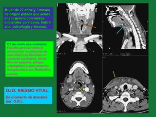
  • 1. Mujer de 27 años y 7 mesesMujer de 27 años y 7 meses de origen polaco que acudede origen polaco que acude a la urgencia con masasa la urgencia con masas bilaterales cervicales, fiebrebilaterales cervicales, fiebre alta. odinofagia y trismus.alta. odinofagia y trismus. CT de cuello con contraste:CT de cuello con contraste: Fascitis cervical bilateral extensa (no necrotizante de momento) con flemones en espacios carotídeos, borde libre de epiglotis, pliegue ariepiglótico y cara posterior laríngea derechos. Mediastino normal. OJO: RIESGO VITAL. De momento no drenable por O.R.L.