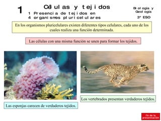
1. 1 Células y tejidos 14 Biología y Geología 3º ESO Presencia de tejidos en organismos pluricelulares En los organismos pluricelulares existen diferentes tipos celulares, cada uno de los cuales realiza una función determinada. Las células con una misma función se unen para formar los tejidos. Las esponjas carecen de verdaderos tejidos. Los vertebrados presentan verdaderos tejidos.
2. 1 Células y tejidos 23 Biología y Geología 3º ESO Células, tejidos, órganos y sistemas Célula muscular Tejido muscular Órgano (Corazón) Sistema circulatorio
3. 1 Células y tejidos 24 Biología y Geología 3º ESO Tejidos, órganos y sistemas EJEMPLOS DE TEJIDOS EJEMPLOS DE ÓRGANOS Y SUS SISTEMAS Muscular Cartilaginoso Nervioso Epitelial Corazón Encéfalo Hueso Estómago Sistema nervioso Sistema óseo Sistema digestivo Sistema circulatorio
5. 1 Células y tejidos 16 Biología y Geología 3º ESO Tejido epitelial (I). Epitelio de revestimiento Reviste la superficie exterior del cuerpo y las cavidades del organismo. Epitelio simple Epitelio estratificado Epitelio pseudoestratificado
6. Slide 5: Gall Bladder Simple columnar epithelium lines the gall bladder. Note the underlying connective tissue with blood vessels. Bar = 100 Microns. Expanded View
7. Slide 7: Male Urethra A pseudostratified columnar epithelium lines most of the penile urethra. Bar = 50 Microns. Expanded View
8. Slide 9: Urinary Bladder The expandible stratified epithelium of the bladder is referred to as transitional epithelium. Note that its surface cells are large rather than flattened as in stratified squamous epithelium. Bar = 50 Microns. Expanded View
9. 1 Células y tejidos 17 Biología y Geología 3º ESO Tejido epitelial (II). Epitelios glandulares Contienen células glandulares especializadas en segregar sustancias con destino al interior o al exterior del cuerpo. Glándulas exocrinas Glándulas endocrinas Glándulas mixtas
10. Slide 15 Pancreas The pancreas, shown here stained with H&E, serves both endocrine and exocrine functions. The round islets of Langerhans are the endocrine portion and serve to identify pancreatic tissue. Bar = 250 Microns Expanded View
11. Slide 8 Thyroid The simple cuboidal epithelium lining the follicles produces the thyroglobulin which is stored in the colloid follicles. Later it is taken back up by these same cells, cleaved, and released as T3 & T4. Notice that the thyroid is the only gland to store its hormones extracellularly. Click here for a more detailed description. Expanded View
12. Slide 6 Simple Tubular The intestinal gland (arrow) is found under the base of the villi. Notice also the goblet cells. Bar = 100 Microns Expanded View
13. 1 Células y tejidos 18 Biología y Geología 3º ESO Tejidos conectivos (I). Tejidos conjuntivo y adiposo Tejido conjuntivo Tejido adiposo Fibroblasto Linfocito Macrófago Fibras de colágeno Núcleo del adipocito Acúmulo de grasa Tejido conjuntivo
14. Slide 5 Elastic Fibers Sheets of elastic fibers, called elastic lamellae, are common in the aorta, shown here. These lamellae give a distinctive refractive appearance when you focus through them. Bar = 100 Microns Expanded View
15. Slide 4 Collagen Fibers The Masson (trichrome) stain leaves collagen green or blue. In the skin, Type 1 collagen predominates, as shown by the thick, wavy bundles. Bar = 250 Microns Expanded View
16. Slide 10 Tendon In cross section, collagen fibers make up the pale pink background. The fine lines separate fiber bundles; numerous fibroblast nuclei can be seen. Bar = 50 Microns Expanded View
18. Slide 1 Hyaline Hyaline cartilage is distinguished by its homogenous matrix surrounding the small nests of chrondrocytes. Notice the perichondrium which surrounds hyaline cartilage. Bar = 250 Microns Expanded View
19. Slide 5 Haversian Canal A higher magnification view of slide 4 clearly shows the concentric circles. After osteoclasts remove old bone, osteoblasts deposit bone in this circular arrangement beginning with the outer ring and working inward. As the osteoblasts become trapped in their own calcified deposits, they are known as osteocytes. Bar = 100 Microns Expanded View
20. 1 Células y tejidos 20 Biología y Geología 3º ESO Tejidos conectivos (III). La sangre Glóbulos blancos o leucocitos Defienden al organismo de cualquier agente causante de enfermedad. Su función es transportar sustancias. Intervienen en la coagulación de la sangre. Transportan oxígeno y dióxido de carbono. Pueden ser: Plasma sanguíneo Glóbulos rojos Endotelio Eosinófilos Monocitos Linfocitos Basófilos Neutrófilos Plaquetas o trombocitos
21.
22. Slide 9: Bone Marrow Notice the granules in the cytoplasm of the promyelocytes (yellow). Plasma cells (blue) are commonly seen in peripheral tissues where they secrete antibodies. Bar = 30
23. Slide 10: Bone Marrow Neutrophil (green) metarubricyte (red) rubricyte (yellow) plasma cell (blue) Bar = 30 Microns Expanded View
24. Slide 12: Bone Marrow Eosinophilic metamyelocyte (Red) Neutrophilic myelocyte (yellow) Metarubricytes (Blue) Bar = 50 Microns Expanded View
25. Slide 2: Bone Marrow Prorubricyte (blue), Band neutrophil (red), Platelets (yellow), Platelets are cell fragments produced by megakaryocytes. You will see a few of them on most of the slides. Bar = 30 Microns Expanded View
26. 1 Células y tejidos 21 Biología y Geología 3º ESO Tejido muscular Tejido muscular estriado esquelético Tejido muscular estriado cardiaco Tejido muscular liso Forma los músculos esqueléticos que se unen a los huesos y permiten el movimiento. Forma el músculo cardiaco. Se encuentra en las paredes del tubo digestivo, el útero, la vejiga y los vasos sanguíneos.
27. Slide 2 Skeletal Muscle At higher magnification, the striations become visible. I-bands (isotropic) are light while A-bands (anisotropic) are dark. Bar = 30 Microns Expanded View
28. Slide 4 Skeletal Muscle In cross section, skeletal muscle is identified by peripheral nuclei and large amounts of cytoplasm. Bar = 50 Microns Expanded View
29. Slide 8 Smooth Muscle Longitudinal smooth muscle is identified by long, thin central nuclei, a more scattered tissue layout, and an absence of striations. Bar = 50 Microns Expanded View
30. Slide 14 Cardiac Muscle Longitudinal cardiac muscle can be identified by centrally placed round to oblong nuclei, striations, branching, and intercalated discs (arrow). Bar = 30 Microns Expanded View
31. Slide 17 Cardiac Muscle The arrows mark organelles of the cardiac myocyte. Specific organelles cannot be distinguished. Bar = 30 Microns Expanded View
32. 1 Células y tejidos 22 Biología y Geología 3º ESO Tejido nervioso Forma el encéfalo, la médula espinal y los nervios. Se compone de dos tipos de células: neuronas y células de glía. NEURONAS CELULAS DE GLÍA Axón Por ejemplo, las células de Schwann que forman las vainas de mielina. Vaina de mielina Axón Núcleo de la célula de Schwann Soma Dendritas
33. Slide 15 Astrocytes Astrocytes are star-shaped glial cells of the CNS that have long processes. Many of these processes extend to blood vessels where they expand and cover much of the external wall. The expanded endings of the astrocyte processes are known as end-feet. While the blood-brain-barrier is formed by tight junctions between endothelial cells, the end-feet function to induce and maintain the blood-brain barrier. In pathology following stroke the relationship of end-feet to the endothelial cells is altered leading to disruption of the blood-brain barrier and subsequent leakage. Bar = 30 Microns Expanded View
34. Slide 7 Dorsal Root Ganglion A higher magnification view shows the surrounding satellite cells (red arrows), the glia of the DRG. Bar = 50 Microns Expanded View