
Recomendados
Más contenido relacionado
La actualidad más candente
La actualidad más candente (7)
Destacado
Destacado (7)
Similar a Bimcv eub heidelberg
Similar a Bimcv eub heidelberg (20)
Más de maigva
Último
Último (20)
Bimcv eub heidelberg
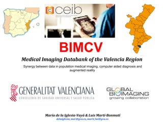
- 1. BIMCV Medical Imaging Databank of the Valencia Region Synergy between data in population medical imaging, computer aided diagnosis and augmented reality María de la Iglesia-Vayá & Luis Martí-Bonmatí delaiglesia_mar@gva.es, marti_lui@gva.es
- 2. Other facilities Single Technology Flagship Node EoI EuBI BIMCV
- 4. BIMCV consortium. https://ceib.cipf.es/bimcv Consortium Members: 1.- CEIB-CS (Regional Ministry of Health) 2.- La Fe Polytechnic University Hospital 3.- CIPF 4.- I3M. U.P.V. 5.- Universidad Valencia 5.- Universidad Alicante 6.- QuiBIM
- 8. P.A.S. community VNA - Vendor Neutral Archive (Regional)VNA - Vendor Neutral Archive (Regional) 2 x CPD (Data Center)2 x CPD (Data Center) Community 1 Arterias Network Arterias Network 1 PAS center 1 PAS center x PAS center x PAS center Community PACS - AD Community PACS - AD Community 2 Arterias Network Arterias Network 1 PAS center 1 PAS center y PAS center y PAS center Community PACS - AD Community PACS - AD Community 3 Arterias Network Arterias Network 1 PAS center 1 PAS center z PAS center z PAS center Community PACS - AD Community PACS - AD ... ... ... Solution. Logical Scheme
- 9. Two levels of image storage (Local and Regional) Two levels of image storage (Local and Regional) BIMCV Image request associated with an ongoing study anonymised Functional DICOM Circuit
- 10. Population Imaging – 24 Departments of Heath ARTERIAS - WAN
- 11. Population Imaging – Arterias WAN PROTECTED ARTERIAS WAN Research LAN VPN Virtual Private Network BIMCVGIMD Q/R
- 12. • Biobanks Repositories of biological samples. • Emerged as a fundamental tool for clinical research and innovation in genomics and personalized medicine through quality control issues for sample collections, standardized pathways for extraction and sophisticated protocols for data protection. • More recently, virtual biobanks, as repositories of digital information, have increased the opportunities for sharing, federating and exploiting biobank’s data. Imaging Biobanks
- 13. • Generate a structured and fully anonymized information, including medical images and relevant clinical and associated biological data and/or samples. ‒ Standard data formats and annotation through ontologies. ‒ Dissociated data will allow traceability of cases in unexpected findings. ‒ Verified quality of the data. • An infrastructure with massive storage and computing capacity. ‒ Large data samples involve establishing case scenarios and determine the universalization of the results. ‒ High performance computing resources to facilitate image processing comparison, standardization and validation. ‒ Integrate resources and services through a platform managing information flow and image processing and extraction ‒ Provide support to users for its utilization. Imaging Biobanks
- 14. What is BIMCV ? BIMCV - Medical Imaging Databank of the Valencia Region … … is an infrastructure with mass storage capacity (through GIMD – Project from the Regional Ministry of Health in the Valencia Region) and high throughput computational modeling capabilities. Aim To transform the Medical Imaging Databank into an environment for translational innovation in healthcare interventions and management.
- 15. • Generate a structured and fully anonymized information, including medical images and relevant clinical and associated biological data and/or samples. Besides, dissociated data will allow traceability of cases in unexpected findings (De- identification). • Standard data formats and annotation through ontologies. • Quality Control. Verified quality of the data. Principal Components of a Imaging Biobank
- 16. • Generate a structured and fully anonymized information, including medical images and relevant clinical and associated biological data and/or samples. Besides, dissociated data will allow traceability of cases in unexpected findings (De- identification). • Standard data formats and annotation through ontologies. • Quality Control. Verified quality of the data. Principal Components of a Imaging Biobank
- 17. • Anonymization of DICOM Headers following the standard http://dicom.nema.org/standard.html • Face anonymization • Anonymization of the text printed on the image Anonymization
- 18. Recepción de normas de anonimización (en bruto) Transformación de las normas en estructuras para la computación DICOM Part 15: Security and System Management Profiles Creación código de anonimización Imagen con cabeceras sin anonimizar Cabeceras DICOM anonimizadas Ejecución de la anonimización. Código de anonimización Anonymization of DICOM Headers
- 19. Population Imaging – Anonymization Implementation of the 10 DICOM Confidentiality Profiles with CTP-RSNA Software are not aware of any tool aimed to develop all confidentiality profiles defined in Part 15 of the DICOM standard in paragraph E ‘Attribute Confidentiality Profiles’ (http://dicom.nema.org/medical/dicom/current/output/html/part15.html#chapter_E). In order to avoid compromising the privacy, we consider crucial to implement secure and robust software modules in the personal data protection framework Within the DICOM standard, ten profiles are defined as listed:
- 20. Population Imaging – Anonymization Implementation of the 10 DICOM Confidentiality Profiles with CTP-RSNA This profile is the most stringent on and removes all information concerning: • The identity as well as identifying and demographic characteristics of the patient • The identity of the authors, persons responsible or family members • The identity of any personnel involved in the procedure • The identity of the organisations involved in ordering or carrying out the method • The information (not relative to the patient) that could be used to find out the identity of the original files not anonymized (eg UID, date and time) • The private attributes (which are not part of the standard). Some manufacturers keep important information for the image, e.g. gradients used in DTI. Basic profile
- 21. Population Imaging – Anonymization o Anonimización de las tags del estándar DICOM del nivel de aplicación básica del perfil de confidencialidad: ▪ DICOM PS3.6 2015a - Data Dictionary. ▪ DICOM PS3.15 2015a - Security and System Management Profiles. ● E Attribute Confidentiality Profiles (which attributes should be anonymized) http://dicom.nema.org/medical/dicom/current/output/ Implementation of the 10 DICOM Confidentiality Profiles with CTP-RSNA
- 23. Text Detection by: • Digital image processing • Feature comparison in the regions of interest Anonymization of the text printed on the image
- 24. • Generate a structured and fully anonymized information, including medical images and relevant clinical and associated biological data and/or samples. Besides, dissociated data will allow traceability of cases in unexpected findings (De- identification). • Standard data formats and annotation through ontologies. • Quality Control. Verified quality of the data. Principal Components of a Imaging Biobank
- 25. Population Imaging. Quality Control Acquisition Time SNRSpatial Resolution vóxel Size susceptibility Artifacts Effect
- 26. Population Imaging. QC Manual stimation by contrast Modification Automatic Stimation
- 27. Population Imaging. QC Translational Movement Rotational Movement The * indicate movements of more than 1 mm 360 2 απ ∗∗∗ = r L
- 28. Population Imaging. QC Frawise Displacement (Power) DVARS iiiiziyixi dddFd γβα ∆+∆+∆+∆+∆+∆= ixxiix ddd −=∆ − )1( ∑ −−= i titi YY I 2 1,, )( 1 DVARS(t) FD and DVARS
- 29. Population Imaging. QC Test FD y DVARS FD and DVARS
- 31. Population Imaging Use cases with BIMCV
- 32. Use case #1.- 10k Project Big Data in Brain Imaging Population Imaging 10 k is a collaborative project with the San Juan & Sagunto Hospitals. Maria de la Iglesia-Vayá, PhD. & Jose Maria Salinas, PhD.
- 33. Population Imaging Use case #1.- 10k Project Big Data in Brain Imaging
- 34. Population Imaging • Cortical thickness, area and volume structure compared with the reference values Use case #1.- 10k Project Big Data in Brain Imaging
- 35. Population Imaging Use case #1.- 10k Project Big Data in Brain Imaging
- 37. Imaging Biomarkers ImagingLab Quibim_Quiron. Spin-off
- 38. Example
- 39. Use case #2.- BrainGIS (Brain Geografic Information System) Population Imaging – Data Mining
- 40. Use case #2.- BrainGIS (Brain Geografic Information System) Population Imaging – Data Mining
- 41. Use case #3.- NeuroBIM-MS (Multiple Sclerosis) Population Imaging Hospital Vega Baja de Orihuela Dr. Santiago Mola - Head of Neurology Hospital General Universitario de Alicante Dr. Angel Pérez - Neurologist specialist Hospital Universitario San Juan de Alicante PhD. Jose María Salinas - Head of Information Technology and associate professor at the University of Alicante Phd. Miguel Angel Cazorla RoVit research group manager
- 42. Use case #4.- MIDAS (Massive Image Data Anatomy Spine) Population Imaging MIDAS is a collaborative project with the Arnau de Villanova Hospital. Traumatology Service. Dr. Julio Domenech Fernandez
- 43. Use case #4.- MIDAS (Massive Image Data Anatomy Spine) Population Imaging
- 44. Use case #4.- MIDAS (Massive Image Data Anatomy Spine) Population Imaging
- 45. Use case #4.- MIDAS (Massive Image Data Anatomy Spine) Population Imaging https://sourceforge.net/projects/spinalcordtoolbox
- 46. Use case #5.- Augmented Reality for Visualization Population Imaging. Visualization Parametric image by Augmented Reality from Gonzalo M. Rojas. Download from Smartphone Play Store, the Prototype for Android: ARiBraiN3T. http://www.aribrain.info
- 47. http://www.esf.org/activities/forward-looks/medical-sciences-emrc/current-forward-looks-in-medical-sciences/personalised-medicine-for-the-european- citizen/more-information.html. Population Imaging as part of Big Data landscape
- 48. Data Sharing: Code, Manage & Collaborate
- 49. Data Sharing - Open minds The Landscape of BIMCV - Open source
- 50. Acknowledgement
- 51. • Luis Martí-Bonmatí, Head of Biomedical Imaging Research Group (GIBI230) at La Fe Polytechnics and University Hospital – La Fe Health Research Institute. • Oscar Zurriga Llorens, Director General of Research, Innovation Technolgy and Quality. Regional Ministry of Health in the Valencia Region. • Carmen Ferrer Ripollés, Deputy Director General of Information Systems for Health. Regional Ministry of Health in the Valencia Region. • Salvador Peiro Moreno, Deputy Director General of Research and Innovation. Regional Ministry of Health in the Valencia Region. • Ignacio Blanquer, Institute of Instrumentation for Molecular Imaging – I3M. Universitat Politècnica de València. • Jacobo Martínez, Director of FISABIO. • Carlos Martinez Riera, Coordinator European Research projects office, University of Valencia. • Jose María Salinas, University Hospital San Juan de Alicante. Head of Information Technology and associate professor at the University of Alicante. • GIMD team. Regional Ministry of Health in the Valencia Region & General Electric. • Rafael de Andrés and Timo Zimmerman, The Spanish representatives in the Euro- BioImaging Interim Board. Acknowledgements
- 52. Thank you so much to my team Members CEIB-CS •María de la Iglesia Vayá, PhD. Team Leader. •Ángel Fernández-Cañada Vilata, MSc. •José Miguel Calderón Terol, MSc. •Jhon Jairo Sáenz Gamboa, MSc. Past Member CEIB-CS •Jorge Isnardo Altamirano, MSc.
Notas del editor
- Expression of Interest in Population Imaging
- The Spanish BIMCV – Medical Imaging Data Bank – Population Imaging Node Candidate in Valencia provides a multi-level storage service and access (Vendor Neutral Archive) on specific projects. The Node integrates cloud-based computational services with access to high performance computers clusters from local and European infrastructures, and provides open access methodologies that will integrate different data types for population imaging and quantitative resources. The Node develops and provides access to a large database of imaging data and corresponding clinical data records. The aims of this Node are to transform the Medical Imaging Databank into an environment for translational innovation in healthcare interventions and management and to facilitate open translational research with a sufficiently representative sample population. BIMCV gives a clear answer to the creation of an infrastructure for large population base, with associated data acquisition, the quantitative analysis of standardized data and access to the image data.
- Specialties and expertise of the Node CandidateBIMCV is a joint initiative of five main institutions in the region of Valencia: La Fe Polytechnic University Hospital, the Valencian Regional Authorities in Health, the Polytechnic University of Valencia and the University of Valencia. This joint endeavor brings together expertise on medical imaging biomarkers, medical databanks and intensive computing infrastructures. The services that will be provided will include: Access to a selected set of medical images and associated information from a population database and according to multiple criteria. A catalogue of Medical Image Biomarkers and processing services to address different research challenges. A mechanism to configure and deploy virtual infrastructures on research computing infrastructures. Genetic correlation with imaging and phenotypic data. Allow data storage under specific projects.
- THAT CONNECT WITH SEVERAL PLATFORMS 1.- XNAT 2.- ???, IM3, QUIBIM platform… etc
- Management of Digital Medical Imaging Project from Regional Ministry of Health. Health information systems are generally structured in layers where all other sub-systems (secuirity, communications, technical support…) are integrated. Applications constitutes the upper layer, divided itself in three main groups: health-care applications dealing with the patients’ clinical information; management applications dealing the logistical process around the health care process; and inteligence application, offering indicators to better control and improve the two other layers. Within the block of health care applications, Radiology Information System (RIS) is of paramount importance in the framework of brain imaging. RIS is the System that manages the radiology tests and the corrsponding reports within the imaging system SISAN. PACS is the other important system that stores, searches and displays medical images generated in the clinical practice. Both systems are complementary.
- P.A.S. (SERVICE ACCESS POINT)
- Motor de integration = Integration engine (Thoughts) Search Engine de pena ;)
- This cover 24 departments of health, with at least one hospital in each one; And all into a PRIVATE and securized WAN – Network, called ARTERIAS
- Query/Retrieve, or Q/R for short, is the DICOM service for searching images on the PACS and getting a copy of them to the workstation where they can be displayed. Every set of data linked to a projects is anonimysed before been extracted from GIMD and only then in stored in BIMCV
- More recently, virtual biobanks, as repositories of digital information, have increased the opportunities for sharing, federating and exploiting biobank’s data.
- Aim To transform the Medical Imaging Databank into an environment for translational innovation in healthcare interventions and management.
- anonimización de la propia imagen: eliminación de rasgos faciales. texto quemado.
- Clinical Trial Procesor (CTP)
- Clinical Trial Procesor (CTP)
- Clinical Trial Procesor (CTP)
- Text Detection by: Digital image processing Feature comparison in the regions of interest
- All the QC is based on scientific reports.
- STRUCTURED QC REPORT A structured report is generated for each analysis, providing additional quantitative information to the radiology workflow.
- In the next slides, I am presenting a few use cases
- FIRST USE CASE IS THE 10 thousand PROJECT
- In project 10k, measurements are made on cortical thickness, area, volume on more than 10,000 MRI brain images. The objective is to derive the reference values to be used in future medical imaging biomarkers.
- In project 10k, measurements are made on cortical thickness, area, volume on more than 1,000 MRI brain images. The objective is to derive the reference values to be used in future medical imaging biomarkers.
- This values of normality may be downloaded from the project Web page
- Why are those values important? they are important because they will be used in future as imaging biomarkers, very much like they are used in any hematology laboratory nowadays.
- The next future –not, present- of Radiology activity services and Dr. Prof. Luis Martí-Bonmatí (that is promoting Imaging Biomarkers in ERS and all the world) is the new academic and he take the chair 13, of Radiology and Radiodiagnostic. Professor Martí-Bonmatí is the first radiologist who occupies the chair of the specialty, created in 1861.
- Here you can see an example of another spin-off showing an imaging biomarker in Multiple Sclerosis Brain MRI biomarkers for improved follow-up of people with Multiple Sclerosis (MS) Brain atrophy Lesion load
- The second use case is the project BrainGIS The aim is to study the relationship between the geographic areas and some parameters obtained from brain parcelation within the project 10k, This is part the philosophy of Euro-Bioimaging “Re-use”
- applying data mining, machine learning and other techniques, as you can see in this slide,
- BIMCV AS A MEETING POINT WITH DIFFERENT HEALTH INSTITUTIONS IN A COLABORATIVES PROJECTS. ---- The third use case is a collaborative project between several hospitals from Alicante to carry out medical imaging studies on Multiple Sclerosis.
- 1.- Last but not least, a study carried out by the Head of the Traumatology Service of the Hospital Arnau de Vilanova, 2.- aims at obtaining overall measurements of the lumbar spine. Low back pain is a prevalent disorder and a frequent cause of disability. It is associated with increased costs for the healthcare system and society in developed countries, affecting 70% of the general population at some point in their lives, with an annual incidence of 40%. Identifying predictors of chronicity has become a priority for researchers on lumbar pathology. Studies evaluating the presence of anatomical or structural changes in the lumbar spine by techniques such as CT, MRI or discography are not able to correlate these anomalies with a bad prognosis in low back pain. The concept that associates chronic pain with structural alterations should be reconsidered taking into account the recent scientific evidence.
- SCT is a comprehensive and open-source library of analysis tools for multi-parametric MRI of the spinal cord.
- Only I would like to remark as conclusion that ….. OMICS, MEDICAL IMAGING AND BIG DATA will be crucial to the clinical decision systems and practice