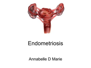
Endometriosis
- 2. Definition • A condition in which actively functioning endometrial tissue and glands which are usually confined to the endometrium are found outside the uterus (ectopic endometrium). The presence of such tissues in ectopic sites elicits inflammatory changes and fibrosis
- 3. Incidence • Prevalence of endometriosis is about 10% of all women between menarche and menopause
- 4. Pathophysiology 1.Retrograde menstruation by Sampson (1922) where there is reflux of the menstrual flow through the fallopian tubes into the peritoneal caivty where it can implant. Proof: Scar endometriosis following classical caesarean section, hysterectomy, myomectomy and episiotomy further supports this view. 2.There is a combination of failure of immune mechanisms associated with stromal cell defect with its increased oestrogen, prostaglandin and progesterone resistance
- 5. 3.Coelomic metaplasia theory (Meyer and Ivanoff 1919) where endometriotic lesions develop when coelomic mesothelial cells of the peritoneum undergo metaplasia 3.The circulation and implantation of ectopic menstrual tissue via the venous or the lymphatic system or both, explains its occurrence at less accessible sites like the umbilicus, pelvic lymph nodes, ureter, rectovaginal septum, bowel wall, and remote sites like the lung, pleura, endocardium and the extremities.
- 6. • Hormonal influence: the initial genesis of endometriosis its further development depends on the presence of hormones mainly estrogen. Pregnancy causes atrophy of endometriosis through high progesterone level. Regression also follows oophorectomy and irradiation. Endometriosis is rarely seen before puberty and it regresses after menopause. Hormones with antiestrogenic activity also suppress endometriosis and are used therapeutically
- 7. Immunological factor • The peritoneal fluid in endometriosis shows the presence of macrophages and natural killer (NK) cells • Impaired T cell and NK cell activity and altered immunology
- 8. Genetic • Familial tendency reported in 15% cases, multifactorial, vaginal or cervical atresia which encourage retrograde spill.
- 9. Sites • Uterine : adenomyosis (50%) • Extrauterine: • Ovary 30% • Pelvic peritoneum 10% • Fallopian tube • Vagina • Bladder and rectum • Pelvic colon • Ligaments
- 12. Endometriosis • Ovary • Cul de sac • Uterosacral ligaments • Broad ligaments • Fallopian tubes • Uterovesical folds • Round ligaments • Vermiform • Vagina • Rectovaginal septum • Rectosigmoid colon • Caecum • Ileum • Inguinal canals • Abdominal scars • Ureters • urinary bladder • Umbilicus • Vulva
- 13. Sites • Pelvic • Extrapelvic – umbilicus – scars (laparotomy) – lungs – pleura – others
- 14. Pathology • Early lesions appear papular and red vesicles are filled with haemorrhagic fluid with surrounding flame like lesions
- 15. • Overtime, these vesicles change colour and endometriotic areas appear as dark red, bluish or black cystic areas adherent to the site
- 16. • Scarring in the endometriosis makes it puckered. Atypical lesions such as non pigmented areas or yellowish white thick plaques have been noticed, which are healed lesions
- 17. • Powder burnt areas are the inactive and old lesions seen scattered over the pelvic peritoneum
- 18. Chocolate cysts • Chocolate cysts of the ovaries represent the most important manifestation of endometriosis. • To the naked eye, the chocolate cyst shows obvious thickening of tunical albuginea, and vascular red adhesions are well marked on the undersurface of the ovary. THe innner surface of the cyst wall is vascular and contains areas of dark brown tissue. THe chocolate cyst lies in the ovary and adherent to lateral pelvic wall
- 21. History • Deep seated dyspareunia- painful sexual intercourse • Severe dysmenorrhoea • Pain at midcycle of menstrual which coincides with ovulation
- 22. • Chronic Pelvic pain is the most common presenting symptoms. Other associated symptoms are back pain, loin pain, dyschezia (ie pain on defaecation) and • Pain with micturition
- 23. • Infertility • Fatigue • There may or may not be abnormal vaginal bleeding • Rarely, cyclical haemoptysis (endometriotic nodule in the lungs) and cyclical haemoaturia (endometitic nodules in the urinary bladder).
- 24. • A patient survey of women in the UK and US who were referred to University based Endometriosis Centres found that 70-71 % presented with pelvic pain, 71-76% with dysmenorrhoea, 44% with dyspareunia • 15-20% with infertility
- 25. Risk factors • First degree relative affected • Short menstual cycles • Long duration of menstrual flow • Low parity • Infertility • Fair complexioned
- 29. Diagnosis • On bimanual pelvic examination, fixed retroverted uterus, bilateral pelvic tenderness, fixed or enlarged ovaries and painful uterosacral nodularity • Depply infiltrating nodules are most reliably detected when clinical examination is performed during menstruation. Adenomyotic uterus is seldom >12 weeks, soft, smooth and tender in contrast to fibroid uterus. • Isolated adenimyoma can be differentiated by presence of localised tenderness
- 30. Examination Findings • Tenderness in the suprapubic region • A tender lower abdominal mass may or may not be present • Pelvic examination (rectovaginal examination included) may reveal a retroverted uterus with restricted mobility and tenderness • Fixed retroverted tender uterus with paindul nodules in POD and uterosacral ligaments that are best assessed during mestruation
- 31. • Painful adnexal mass • Painful nodules in the Pouch of Douglas or uterosacral ligaments • These nodules are nest palpated and appreciated with the examinaition is done during menstruation
- 33. Investigations • Imaging: US of the pelvis and abdomen (transvaginal US) is of limited value in the diagnosiis of early pelvic endometriosis. • In advanced disease where endometrioma and pelvic mass are present pelvic US produce typical images. • The kidneys should be imaged for evidences of obstructive uropathy and hydronephrosis if there is severe pelvic disease.
- 34. Ultrasonography • Endometriotic cysts (oval or round)- capsulated fine homogenous, uniform, granular echoes, anechoic, single or multiple, unilateral or bilateral • On doppler: no vascularity within the mass • Ovarian adhesion to uterus • Free floating fimbria or sonosalphingography
- 35. Laparoscopy • Both diagnostic and therapeutic • Gold standard • It should not be performed within 3 mnths of hormonal treatment to prevent under diagnosis
- 36. Appearance • Match stick head or blackened spot like lesions oveer the ovaries, serosal surface and peritoneum • Red implants (petechial, vesicular, polypoid, haemorrhagic , red flamelike) which are atypical with areas of fibrosis • Vesicles (serous and clear) • Peritoneal defects (scarring and yellow brown peritoneal discolouration) • Endometriomas (ovarian cysts containing stale blood which appears like tar. Widely referred to as chocolate cysts. Obliteration of the ovarian fissa can slso taje place Laparoscopy, showing minimal endometriosis, in the form of " powder-burn" deposits.
- 37. • Powder burn or black lesions • White opacifiied peritoneum • Glandular excrescences • Flame like red lesions • Peritoneal pockets or windows
- 38. • Clear vesicles • Yellow brown patches • Unexplained adherence of ovary to peritoneum of ovarian fossa • Encysted collection of thick chocolate coloured or tarry fluid • Adhesions to posterior lip of broad ligaments/ other pelvic structure
- 40. Endometriotic cyst of the left ovary (typical laparoscopic image).
- 41. A cluster of chocolate cysts
- 43. Dense Adhesions
- 44. MRI • When endometriosis is thought to have a deeply invasive component (bowel and bladder invasion), ancillary tests such as colonoscopy, cystoscopy, rectal ultrasonography and MRI may be required.
- 45. • Endometriotic deposits in rectovaginal septum seen as as high intensity signal in MRI image
- 46. CA 125 • May be elevated in severe endometriosis
- 47. Histological Confirmation • Visual inspection is usually adequate but histological confirmation of at least one lesiion is ideal • In cases of ovarian endometrioma >3cm in diameter and in deeply infiltrating disease histology is a must to rule out malignancy
- 48. American Society for Reproductive Medicine revised classification of endometriosis (American Fertility Society AFS grading)
- 50. In SHORT • Score 1-5: Stage I: minimal disease • Score 6-15: Stage II: mild disease • Score 16-40: Stage III: moderate disease • Score >40: Stage IV: severe disease
- 51. • -Grade 1: Possible endometriosis - Peritoneal vesicles, red polyps, yellow polyps, hypervascularity, scar, adhesions. • Grade 2: Suggestive of endometriosis. Chocolate cyst with free flow of • chocolate fluid • Grade 3: Consistent with endometriosis - Dark scarred (puckered pigmented or mixed color) lesions, red lesion on fibrous scarred background,chocolate cyst with mottled red and dark areas on white background. • -Grade 4: Endometriosis. Dark, scarred (or puckered, pigmented) lesions at first surgery
- 52. Treatment • Treatment of endometriosis can be either medical, surgical or combination of both • The medical mangement can be for pain control or prevention of menstruation and therefore restric progressive ectopic endometrial profileration.
- 53. • Surgical management is more definitive and can also leaf to reduction of sumptoms of dysmenorrhoea and increase pregnancy rate
- 54. • If fertility is the main issue, the management should be geared towards surgical excision/ ablation of the endometriotic lesions and iUI or IVF
- 55. Medical Management • Recognise goals – Pain management – Preservation/ Restoration of fertility • Discuss with the patient – Disease may be chronic and not curable • Curable – Optimal treattment unproven or nonexistent
- 56. • Empirical treatment of pain symptoms without definitive diagnosis of endometriosis, a therapeutic trial of hormonal drug to reduce menstrual flow is appropiate • Medical therapy for endometriosis can be used either as primary therapy or in conjunction with surgery preoperatively or postoperatively sandwich therapy
- 57. NSAIDS for pain Management • There is inconclusive evidence to show whether NSAIDs (specifically Naproxen) are effective in managing pain caused by endometriosis. THis should not be taken at the time of ovulation in women who want to get pregnant as this can inhibit the process of ovulation.
- 59. • T. Ponstan 500mg TDS for 5 days
- 60. Hormones • COmbined oral contraceptives (COCs) • Prevention of withdrawal bleeding by taking COC continuously can prevent retrograde menstruation, hence this ethod is said to be effective pain relief.
- 61. • To reduce the frequent prolonged bleeding not recommended in infertility endometriotic women • However COCs are the only effective prophylaxis in against endometriosis
- 63. Medroxyprogesterone • Medroxyprogesterone especially the depot form, may be effective in reducing pain symptoms but long term usage may reduce bone mineral density
- 64. • Progesterone: pseudo pregnancy (Kristner's regime) state • Acts by decidualisation and atrophy of the estrogen dependent endometriotic foci • COmmon progesterone: medroxyprogesterone acetate, norethisterone, dydrogesterone • DMPA- cost effective, readily available 66% complete resolution
- 65. • Side effects: Irregular bleeding, weight gain, fluid retention,m breast tenderness, mood changes
- 66. Danazol and Gestrinone • Weak angrogens, progestogenic and anti estrogenic. • May have many androgen induced side effectis which limit their usage. • Low dose regimens and vaginal usage have been proposed.
- 67. What is gestrinone? • AN ANTI-PROGESTIN • Gestrinone 1-25-2-5mg biweekly • Side effects: similar to danazol
- 68. GnRH agonist • This can induce hypoestrogenism and therby reduce not only pain but also size of lesions. THe only drawbacks of this form of therapy is that it can't be used longterm due to effects of hypoestrogenism and reduction in bone mineral density. Add back therapy with estrogen and progesterone enables the usage of this drug for up to 12months
- 69. Aromatase Inhibitor • Associated with bone loss
- 70. Surgical Management • Indications • Mild endometriosis associated with infertility • Endometrioma >4 cm in diameter • Endometriosis of rectovaginal septum or rectal wall • Failed medical therapy • Intolerable side effects of medical therapy • Endometriosis with other surgically correctable infertility factors
- 71. • Surggical removeal is preferred as it has been proven that surgical ablation reduces the dysmenorrhoea caused by endometriosi. Even in deeply infiiltrating diease, the removal of the lesions in entirety reduces pain symptoms. • ENdometriomas more than 4 cm are best treated by laparoscopyc ovarian cystectomy
- 72. • Combination of ablation of endometriotic lesions and adhesiolysis improves the rates of fertility in mild to moderate endometrriosis • THeremay be no role for just performing laparoscopic nerve ablation (without any ablation of endometriotic deposits) for as there is no proven reducition in pain
- 73. • In patients with mild to moderate endometriosis, IUI improves fertility • Women treated with GnRH agonists for duration of 3-6 months with addback therapy prior to IVF have shown to have higher rates of clinical pregnancy
- 74. Pre-operative assessment • MRI or USS with or without IVP, Barium enema, sigmoidoscopy • Pre- op and postop medical managemnent • GnRH agonist like goserelin for 3 months preop reduces the size and AFS score. • Postop therapy gives longer periods of remission.
- 75. • Primary operation is the best opportinity • Best outcome by excision of the lesion • COmplete excision has lowest recurrence of 19% • Adhesions require excision rather than simple diivision
- 76. Endometriosis and fertility • 20-25% of women undergoing laparoscopy for infertility or for chronic pelivic pain demonstrate underlying endometriosis
- 77. • Ovarian follicles have abnormal growth rates and are defective in their function • The ovulation process itself is affected where the mature follicle fails to ruepture and release the ooctye and gets luuteiniised instead
- 78. • Local inflammation in the pelvis and the presence of excess peritoneal fluid with large amounts of macrrophages may dirsrupt ovarian function, capture of the ovum by the imbriae, affct sprm and also the process of fertilisation. • This is an environment which is not conducive to conception.
- 79. • Early stage disease: laparoscopic excision or ablation with adhesiolysis • Moderate to severe endometrosis: role of surgery is uncertain (overactive excision may reduce fertility)
- 80. • Endometrioma: laparoscopic cystectomy better than drainage and coagulation • Post operative hormonal treatment has no beneficial effect on pregnancy rates after surgery • Tubal flushing improves pregnancy rates
- 81. Medical Management of Infertility due to endometriosis • There is no evidence to show that suppression of ovarian function using drugs like medroxyprogesterone, danazol etc having any beneficial effect on fertilty in mild to moderate endometriosis • It is not effective in more severe forms of endometriosis
- 82. • There is however an improvement in pregnancy rates in women with endometriosis who are treated with GnRH agionist suppression therapy with hormonal replacement add therapy for 3-6 months prior to iVF
- 83. Surgical Management of infertility due to endometriosis • Ablation of endometriotic lesions and adhesiolysis improves the rates of pregnancy in mild to moderate endometriosis • It is of uncertain benefit in moderate to severe disease • Endometriomas more than 4cm are best treated by laparoscopic ovarian cystectomy
- 84. ADENOMYOSIS
- 85. Introduction • A benign condition of the uterus • Bears close clinical similarities with leiyomyoma • Mainly confined to body of the uterus occurring as discrete lesions or more extensively • Rarely seen in the cervical part • It is considered as an extension of endometriosis wherein the endometrial glands grow inside the uterine musculature
- 86. Incidence • Commonly seen in women above 40 years with an overall incidence of 10% • Occurrence is more likely to be seen in women who are parous and have had termination of pregnancy, spontaneous miscarriages and endometriosis
- 87. Diagnosis • 30% of women are asymptomatic, heavy menstruation (40-50%) and dysmenorrhea (15-20%) in parous women between 40-50 years with a globularly enlarged uterus • Clinical features are similar to leiyomyoma • Adenomyosis can be asymptomatic, it is frequently diagnosed after hysterectomy • Heavy menstruation if present will be progressive over time
- 88. • On examination, it might be difficult to differentiate from leiyomyoma when the uterus is uniformly enlarged • Suspician is aroused when: - the enlarged uterus rarely exceeds 12-14 weeks size - enlargement is regular compared to the nodular enlargement of leiyomyoma
- 89. Reasons for menorrhagia 1 . Increase in surface area due to enlarged uterus 2 . Increased vascularity to support the enlarged uterus 3 . Impaired contraction due to glandular tissue in the myometrium 4 . Probable association of endometrial hyperplasia
- 90. Investigations 1. Ultrasonography - TVS and TAS: reveal a decreased echogenicity/heterogenicity in the myometrium - reason for heterogenicity is mainly due to the presence of cystic glands amongst smooth muscles 2. MRI - significant when differentiating between leiyomyoma and adenomyosis cannot be ascertained clinically
- 91. Management 1. Medical management - menorrhagia & dysmenorrhea: Tranexamic acid (500mg TDS) and Mefenamic acid (500mg TDS) - women <35 years, combined oral pills - women >35 years, progestins are administered - GnRH analogues reduces symptoms of adenomyosis and uterine size - IUS Levonorgestrel, Leuprolide - low dose of Mifeprostone for periods of 30 days
- 92. 2. Surgical management - hysterectomy - hysteroscopic resection: superficial adenomyosis ( <1mm myometrial invasion) - Uterine artery embolisation (UAE) and MRI guided focused ultrasound surgery
