
Recomendados
Recomendados
Más contenido relacionado
La actualidad más candente
La actualidad más candente (20)
Similar a Endometrial cancer
Similar a Endometrial cancer (20)
Más de Danilo Baltazar Chacon
Más de Danilo Baltazar Chacon (20)
Último
Último (20)
Endometrial cancer
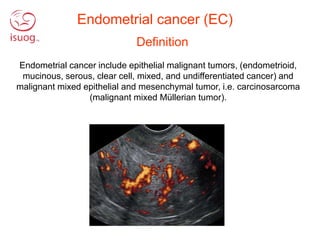
- 1. Definition Endometrial cancer include epithelial malignant tumors, (endometrioid, mucinous, serous, clear cell, mixed, and undifferentiated cancer) and malignant mixed epithelial and mesenchymal tumor, i.e. carcinosarcoma (malignant mixed Müllerian tumor). Endometrial cancer (EC)
- 2. Macroscopic appearance Figure 1a. Macroscopic appearance of a tumour originating in the endometrium of the corpus uteri, infiltrating into the myometrium
- 3. Macroscopic appearance Figure 1b. Macrosocpic apperence of endometrial cancer that originates in the lower uterine segment.
- 4. Lägg FIGO 2 kolla om Lägg till Mucinous? Adenosquamous? Microscopic appearance of endometrioid cancers Figure 2 a-c Lägg FIGO 3 kolla om FIGO I och II är OK Lägg till Mucinous? Adenosquamous? Endometrioid-FIGO 3, Solid growth Endometrioid with mucinous differentiation
- 5. Microscopic appearance of the MELF (Microcystic, Elongated structures or Fragmented solid cells) - growth pattern Figure 3: MELF growth pattern, associated with adverse prognosis
- 6. Microscopic appearance– Non-endometrioid tumours Figure 4b Figure 4c Figure 4a
- 7. Assessment of endometrial gray-scale morphology according to IETA (Leone et al UOG 2010) IRREGULAR INTERRUPTED NON-UNIFORM ECHOGENICITY ENDOMETRIAL – MYOMETRIAL JUNCTION UNIFORM ECHOGENICITY ENDOMETRIAL ECHOGENICITY REGULAR Figure 5a
- 8. Assessment of endometrial vascularity according to IETA (Leone et al UOG 2010) VESSEL PATTERN MULTIPLE FOCAL MULTIPLE MULTIFOCAL 1 2 3 4 COLOR SCORE SINGLE SINGLE BRANCHING SCATTERED CIRCULAR Figure 5b
- 9. Sonographic vessel pattern may reflect tumour growth pattern (Epstein et al UOG 2011) Focal vessel- pattern associated with exophytic growth Multiple multifocal vessel pattern associated with invasive growth Figure 6
- 10. Morphology of endometrioid endometrial cancer (EEC) dependents on grade and stage (Epstein et al UOG 2018) Stage 1A, grade 1 Stage 1A, grade 3 Stage 1B, grade 1 Stage II, grade 3 Low risk tumors High risk tumors Figure 7
- 11. Ultrasound characteristics LOW RISK CANCER Regular endometrial- myometrial junction Uniform Echogenicity Single branching, or Multiple focal vessels Sparse vascularization Figure 8
- 12. Ultrasound characteristics HIGH RISK CANCER Irregular endometrial- myometrial junction Non-uniform echogenicity Multiple, multifocal vessel pattern Color score 3-4 Figure 9
- 13. Endometrial Morphology – non-endometroid tumours (Epstein et al UOG 2018) Non-endometrioid tumours are all high-risk tumours Carcinosarcoma Clear cell Mixed Serous Figure 10
- 14. VIDEO – typical case low risk endometrial cancer No cervical stromal invasion Myometrial invasion < 50% Figure 11
- 15. VIDEO – Typical case high risk endometrial cancer Cervical stromal invasion present Myometrial invasion present Figure 12
- 16. Differential diagnosis Endometrial Stromal Sarcoma Uterus completely infiltrated by colorectal cancer metastasis Benign Endometrial polyp Figure 13 Cervical cancer