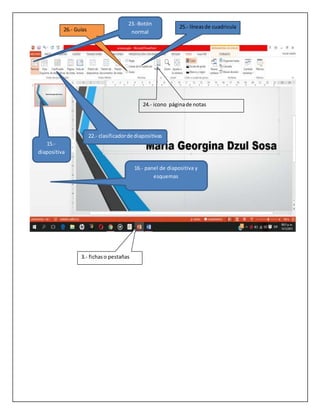
Este documento proporciona instrucciones para un ejercicio en el que los estudiantes deben identificar y capturar las opciones desplegadas al seleccionar cada pestaña en Microsoft PowerPoint. También deben señalar el nombre de 4 iconos en cada pestaña y comparar las similitudes y diferencias entre PowerPoint y Microsoft Word.
Nano-adjuvanted polio vaccine: Preparation and characterization of chitosan ...Nanomedicine Journal (NMJ)
Abstract
Objective(s):
It is proposed that particulate antigens could better interact with the antigen presenting cells (APCs). A fast, simple and scalable process for preparation of polymeric nanoparticles (NPs) is coating of charged antigenic particles, like viruses, with oppositely charged polymers. A second coating with a charged polymer could increase the stability and modify the immunomodulatory potentials of NPs.
Materials and Methods:
Negatively charged inactivated polio virus (IPV) was coated with cationic polymers, chitosan (CHT) and trimethylchitosan (TMC) by a simple incubation method. CHT: IPV and TMC: IPV NPs were coated by anionic polymer, sodium alginate (ALG). Physical characteristics and stability of NPs were studied. Cytocompatibility of NPs was checked with MTT assay. DC maturation study was used for evaluation of the NPs potential in interaction with DCs.
Results:
Among the various polymer to antigen ratios tested, the least size and PDI and the highest ZP was seen in TMC: IPV (2:1), CHT: IPV (2:1), ALG: TMC: IPV (2:2:1) and ALG: CHT: IPV (4:2:1). The physical stability of TMC: IPV and CHT: IPV was preserved until 15 days. After an early de-association of some part of coated alginate, ALG: CHT: IPV and ALG: TMC: IPC NPs were stable until the end of study (25th day). No one of the NPs formulations had a negative effect on cell viability. Compared with plain IPV, nanoparticulate IPV formulations failed to increase the expression of CD40 and CD86 markers of DCs.
Conclusion:
NPs prepared with simple and scalable method, had reasonable physical characteristics, stability and cytocompatibility and could be tested in vivo for their immunoadjuvant potential.
Geza Vermer
Makó, Hungría, 22 de junio de 1924 - 8 de mayo de 2013), fue un historiador de las religiones. doctor en Teología y Letras, fue profesor en la Universidad de Oxford y escritor de numerosos estudios sobre la relación entre judaísmo y cristianismo, siendo considerado especialista sobre los
esenios, los textos arameos y Jesús de Nazaret. Se le estima como uno de los más eminentes conocedores de la vida y doctrina de Jesús,cuya narración evangélica ha situado en su contexto judío.
El profesor Vermes fue uno de los primeros especialistas en examinar los Manuscritos del mar Muerto poco después de su descubrimiento en 1947 y es el autor de su traducción al inglés.
Geza Vermes era, al momento de su muerte, profesor emérito de estudios judíos y Fellow emérito del Wolfson College de Oxford y continúa enseñando en el Instituto Oriental de Oxford. Es el Redactor Jefe del Journal of Jewish Studies Desde 1971 a 1991 dirigió el Oxford Forum for Qumran Research en el Centro de Estudios Hebraicos y Judíos de Oxford.
Abstract
Objective(s):
Biosynthesis of gold nanoparticles (NGPs) is environmentally safer than chemical and physical procedures. This method requires no use of toxic solvents and synthesis of dangerous products and is environmentally safe. In this study, we report the biosynthesis of NGPs using Streptomyces djakartensis
isolate B-5.
Materials and Methods:
NGPs were biosynthesized by reducing aqueous gold chloride solution via a Streptomyces isolate without the need for any additive for protecting nanoparticles from aggregation. We characterized the responsible Streptomycete; its genome DNA was isolated, purified and 16S rRNA was amplified by PCR. The amplified isolate was sequenced; using the BLAST search tool from NCBI, the microorganism was identified to species level.
Results:
Treating chloroauric acid solutions with this bacterium resulted in reduction of gold ions and formation of stable NGPs. TEM and SEM electro micrographs of NGPs indicated size range from 2- 25 nm with average of 9.09 nm produced intracellular by the bacterium. SEM electro micrographs revealed morphology of spores and mycelia. The amplified PCR fragment of 16S rRNA gene was cloned and sequenced from both sides; it consisted of 741 nucleotides. According to NCBI GenBank, the bacterium had 97.1% homology with Streptomyces djakartensis strain RT-49. The GenBank accession number for partial 16S rRNA gene was recorded as JX162550.
Conclusion:
Optimized application of such findings may create applications of Streptomycetes for use as bio-factories in eco-friendly production of NGPs to serve in demanding industries and related biomedical areas. Research in this area should also focus on the unlocking the full mechanism of NGPs biosynthesis by Streptomycetes.
Epic Research has proven itself as one of the primary platforms for share market tips. Our opinion are based on Indian share market fundamental, technical & equity research. we offer wide range of service packs related to these segment.
Subacute dermal toxicity investigation of nanosilver on serum chemical biomar...Nanomedicine Journal (NMJ)
Abstract
Objective(s):
Nanosilver is one of the most widely used nanomaterials due to its strong antimicrobial activity. Thus, because of increasing potential for exposure of human to nanosilver, there is an increasing concern about possible side effects of these nanoparticles. In this study, we tested the potential dermal toxicity of nanosilver bandage on serum chemical biomarkers in mice.
Materials and Methods:
In this study, 20 male BALB/c mice were randomly allocated into the treatment and control groups (n=10). After general anesthesia and shaving the back of all animals in near the vertebral column, in the nanosilver group, a volume of 50μl of 10 μg/ml of nanosilver solution (40 nm), and in the control group the same amount of distilled water was added to the sterile bandage of mice, then the bandages were fixed on the skin surface with cloth glue. After 3 and 7 days, the bandages were opened and serum levels of blood urea
nitrogen (BUN), creatinine (Cr), alanine aminotransferase (ALT) and aspartate aminotransferase (AST) were measured by using standard kits for two groups of mice.
Results:
In treatment group, a significant increase in ALT, AST and BUN levels were observed compared with control group during experiment periods (p<0.05),>0.05).
Conclusion:
The present results indicated that the dermal absorption of 10 μg/ml nanosilver (40 nm) can lead to hepatotoxicity and renal toxicity in mice.
Abstract
Objective(s):
The field of nanotechnology is rapidly expanding .The development quantum dots quantum dot (QDs), show great promise for treatment and diagnosis of cancer and targeted drug delivery little data on the toxicity of QDs, especially for in vivo applications, are available. As a result, concerns exist over their toxicity for in vivo applications. Then, cytotoxic effects of cadmium selenide (CdSe) quantum dots on organs development before maturity were studied in this study.
Materials and Methods:
One month old male Mice treated by injection of CdSe at the doses of 10, 20 and 40 mg/kg. Structural and optical properties of quantum dots were studied by XRD, UV-Vis absorption spectrum and Scanning Tunneling Microscopy and the number of cells in seminiferous tubes of various groups were analyzed using SPSS 16 program (one way ANOVA test).
Results:
Histological studies of testis tissue showed high toxicity of cdse in the dose of 40 mg/kg which followed by decrease in lamina propria thickness, destruction in interstitial tissue, deformation of seminiferoustubes, and reduction in number cells. Also histological study of lung tissue showed in 20 and 40 mg/kg doses destruction in interstitial and epithelium tissues.
Conclusion:
On the whole, this study showed high toxicity of cdse on development of testis and lung tissues, even in low doses considering lack of literature review in this field, this study can be an introduction to researches about toxicity effect of quantum dots on development of organs.
2. 1-Barra de título
2.-Cintade opciones
5.- grupos
7.- zoom
11.- barra de desplazamientovertical
9.- reglavertical
10.- reglahorizontal
4-Barra de herramientasde accesorápido
27.- botón presentacióncon
diapositivas
12 barra de estado 13.- vistadel documento
14.- ayuda de Microsoft(?)
6.- iniciador
18.- icono nueva
diapositiva
3. 25.- líneasde cuadricula
24.- icono páginade notas
26.- Guías
3.- fichaso pestañas
23.-Botón
normal
22.- clasificadorde diapositivas
15.-
diapositiva
16.- panel de diapositiva y
esquemas
6. Cinta de opciones
Para cada una de las fichas deberás capturar y recortar las opciones desplegadas al ser
seleccionada, así como señalar el nombre de 4 iconos que contengan.
7. Semejanzas diferencas word ,power point
semejanzas diferencias
Son procesadoresde textos En power, espara hacer presentaciones
Tiene lasmismasopcionesde texto En word espara imprimirundocumento
formal
Las caracteristicaspara editarsonparecidas Powertiene opcionesrelacionadascon
presentacionescomotransicionesyefectosen
lasimágenesytextos.