Ecmo
•Descargar como PPT, PDF•
101 recomendaciones•11,826 vistas

ECMO, DEFINITION, ETIOLOGY, INDICATION, CONTRAINDICATION, TYPES OF ECMO, VENOVENOUS ECMO, VENO ARTERIAL ECMO, NURSING CARE OF PATIENT ON ECMO, WEANING FROM ECMO,

Recomendados
Más contenido relacionado
La actualidad más candente
La actualidad más candente (20)
Destacado
Destacado (20)
Similar a Ecmo
Similar a Ecmo (20)
Último
Último (18)
Ecmo
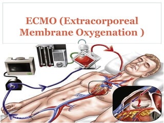
- 1. ECMO (Extracorporeal Membrane Oxygenation ) AVELIN D'SOUZA
- 2. When I think about Ecmo, I think … AVELIN D'SOUZA
- 3. Introduction ECMO is instituted for the management of life threatening pulmonary or cardiac failure (or both), when no other form of treatment has been or is likely to be successful. ECMO is essentially a modification of the cardiopulmonary bypass circuit which is used routinely in cardiac surgery. AVELIN D'SOUZA
- 4. Introduction Instituted in an emergency or urgent situation after failure of other treatment modalities. It is used as temporary support, usually awaiting recovery of organs. AVELIN D'SOUZA
- 5. History In 1965, Rashkind and coworkers were the first to use a bubble oxygenator as support in a neonate dying of respiratory failure. In 1969, Dorson and colleagues reported the use of a membrane oxygenator for cardiopulmonary bypass in infants. In 1970, Baffes et al reported the successful use of extracorporeal membrane oxygenation as support in infants with congenital heart defects who were undergoing cardiac surgery. In 1975, Bartlett et al were the first to successfully use ECMO in neonates with severe respiratory distress. AVELIN D'SOUZA
- 6. Parts of ECMO AVELIN D'SOUZA
- 7. Definition ECMO or Extra Corporeal Membrane Oxygenation is a form of extracorporeal life support where an external artificial circuit carries venous blood from the patient to a gas exchange device (oxygenator) where blood becomes enriched with oxygen and has carbon dioxide removed. This blood then reenters the patient circulation. ECMO circuit blood flow is optimised to provide adequate patient support in the absence of native lung or heart function. AVELIN D'SOUZA
- 8. Configurations for ECMO ECMO can be inserted in 2 configurations: Veno-venous Veno-arterial AVELIN D'SOUZA
- 9. The Mode of ECMO is defined by the position of the access and return cannulae. There are three modes of ECMO: Veno-Venous (VV) Veno-Arterial (VA) and Veno-Pulmonary Artery (V-PA) VV and VA modes of ECMO have a number of configurations to best suit patient needs. AVELIN D'SOUZA
- 10. Veno-arterial (VA) configuration Blood being drained from the venous system and returned to the arterial system. Provides both cardiac and respiratory support. Achieved by either peripheral or central cannulation AVELIN D'SOUZA
- 11. Peripheral ECMO Cannulation AVELIN D'SOUZA
- 12. Veno-Venous (VV) configuration Provides oxygenation Blood being drained from venous system and returned to venous system. Only provides respiratory support Achieved by peripheral cannulation, usually of both femoral veins. AVELIN D'SOUZA
- 13. a. Veno-Venous ECMO: Venous blood is accessed from the large central veins, pumped through the oxygenator and returned to the venous system near the right atrium. It provides support for severe respiratory failure. There are 4 configurations of VV ECMO i. Femoro-Femoral (Fem/Fem) ii. High-Flow iii. Femoro-Jugular iv. Dual lumen/Two stage single cannula (Avalon) In all cases, ECMO blood flow travels from the vena cavae to the atria (Cavo-Atrial Flow) to minimise recirculationAVELIN D'SOUZA
- 14. a.Femoro-Femoral: i. Two long “venous” cannulae are used ii. Direction of flow is cavo-atrial iii. Access cannula (single stage, or multistage) is inserted via the femoral vein. Usual sizes 21-25 F iv. Return cannula (single stage) is inserted via the contralateral femoral vein with the tip sited within the right atrium. If the tip is advanced too far it will impinge on the inter-atrial septum. Usual sizes 21-25 F v. The tip of the access cannula is positioned 10-15cm lower than the tip of the return cannula to minimise recirculation. Advantages: Quick and safe to insert; easy to secure cannulae; Disadvantages: Limited maximum flow rates so often requires conversion to a high-flow configuration. Patient remains bed bound AVELIN D'SOUZA
- 15. AVELIN D'SOUZA
- 16. b. High-flow: i. Uses the same bi-femoral cannulation as femoro-femoral ii. An additional short access cannula (“arterial”) is inserted via the right internal jugular vein with the tip sited in the superior vena cava. The optimal position of the tip is established after commencing full circuit blood flow. The tip is withdrawn sufficiently to prevent visible recirculation. Usual size 17-19 F iii. Direction of flow is bi-cavo-atrial to minimise recirculation Advantages: Allows higher circuit blood flows as two access cannulae draw patient blood from the great veins . Can provide maximal oxygen delivery if configured correctly Disadvantages: Occupies 3 veins. Relatively complex to secure and dress the jugular cannula. Patient remains bed bound. AVELIN D'SOUZA
- 17. c. Femoro-Jugular: i. Direction of flow is cavo-atrial to minimise recirculation ii. Access cannula (multi-stage) is inserted via the femoral vein with the tip sited just below the inferior cavo-atrial junction. Usual size 21-25 F iii. Return short cannula (“arterial”) is inserted into the right internal jugular vein with the tip sited in the lower superior vena cava. Blood returning in this direction preferentially flows towards the tricuspid valve and right ventricle, which minimises recirculation. Usual sizes 19-23 F iv. Advantages: Nearly always can provide adequate support (5-7 L/min) without large recirculation, only two veins occupied Disadvantages: Relatively difficult to secure and dress the jugular return cannula. Requires two sterile fields to be during ECMO cannulation. Access insufficiency can be more difficult to identifyin the early stages without negative pressure monitoring Patient remains bed bound AVELIN D'SOUZA
- 18. d. Dual lumen/Two stage single cannula (Avalon): i. Direction of flow is bi-cavo-atrial to minimise recirculation ii. Single cannula with two lumens for access and return inserted via the right internal Jugular vein. iii. Two access stages (SVC and IVC) iv. Return port emerges between the two access ports and is positioned at the level of the tricuspid valve. Advantages: Single vein cannulation. Allows movement from bed and potentially ambulation Disadvantages: Care on insertion to avoid right ventricular placement/rupture and hepatic vein cannulation.Large cannula to insert (27F or 31F for adults). Difficult to position return port towards the tricuspid valve. AVELIN D'SOUZA
- 19. AVELIN D'SOUZA
- 20. a. Standard Femoro-Femoral i. Access Cannula (multistage) is inserted via the femoral vein with the tip sited within the right atriumwithout impinging on the interatrial septum. Usual size 21-25 F ii. Return cannula is a short arterial cannula inserted via the common femoral artery. Usual size 17-21 F Advantages: Provides full or partial cardiac support. Disadvantages: Risk of differential hypoxia may need conversion to high flow configuration if native cardiac function improves in the setting of significant respiratory failure AVELIN D'SOUZA
- 21. Central: Specialised cannulae i. Uses specialised surgical cannulae. Access cannula is wire reinforced and malleable and is sited within the right atrium via the atrial appendage. Cannula is then tunnelled out of the chest and t he sternum closed. Usual size >30 F ii. Return cannula is Dacron tipped and sewn directly onto the proximal aorta. Cannula is then tunnelled out of the chest and the sternum closed. Usual size >30 F iii. Advantages: Can provide full cardiac and respiratory support and is not associated with differential hypoxia in the setting of combined cardiac and respiratory failure. Optimal support for severe cardiac and respiratory failure in the immediate post cardiotomy setting. Allows sternum to be closed and facilitates standard patient pressure area care. Can provide support for upto 2 weeks iv.Disadvantages Requires sternotomy for institution and re-sternotomy for decannulation. Bleedingmore common than in femoro-femoral configuration AVELIN D'SOUZA
- 22. Central ECMO Cannulation AVELIN D'SOUZA
- 23. AVELIN D'SOUZA
- 24. Central vs. Peripheral Cannulation Advantages Flow from Central ECMO is directly from the outflow cannula into the aorta provides antegrade flow to the arch vessels, coronaries and the rest of the body In contrast, the retrograde aortic flow provided by peripheral leads to mixing in the arch. AVELIN D'SOUZA
- 25. Disadvantages Previously insertion of central ECMO required leaving chest open to allow the cannulae to exit. Increased the risk of bleeding and infection Newer cannulae are designed to be tunneled through the subcostal abdominal wall allowing the chest to be completely closed. Central cannula are costly (approximately 4 times as much as peripheral) AVELIN D'SOUZA
- 26. The Configuration of ECMO refers to the cannula insertion site, type, tip position and size used in a particular mode. Cannulae Definitions: Access Cannulae: drain blood from the venous system into the ECMO circuit. Single stage access cannulae drain blood via a short region near the tip only Multi-stage access cannulae drain blood through side holes over a long length of the cannula in addition to the tip. AVELIN D'SOUZA
- 27. AVELIN D'SOUZA
- 28. b) Return Cannulae: deliver blood back to the patient from the ECMO circuit and only ever expel blood at the cannula tip (single stage). c) Distal Perfusion Cannula: Deliver blood antegradely into the femoral artery distal to the ECMO return cannula to maintain perfusion to the leg. d) d. Double-lumen Cannulae single cannula partitioned into two lumens with both access blood flow and return blood flow, similar to vascular access cannulae used in renal replacement therapy. AVELIN D'SOUZA
- 29. e) Cannula Length i. Long cannulae (55 cm) are labelled by manufacturers as “venous” and are designed for use in the venous system. ii. Short cannulae (15-25cm) are labelled by manufacturers as “arterial”. They are used to return blood in both VA ECMO and some VV ECMO configurations AVELIN D'SOUZA
- 30. Dynamics of ECMO Blood is removed from the venous system either peripherally via cannulation of a femoral vein or centrally via cannulation of the right atrium, Oxygenate Extract carbon dioxide Blood is then returned back to the body either peripherally via a femoral artery or centrally via the ascending aorta. AVELIN D'SOUZA
- 31. Indications for ECMO Divided into two type Cardiac Failure Respiratory Failure AVELIN D'SOUZA
- 32. Indications – Cardiac Failure Post-cardiotomy when unable to get pt off cardiopulmonary bypass following cardiac surgery Post-heart transplant usually due to primary graft failure Severe cardiac failure due to almost any other cause Decompensated cardiomyopathy Myocarditis Acute coronary syndrome with cardiogenic shock Profound cardiac depression due to drug overdose or sepsis AVELIN D'SOUZA
- 33. Indications – Respiratory Failure Adult respiratory distress syndrome (ARDS) Pneumonia Trauma Primary graft failure following lung transplantation. ECMO is also used for neonatal and pediatric respiratory support This is where most of the research on ECMO has been done AVELIN D'SOUZA
- 34. Decision to Institute ECMO Several considerations must be weighed: Likelihood of organ recovery.: only appropriate if disease process is reversible with therapy and rest on ECMO Cardiac recovery: to either wait for further cardiac recovery to allow implant of device (LVAD) or to list for transplantation. Disseminated malignancy Advanced age Graft vs. host disease Known severe brain injury Unwitnessed cardiac arrest or cardiac arrest of prolonged duration. Technical contraindications to consider: aortic dissection or aortic incompetence AVELIN D'SOUZA
- 35. AVELIN D'SOUZA
- 36. AVELIN D'SOUZA
- 37. Things to Think About Mechanical ventilation must be continued during ECMO support to try to maintain oxygen saturation of blood ejected from the left ventricle to at least above 90%. ECMO flow can be very volume dependent ECMO flow will drop: Hypovolemia Cannula malposition Pneumothorax Pericardial tamponade. AVELIN D'SOUZA
- 38. Weaning of ECMO – VV ECMO Actual ECMO flows do not need to be altered to assess native respiratory function Done by altering gas flow through the ECMO circuit Pt may be weanable: Gas exchange is able to be maintained with a low FiO2 (<30%) Low fresh gas flow rates into the circuit (<2 L/min) Caveat: RR and PEEP set on ventilator are not too high (e.g. <25 breaths/min and <15cmH2O, respectively). AVELIN D'SOUZA
- 39. Weaning of ECMO – VA ECMO Depends on cardiac recovery, Factors: Increasing blood pressure Return or increasing pulsatility on the arterial pressure waveform Falling pO2 by a right radial arterial line indicating more blood is being pumped through the heart which may be less well oxygenated, Falling central venous and/or pulmonary pressures. It is important to note that cardiac outputs from pulmonary artery catheter are inaccurate on ECMO Most of the circulating blood volume is bypassing the pulmonary circulation AVELIN D'SOUZA
- 40. Complications Falls into one of three major categories 1) Bleeding associated with heparinization 2) technical failure 3) neurologic sequelae AVELIN D'SOUZA
- 41. Complications of ECMO Bleeding/Hemolysis Out of proportion to the degree of coagulopathy and patient platelet count Coagulopathy Continuous activation of contact and fibrinolytic systems by the circuit Consumption and dilution of factors within minutes of initiation of ECMO AVELIN D'SOUZA
- 42. Complications of ECMO Thrombocytopenia Platelets adhere to surface fibrinogen and are activated Resultant platelet aggregation and clumping causes numbers to drop Non-pulsatile perfusion to end organs Kidneys Splanchnic circulation seems to be particularly susceptible GI bleeding, ulceration and perforation Liver impairment AVELIN D'SOUZA
- 43. Complications of ECMO Mechanical Complications Tubing rupture Pump malfunction Cannula related problems Local complications: Leg ischemia Particularly at peripheral insertion site of VA Air embolism/Thromboembolism Neurological: Intracerebral bleeds Largely associated with sepsis Manifest as seizures or brain death AVELIN D'SOUZA
- 44. Management of Complications Regular measurements of blood tests (Q6-Q8h) Coagulation Profile Platelet Count Hemoglobin Creatinine to evaluate for renal insufficiency Aggressive replacement of clotting factors, electrolytes, PRBC AVELIN D'SOUZA
- 45. Outcomes of ECMO Good quality RCT of ECMO outcomes in adult population are lacking There are very promising studies in the Pediatric populations, however it is hard to know if this translates into the adult population. Completed yet unpublished CESAR Trial shows some potential impact in ECMO research AVELIN D'SOUZA
- 46. ECMO Observations and Documentation Distal Perfusion (“Backflow”) cannula and leg vascular observations must also be performed each hour for patients on peripheral VA ECMO. In the event that blood flow through the distal perfusion circuit is occluded, blood within the back flow tubing will separate into plasma and cells, which is obvious on inspection. Normal blood flow can be seen under torchlight. AVELIN D'SOUZA
- 47. ECMO dressings and line position monitoring The bedside nurse is responsible for maintaining cannula dressings and redressing soiled or inadequate dressings. Line position monitoring is performed at least once per shift for patients with peripheral cannulae skin markings. Pressure area care: Patient Arterial Blood Gas (ABG) Ensure that heater water level is approximately 75-80% full Routine Investigations: CXR, Mg; PO4; LFT; APTT, INR, Fibrinogen and D-dimers AVELIN D'SOUZA
- 48. APTT is measured 6 hourly while the patient is on ECMO if they are receiving systemic anticoagulation. Peripheral VA ECMO patients require vascular ultrasound studies to be performed on lower limbs on day 1 and additionally as clinically indicated. For VV ECMO target blood flows must provide adequate arterial oxygenation while allowing non- injurious lung ventilation. Anticoagulation and prevention of bleeding. AVELIN D'SOUZA
- 49. All ECMO lines must be secured at 2 points with properly adherent skin dressings. Sterility must be maintained and insertion sites kept unsoiled. A designated staff member must secure ECMO circuit lines to prevent tension or torsion during patient moves. Do not allow water to enter the ECMO drive unit Alcohol Containing Cleaning solutions (including triclosan) should not come in to contact with the ECMO circuit as they may cause cracking of some circuit componentsAVELIN D'SOUZA
- 50. WEANING VV ECMO weaning: i. Circuit flow need not be reduced at any stage for weaning and therefore, no additional heparin is required ii. Weaning VV ECMO is achieved by progressively reducing the Fresh Gas Flow to the oxygenator. An increase in lung ventilation is required to ensure adequate CO2 clearance. iii. In normal circumstances there is no requirement to wean the blender FiO2 as part of the weaning process iv. It is usual to observe the patient to be stable for 4-24 hours with the Fresh Gas Flow to the ECMO circuit at 0 L/min . v. Echocardiography is not required AVELIN D'SOUZA
- 51. VA ECMO weaning: Formal weaning studies must be performed to assess the heart’s ability to manage the circulation without VA ECMO support. Circuit flow must be reduced to assess native heart function in the setting of an increased venous return. Flow is reduced from 2.5 L/min in a series of 0.5 L/min increments while haemodynamic and echocardiographic data are collected ii. Lung ventilation must be increased and oxygenator Fresh Gas Flow reduced during the weaning study iii. The reduced flow during the VA weaning process increases the risk of stasis and clotting within the circuit. Additional heparin is required to reduce the risk of clotting in this setting iv. Echocardiography is essential to assessing cardiac function during weaning from ECMO v. Once cardiac function has improved and ECMO decannulation is planned, ECMO flows should be maintained above 2.5L until decannulation takes place.AVELIN D'SOUZA
- 52. Cannula Removal and Circuit Disposal Cannula Removal: Heparin should be ceased for at least 2 hours prior to decannulation. The patient should be adequately sedated or have received an explanation of the procedure prior to commencement. After dressings are removed, all access and return lines are clamped, the ECMO console powered off and the cannulae removed simultaneously with immediate adequate pressure applied to the vessel puncture sites with sterile gauze. Returning the blood held within the ECMO circuit to the patient prior to cannula removal carries additional risk of air and thrombus entering the patient’s circulation and volume overload. It should only be performed if there is a clinical imperative to conserve blood. AVELIN D'SOUZA
- 53. Cannula Removal and Circuit Disposal Bleeding control: After percutaneously placed venous cannulae are removed the venous puncture site is compressed for 20 minutes by ECMO trained medical staff. The patient must remain supine and still for approximately 4 hours post cannula removal and the site monitored for re- bleeding d. Circuit disposal: The circuit should be disposed of in a biohazard bag/bin with cytotoxic precautions where appropriate. Reusable (metal) ECMO Clamps MUST not be discarded. AVELIN D'SOUZA
- 54. Summary ECMO is instituted for the management of life threatening pulmonary or cardiac failure (or both), when no other form of treatment has been or is likely to be successful. ECMO is essentially a modification of the cardiopulmonary bypass circuit which is used routinely in cardiac surgery. ECMO can be inserted in 2 configurations: Veno- venous & Veno-arterial Completed yet unpublished CESAR Trial shows some potential impact in ECMO research AVELIN D'SOUZA
- 56. AVELIN D'SOUZA