SlideShare una empresa de Scribd logo
1 de 48
COMPLEJO HOSPITALARIO DE NAVARRA
           HOSPITAL VIRGEN DEL CAMINO
       SERVICIO DE NEUROFISIOLOGÍA CLÍNICA


ELECTROENCEFALOGRAFÍA.

         BASES NEUROFISIOLÓGICAS
                                         Silvano G. Pino Zacarías.
                                              Residente de 2° año.
                                           Neurofisiología Clínica.


  Pamplona, jueves 10 de mayo de 2012.
ELECTROENCEFALOGRAFÍA

CONSIDERACIONES ANATÓMICAS
                                                                        Circunvolución del cíngulo
              Núcleo Caudado

                                                                                                      Lóbulo Parietal
              Ventrículo

               Opérculo
                Tálamo


                                                                                                                         Lóbulo Temporal

 Ventrículo                                                                                                                              Lóbulo
                                                                                                                                         Occipital
                                                                                Lóbulo Temporal
 Lóbulo
 Frontal


                           Amaral, D (2000). The Anatomical Organization of the Central Nervous System. In: Principles of Neural Science, 4th ed.. McGraw-Hill: 319-336.
ELECTROENCEFALOGRAFÍA

CONSIDERACIONES ANATÓMICAS


                                                        Sustancia Gris                       Sustancia Blanca


            Amaral, D (2000). The Anatomical Organization of the Central Nervous System. In: Principles of Neural Science, 4th ed.. McGraw-Hill: 319-336.
ELECTROENCEFALOGRAFÍA

CONSIDERACIONES ANATÓMICAS


      Snell, R. (2010). The Structure and Functional Localization of Cerebral Cortex. In: Clinical Neuroanatomy, 7th ed. Wolters Kluwer/Lippincott Williams & Wilkins: 284-303.
ELECTROENCEFALOGRAFÍA

CONSIDERACIONES ANATÓMICAS


         I                                                                        Capa Molecular

         II                                                                       Capa Granulosa Externa


       III                                                                        Capa Piramidal Externa


       IV                                                                         Capa Granulosa Interna

        V                                                                         Capa Piramidal Interna


       VI                                                                         Capa Polimórfica


              Amaral, D (2000). The Anatomical Organization of the Central Nervous System. In: Principles of Neural Science, 4th ed.. McGraw-Hill: 319-336.
ELECTROENCEFALOGRAFÍA

CONSIDERACIONES ANATÓMICAS


         I                   Capa Molecular

         II                  Capa Granulosa Externa


       III
                             Capa Piramidal Externa

        IV
                             Capa Granulosa Interna
        V
  VI                         Capa Piramidal Interna


                             Capa Polimórfica


                              Barea, R (2000). Electroencefalografía., 4th ed.. McGraw-Hill: 319-336.
ELECTROENCEFALOGRAFÍA

CONSIDERACIONES ANATÓMICAS


            Amaral, D (2000). The Anatomical Organization of the Central Nervous System. In: Principles of Neural Science, 4th ed.. McGraw-Hill: 319-336.
ELECTROENCEFALOGRAFÍA

POTENCIALES DE MEMBRANA


         “Una Célula excitable es toda célula capaz de

       responder ante la aplicación de un estímulo con un

            cambio de su potencial de membrana.”
ELECTROENCEFALOGRAFÍA

POTENCIALES DE MEMBRANA


   • Potencial de Membrana en Reposo.

   • Potencial de Acción.

   • Potenciales Locales.

      • Potenciales Post-sinápticos.

      • Potencial de Receptor.

      • Potencial de Placa Motora.
ELECTROENCEFALOGRAFÍA

POTENCIALES DE MEMBRANA

                                                  Amplificador

               -   +
               -   +
               -   +
               -   +
               -   +
               -   +
               -   +
               -   +
               -   +
               -   +
               -   +
    Dendrita   -   +
               -   +
               -   +
               -   +
                                            +

                          Potencial (mV)
               -   +


                                            0
    Soma
                                            -
                                           -70

    Axón                                                   Tiempo
ELECTROENCEFALOGRAFÍA

POTENCIALES DE MEMBRANA


                                                                                    Lado
   Electroneutro
                                                                                 Extracelular
     + - =


                   Membrana Plasmática


   Electroneutro                                                                     Lado
                                                                                  Intracelular
     + - =


                        Barret, K (2010). Excitable Tissue: Nerve. In: Ganong’s Review of Medical Physiology, 23rd ed. McGraw-Hill. 79-92.
ELECTROENCEFALOGRAFÍA

POTENCIALES DE MEMBRANA
                           + 60


                           + 40
                                                          Inversión de la Polaridad
                           + 20


                             0
   Potencial de Membrana
        (milivoltios)


                           - 20


                           - 40
                                                          Despolarización

                           - 60


                           - 80                           Hiperpolarización

                      - 100
                                  0   1   2   3   4   5       6      7       8      9      10
                                                                         Tiempo (milisegundos)
ELECTROENCEFALOGRAFÍA

POTENCIALES DE MEMBRANA
                      + 40


                      + 20


                             0
   Potencial de Membrana


                           - 20
        (milivoltios)


                           - 40


                           - 60


                           - 80


                    - 100

                                  0   1   2   3   4   5    6   7       8      9     10
                                                                   Tiempo (milisegundos)
ELECTROENCEFALOGRAFÍA

POTENCIALES DE MEMBRANA


                   Barret, K (2010). Excitable Tissue: Nerve. In: Ganong’s Review of Medical Physiology, 23rd ed. McGraw-Hill. 79-92.
ELECTROENCEFALOGRAFÍA

POTENCIALES DE MEMBRANA


         LOS REGISTROS OBTENIDOS

     EN EL ELECTROENCEFALOGRAMA

       NO SON POTENCIALES DE ACCIÓN
ELECTROENCEFALOGRAFÍA

POTENCIALES DE MEMBRANA


        POTENCIALES POST-SINÁPTICOS


     EXCITATORIOS           INHIBITORIOS

        (PPSE)                  (PPSI)
ELECTROENCEFALOGRAFÍA

POTENCIALES DE MEMBRANA

                           +20


                             0
                                             Potencial Post-sináptico Excitatorio
                                                           (PPSE)
   Potencial de Membrana


                           -20
        (milivoltios)


                           -40


                           -60


                           -80


                                 0   1   2    3         4             5            6            7             8            9   10
                                                                                                                          Tiempo
                                                                                                                   (milisegundos)

                                                   Barret, K (2010). Excitable Tissue: Nerve. In: Ganong’s Review of Medical Physiology, 23rd ed. McGraw-Hill. 79-92.
ELECTROENCEFALOGRAFÍA

POTENCIALES DE MEMBRANA

                           +20


                             0
                                             Potencial Post-sináptico Inhibitorio
                                                           (PPSI)
   Potencial de Membrana


                           -20
        (milivoltios)


                           -40


                           -60


                           -80


                                 0   1   2    3         4             5            6            7             8            9   10
                                                                                                                          Tiempo
                                                                                                                   (milisegundos)

                                                   Barret, K (2010). Excitable Tissue: Nerve. In: Ganong’s Review of Medical Physiology, 23rd ed. McGraw-Hill. 79-92.
ELECTROENCEFALOGRAFÍA

POTENCIALES DE MEMBRANA


                 Aferencias
                 Excitatorias


                                                              Cono de
                                                              Arranque


           Aferencias
          Inhibitorias


                                Barret, K (2010). Excitable Tissue: Nerve. In: Ganong’s Review of Medical Physiology, 23rd ed. McGraw-Hill. 79-92.
ELECTROENCEFALOGRAFÍA

POTENCIALES DE MEMBRANA
ELECTROENCEFALOGRAFÍA

DIPOLOS


                      -   +
                      -   +
                      +
                      -   -
                          +
                     ++
                      -   -
                          +   -
                      +
                      -   -
                          +                      Aferencia Excitatoria
                      -   +
                      -   +
                      -   +
                      -   +
                                     Sumidero (-)
          Dendrita   --
                      -
                          +
                          +
                              +
                      -   +
                      -   +
                      -   +
                      -   +         Fuente (+)
                      -   +


          Soma


          Axón
ELECTROENCEFALOGRAFÍA

DIPOLOS
                     Registro                                                                            Registro
                   Intracelular                                                                         Extracelular


                                                                                                      Sumidero (-)


                   Dendrita


                                                                                                       Fuente (+)


                     Soma


                     Axón


          Swanson, T (2000). Basic Cellular and Synaptic Mechanisms Underlying the EEG. In: Comprehensive Clinical Neurophysiology. W.B. Saunders Company. Pag: 349-357.
ELECTROENCEFALOGRAFÍA

DIPOLOS


                    -   +
                    -   +
                   --   +
                 - --   +   +
                   --   +                      Aferencia Inhibitoria
                    -   +
                    -   +
                    -   +
                    -   +         Fuente (+)
                 +-     +   -
                    -   +
      Dendrita      -   +         Sumidero (-)
                    -   +
                    -   +
                    -   +
                    -   +


      Soma


          Axón
ELECTROENCEFALOGRAFÍA

DIPOLOS
                    Registro                                                                                 Registro
                  Intracelular                                                                              Extracelular


                                                                                                             Fuente (+)

                Dendrita
                                                                                                             Sumidero (-)


                  Soma


                   Axón


          Swanson, T (2000). Basic Cellular and Synaptic Mechanisms Underlying the EEG. In: Comprehensive Clinical Neurophysiology. W.B. Saunders Company. Pag: 349-357.
ELECTROENCEFALOGRAFÍA

DIPOLOS

           Líneas de Campo Eléctrico


                Líneas Isopotencial
ELECTROENCEFALOGRAFÍA

DIPOLOS
ELECTROENCEFALOGRAFÍA

DIPOLOS


          Westbrook, G.(2000). Seizures and Epilepsy. In: Principles of Neural Science, 4th ed.. McGraw-Hill: 910-937..
ELECTROENCEFALOGRAFÍA

DIPOLOS

              Registros Negativos               Registros Positivos


                          -                                 +
    PPSEs
                          +          PPSEs                  -
  Superficiales
                                    Profundos


                          -
                                                            +

    PPSIs                              PPSIs                -
                          +
  Profundos                         Superficiales
ELECTROENCEFALOGRAFÍA

DIPOLOS


                                                  Aferencias Excitatorias
                -


                +


                                          Aferencias Provenientes de la
                                          Corteza cerebral Contralateral

          Westbrook, G.(2000). Seizures and Epilepsy. In: Principles of Neural Science, 4th ed.. McGraw-Hill: 910-937..
ELECTROENCEFALOGRAFÍA

DIPOLOS


                             +


   Aferencias Excitatorias   -


  Aferencias Provenientes
           del Tálamo

                                 Westbrook, G.(2000). Seizures and Epilepsy. In: Principles of Neural Science, 4th ed.. McGraw-Hill: 910-937..
ELECTROENCEFALOGRAFÍA

DIPOLOS


                                                              Potencial de Campo
                                                                    Local (LFP)
                                      - 10 mV

                               - 100 µV                    - 100 µV
                    - 100 µV                                        - 100 µV
  - 100 µV - 100 µV                          - 100 µV                                 - 100 µV
                          - 100 µV-              -             -             - 100 µV
          - 100-µV      -                          - 100 µV             -                 -
      -                             - 100 µV     -
                           -                                                   -
            -                     +       -    -     -       +         +
     +          +      +                       +                                        +
                       + + 100 µV                   + 100 µV
                                                             + 100 µV +
            + µV+ 100 µV
  + 100 µV + 100                 +     + 100 µV +                            + 100 µV
                    + 100 µV                                         + 100 µV
         + 100 µV             + 100 µV       + 100 µV
ELECTROENCEFALOGRAFÍA

AMPLITUD DE LOS POTENCIALES REGISTRADOS

   • Factores que influyen en la Amplitud del Potencial Registrado:
       • Amplitud del Potencial Neuronal.
       • Área de Corteza Involucrada.
       • Sincronización de los potenciales.
       • Orientación de los dipolos producidos.
            • Ángulo Plano.
            • Ángulo Sólido
       • Atenuación causada por los elementos interpuestos


            Swanson, T (2000). Basic Cellular and Synaptic Mechanisms Underlying the EEG. In: Comprehensive Clinical Neurophysiology. W.B. Saunders Company. Pag: 349-357.
ELECTROENCEFALOGRAFÍA

AMPLITUD DE LOS POTENCIALES REGISTRADOS


               -          +
ELECTROENCEFALOGRAFÍA

AMPLITUD DE LOS POTENCIALES REGISTRADOS
    Negatividad
       Alta                          Negatividad
                                        Baja


                      -
                                                  Neutral


                      +

                                    Positividad
       Positividad                     Baja
          Alta
ELECTROENCEFALOGRAFÍA

AMPLITUD DE LOS POTENCIALES REGISTRADOS


       Swanson, T (2000). Basic Cellular and Synaptic Mechanisms Underlying the EEG. In: Comprehensive Clinical Neurophysiology. W.B. Saunders Company. Pag: 349-357.
ELECTROENCEFALOGRAFÍA

AMPLITUD DE LOS POTENCIALES REGISTRADOS


       Swanson, T (2000). Basic Cellular and Synaptic Mechanisms Underlying the EEG. In: Comprehensive Clinical Neurophysiology. W.B. Saunders Company. Pag: 349-357.
ELECTROENCEFALOGRAFÍA

AMPLITUD DE LOS POTENCIALES REGISTRADOS


                                   El área de la atmósfera

                       circunscrita por el ángulo sólido

                    formado por el sol es igual al área

                       circunscrita por el ángulo sólido

                                       formado por la luna.


       Swanson, T (2000). Basic Cellular and Synaptic Mechanisms Underlying the EEG. In: Comprehensive Clinical Neurophysiology. W.B. Saunders Company. Pag: 349-357.
ELECTROENCEFALOGRAFÍA

AMPLITUD DE LOS POTENCIALES REGISTRADOS


       Swanson, T (2000). Basic Cellular and Synaptic Mechanisms Underlying the EEG. In: Comprehensive Clinical Neurophysiology. W.B. Saunders Company. Pag: 349-357.
ELECTROENCEFALOGRAFÍA

AMPLITUD DE LOS POTENCIALES REGISTRADOS


           ±e                                                                                                     Ω
  • P=                      Ω
           4π


         Swanson, T (2000). Basic Cellular and Synaptic Mechanisms Underlying the EEG. In: Comprehensive Clinical Neurophysiology. W.B. Saunders Company. Pag: 349-357.
ELECTROENCEFALOGRAFÍA

AMPLITUD DE LOS POTENCIALES REGISTRADOS


                                 ΩA                                                                                              ΩB


       +   +        +       +         +                                                   +              +          +          +          +
   +   +                              +       +                           +              +                                                    +           +
           +        +       +                                             -                          +              +           +                         -
   -   -                              -       -                                          -                                                    -
           -        -       -                                                                        -              -          -

               A                                                                                              B


           Swanson, T (2000). Basic Cellular and Synaptic Mechanisms Underlying the EEG. In: Comprehensive Clinical Neurophysiology. W.B. Saunders Company. Pag: 349-357.
ELECTROENCEFALOGRAFÍA

AMPLITUD DE LOS POTENCIALES REGISTRADOS


                                 ΩA                                                                                          ΩB


                                                                                               + +                  +       + +
                                                                                     +         + +                                           +
       +   +        +       +         +                                              -                              +       + +
   +                                          +                                                - --                                          -
       +   +        +       +         +                                                                             -       - - -
   -   -                              -       -
           -        -       -

               A                                                                                              B


           Swanson, T (2000). Basic Cellular and Synaptic Mechanisms Underlying the EEG. In: Comprehensive Clinical Neurophysiology. W.B. Saunders Company. Pag: 349-357.
ELECTROENCEFALOGRAFÍA

AMPLITUD DE LOS POTENCIALES REGISTRADOS


                                 ΩA                                         ΩB


       +   +        +       +         +                                                                        + +  + +                              +
   +   +                              +       +                                                      +         + +                                   +       +
           +        +       +                                                                                       + +
   -   -                              -       -                                                      -         - -                                   -       -
           -        -       -                                                                                      - -

               A                                                                                                         B


           Swanson, T (2000). Basic Cellular and Synaptic Mechanisms Underlying the EEG. In: Comprehensive Clinical Neurophysiology. W.B. Saunders Company. Pag: 349-357.
ELECTROENCEFALOGRAFÍA

AMPLITUD DE LOS POTENCIALES REGISTRADOS


       Swanson, T (2000). Basic Cellular and Synaptic Mechanisms Underlying the EEG. In: Comprehensive Clinical Neurophysiology. W.B. Saunders Company. Pag: 349-357.
ELECTROENCEFALOGRAFÍA

AMPLITUD DE LOS POTENCIALES REGISTRADOS

   • Elementos que atenúan los Potenciales extracelulares:
       • Líquido Intersticial.
       • Líquido Cefalorraquídeo.
       • Meninges.
       • Huesos del Cráneo.
       • Músculos.
       •   Cuero Cabelludo.


            Swanson, T (2000). Basic Cellular and Synaptic Mechanisms Underlying the EEG. In: Comprehensive Clinical Neurophysiology. W.B. Saunders Company. Pag: 349-357.
ELECTROENCEFALOGRAFÍA

AMPLITUD DE LOS POTENCIALES REGISTRADOS

               P = 1 mV / 5 000                                                                             P = 1 mV / 2


                                  P = 1 mV                                                                                 P = 1 mV

           +                                                                                        +


           -                                                                                        -


       Swanson, T (2000). Basic Cellular and Synaptic Mechanisms Underlying the EEG. In: Comprehensive Clinical Neurophysiology. W.B. Saunders Company. Pag: 349-357.
“El cerebro humano es como una
                                          máquina de acuñar monedas. Si
                                            echas en ella metal impuro
                                          obtendrás escoria; pero si echas
                                         oro, obtendrás monedas de ley.”
   Santiago Ramón y Cajal (1852 – 1934)
Premio Nobel en Fisiología y Medicina 1906.

Más contenido relacionado

La actualidad más candente (20)

Neuroestimulacion
NeuroestimulacionNeuroestimulacion
Neuroestimulacion
 
Tabla de neurotransmisores
Tabla de neurotransmisoresTabla de neurotransmisores
Tabla de neurotransmisores
 
Eeg neonatal
Eeg neonatalEeg neonatal
Eeg neonatal
 
Migración neuronal
Migración neuronalMigración neuronal
Migración neuronal
 
Ganglios de la base (JC)
Ganglios de la base (JC)Ganglios de la base (JC)
Ganglios de la base (JC)
 
Exposicion parkinson
Exposicion parkinsonExposicion parkinson
Exposicion parkinson
 
Sinapsis
SinapsisSinapsis
Sinapsis
 
Psicofarmacologia
PsicofarmacologiaPsicofarmacologia
Psicofarmacologia
 
ECG. Día 5. Síndrome Coronario Agudo
ECG. Día 5. Síndrome Coronario AgudoECG. Día 5. Síndrome Coronario Agudo
ECG. Día 5. Síndrome Coronario Agudo
 
Taquicardia supraventricular
Taquicardia supraventricularTaquicardia supraventricular
Taquicardia supraventricular
 
10- Sueño
10- Sueño10- Sueño
10- Sueño
 
Anatomia y fisiologia de los ganglios de la base (basico)
Anatomia y fisiologia de los ganglios de la base (basico)Anatomia y fisiologia de los ganglios de la base (basico)
Anatomia y fisiologia de los ganglios de la base (basico)
 
Compartimientos liquidos del sistema nervioso
Compartimientos liquidos del sistema nerviosoCompartimientos liquidos del sistema nervioso
Compartimientos liquidos del sistema nervioso
 
Antidepresivos. psicofarmacologia stahl 2013
Antidepresivos. psicofarmacologia stahl 2013Antidepresivos. psicofarmacologia stahl 2013
Antidepresivos. psicofarmacologia stahl 2013
 
Cerebelo fisiologia
Cerebelo fisiologiaCerebelo fisiologia
Cerebelo fisiologia
 
Ley de todo o nada fisiología..
Ley de todo o nada  fisiología..Ley de todo o nada  fisiología..
Ley de todo o nada fisiología..
 
Fisiologia Sistema Motor
Fisiologia Sistema MotorFisiologia Sistema Motor
Fisiologia Sistema Motor
 
Neurotransmisores!
Neurotransmisores!Neurotransmisores!
Neurotransmisores!
 
Clasificación de la Herniacion cerebral.
Clasificación de la Herniacion cerebral.Clasificación de la Herniacion cerebral.
Clasificación de la Herniacion cerebral.
 
Eeg basics, part 1
Eeg basics, part 1Eeg basics, part 1
Eeg basics, part 1
 

Bases neurofisiológicas del eeg parte 1

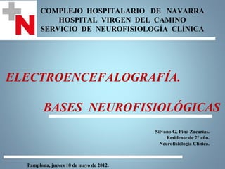
  • 1. COMPLEJO HOSPITALARIO DE NAVARRA HOSPITAL VIRGEN DEL CAMINO SERVICIO DE NEUROFISIOLOGÍA CLÍNICA ELECTROENCEFALOGRAFÍA. BASES NEUROFISIOLÓGICAS Silvano G. Pino Zacarías. Residente de 2° año. Neurofisiología Clínica. Pamplona, jueves 10 de mayo de 2012.
  • 2. ELECTROENCEFALOGRAFÍA CONSIDERACIONES ANATÓMICAS Circunvolución del cíngulo Núcleo Caudado Lóbulo Parietal Ventrículo Opérculo Tálamo Lóbulo Temporal Ventrículo Lóbulo Occipital Lóbulo Temporal Lóbulo Frontal Amaral, D (2000). The Anatomical Organization of the Central Nervous System. In: Principles of Neural Science, 4th ed.. McGraw-Hill: 319-336.
  • 3. ELECTROENCEFALOGRAFÍA CONSIDERACIONES ANATÓMICAS Sustancia Gris Sustancia Blanca Amaral, D (2000). The Anatomical Organization of the Central Nervous System. In: Principles of Neural Science, 4th ed.. McGraw-Hill: 319-336.
  • 4. ELECTROENCEFALOGRAFÍA CONSIDERACIONES ANATÓMICAS Snell, R. (2010). The Structure and Functional Localization of Cerebral Cortex. In: Clinical Neuroanatomy, 7th ed. Wolters Kluwer/Lippincott Williams & Wilkins: 284-303.
  • 5. ELECTROENCEFALOGRAFÍA CONSIDERACIONES ANATÓMICAS I Capa Molecular II Capa Granulosa Externa III Capa Piramidal Externa IV Capa Granulosa Interna V Capa Piramidal Interna VI Capa Polimórfica Amaral, D (2000). The Anatomical Organization of the Central Nervous System. In: Principles of Neural Science, 4th ed.. McGraw-Hill: 319-336.
  • 6. ELECTROENCEFALOGRAFÍA CONSIDERACIONES ANATÓMICAS I Capa Molecular II Capa Granulosa Externa III Capa Piramidal Externa IV Capa Granulosa Interna V VI Capa Piramidal Interna Capa Polimórfica Barea, R (2000). Electroencefalografía., 4th ed.. McGraw-Hill: 319-336.
  • 7. ELECTROENCEFALOGRAFÍA CONSIDERACIONES ANATÓMICAS Amaral, D (2000). The Anatomical Organization of the Central Nervous System. In: Principles of Neural Science, 4th ed.. McGraw-Hill: 319-336.
  • 8. ELECTROENCEFALOGRAFÍA POTENCIALES DE MEMBRANA “Una Célula excitable es toda célula capaz de responder ante la aplicación de un estímulo con un cambio de su potencial de membrana.”
  • 9. ELECTROENCEFALOGRAFÍA POTENCIALES DE MEMBRANA • Potencial de Membrana en Reposo. • Potencial de Acción. • Potenciales Locales. • Potenciales Post-sinápticos. • Potencial de Receptor. • Potencial de Placa Motora.
  • 10. ELECTROENCEFALOGRAFÍA POTENCIALES DE MEMBRANA Amplificador - + - + - + - + - + - + - + - + - + - + - + Dendrita - + - + - + - + + Potencial (mV) - + 0 Soma - -70 Axón Tiempo
  • 11. ELECTROENCEFALOGRAFÍA POTENCIALES DE MEMBRANA Lado Electroneutro Extracelular + - = Membrana Plasmática Electroneutro Lado Intracelular + - = Barret, K (2010). Excitable Tissue: Nerve. In: Ganong’s Review of Medical Physiology, 23rd ed. McGraw-Hill. 79-92.
  • 12. ELECTROENCEFALOGRAFÍA POTENCIALES DE MEMBRANA + 60 + 40 Inversión de la Polaridad + 20 0 Potencial de Membrana (milivoltios) - 20 - 40 Despolarización - 60 - 80 Hiperpolarización - 100 0 1 2 3 4 5 6 7 8 9 10 Tiempo (milisegundos)
  • 13. ELECTROENCEFALOGRAFÍA POTENCIALES DE MEMBRANA + 40 + 20 0 Potencial de Membrana - 20 (milivoltios) - 40 - 60 - 80 - 100 0 1 2 3 4 5 6 7 8 9 10 Tiempo (milisegundos)
  • 14. ELECTROENCEFALOGRAFÍA POTENCIALES DE MEMBRANA Barret, K (2010). Excitable Tissue: Nerve. In: Ganong’s Review of Medical Physiology, 23rd ed. McGraw-Hill. 79-92.
  • 15. ELECTROENCEFALOGRAFÍA POTENCIALES DE MEMBRANA LOS REGISTROS OBTENIDOS EN EL ELECTROENCEFALOGRAMA NO SON POTENCIALES DE ACCIÓN
  • 16. ELECTROENCEFALOGRAFÍA POTENCIALES DE MEMBRANA POTENCIALES POST-SINÁPTICOS EXCITATORIOS INHIBITORIOS (PPSE) (PPSI)
  • 17. ELECTROENCEFALOGRAFÍA POTENCIALES DE MEMBRANA +20 0 Potencial Post-sináptico Excitatorio (PPSE) Potencial de Membrana -20 (milivoltios) -40 -60 -80 0 1 2 3 4 5 6 7 8 9 10 Tiempo (milisegundos) Barret, K (2010). Excitable Tissue: Nerve. In: Ganong’s Review of Medical Physiology, 23rd ed. McGraw-Hill. 79-92.
  • 18. ELECTROENCEFALOGRAFÍA POTENCIALES DE MEMBRANA +20 0 Potencial Post-sináptico Inhibitorio (PPSI) Potencial de Membrana -20 (milivoltios) -40 -60 -80 0 1 2 3 4 5 6 7 8 9 10 Tiempo (milisegundos) Barret, K (2010). Excitable Tissue: Nerve. In: Ganong’s Review of Medical Physiology, 23rd ed. McGraw-Hill. 79-92.
  • 19. ELECTROENCEFALOGRAFÍA POTENCIALES DE MEMBRANA Aferencias Excitatorias Cono de Arranque Aferencias Inhibitorias Barret, K (2010). Excitable Tissue: Nerve. In: Ganong’s Review of Medical Physiology, 23rd ed. McGraw-Hill. 79-92.
  • 21.
  • 22. ELECTROENCEFALOGRAFÍA DIPOLOS - + - + + - - + ++ - - + - + - - + Aferencia Excitatoria - + - + - + - + Sumidero (-) Dendrita -- - + + + - + - + - + - + Fuente (+) - + Soma Axón
  • 23. ELECTROENCEFALOGRAFÍA DIPOLOS Registro Registro Intracelular Extracelular Sumidero (-) Dendrita Fuente (+) Soma Axón Swanson, T (2000). Basic Cellular and Synaptic Mechanisms Underlying the EEG. In: Comprehensive Clinical Neurophysiology. W.B. Saunders Company. Pag: 349-357.
  • 24. ELECTROENCEFALOGRAFÍA DIPOLOS - + - + -- + - -- + + -- + Aferencia Inhibitoria - + - + - + - + Fuente (+) +- + - - + Dendrita - + Sumidero (-) - + - + - + - + Soma Axón
  • 25. ELECTROENCEFALOGRAFÍA DIPOLOS Registro Registro Intracelular Extracelular Fuente (+) Dendrita Sumidero (-) Soma Axón Swanson, T (2000). Basic Cellular and Synaptic Mechanisms Underlying the EEG. In: Comprehensive Clinical Neurophysiology. W.B. Saunders Company. Pag: 349-357.
  • 26. ELECTROENCEFALOGRAFÍA DIPOLOS Líneas de Campo Eléctrico Líneas Isopotencial
  • 28. ELECTROENCEFALOGRAFÍA DIPOLOS Westbrook, G.(2000). Seizures and Epilepsy. In: Principles of Neural Science, 4th ed.. McGraw-Hill: 910-937..
  • 29. ELECTROENCEFALOGRAFÍA DIPOLOS Registros Negativos Registros Positivos - + PPSEs + PPSEs - Superficiales Profundos - + PPSIs PPSIs - + Profundos Superficiales
  • 30. ELECTROENCEFALOGRAFÍA DIPOLOS Aferencias Excitatorias - + Aferencias Provenientes de la Corteza cerebral Contralateral Westbrook, G.(2000). Seizures and Epilepsy. In: Principles of Neural Science, 4th ed.. McGraw-Hill: 910-937..
  • 31. ELECTROENCEFALOGRAFÍA DIPOLOS + Aferencias Excitatorias - Aferencias Provenientes del Tálamo Westbrook, G.(2000). Seizures and Epilepsy. In: Principles of Neural Science, 4th ed.. McGraw-Hill: 910-937..
  • 32. ELECTROENCEFALOGRAFÍA DIPOLOS Potencial de Campo Local (LFP) - 10 mV - 100 µV - 100 µV - 100 µV - 100 µV - 100 µV - 100 µV - 100 µV - 100 µV - 100 µV- - - - 100 µV - 100-µV - - 100 µV - - - - 100 µV - - - - + - - - + + + + + + + + + 100 µV + 100 µV + 100 µV + + µV+ 100 µV + 100 µV + 100 + + 100 µV + + 100 µV + 100 µV + 100 µV + 100 µV + 100 µV + 100 µV
  • 33.
  • 34. ELECTROENCEFALOGRAFÍA AMPLITUD DE LOS POTENCIALES REGISTRADOS • Factores que influyen en la Amplitud del Potencial Registrado: • Amplitud del Potencial Neuronal. • Área de Corteza Involucrada. • Sincronización de los potenciales. • Orientación de los dipolos producidos. • Ángulo Plano. • Ángulo Sólido • Atenuación causada por los elementos interpuestos Swanson, T (2000). Basic Cellular and Synaptic Mechanisms Underlying the EEG. In: Comprehensive Clinical Neurophysiology. W.B. Saunders Company. Pag: 349-357.
  • 35. ELECTROENCEFALOGRAFÍA AMPLITUD DE LOS POTENCIALES REGISTRADOS - +
  • 36. ELECTROENCEFALOGRAFÍA AMPLITUD DE LOS POTENCIALES REGISTRADOS Negatividad Alta Negatividad Baja - Neutral + Positividad Positividad Baja Alta
  • 37. ELECTROENCEFALOGRAFÍA AMPLITUD DE LOS POTENCIALES REGISTRADOS Swanson, T (2000). Basic Cellular and Synaptic Mechanisms Underlying the EEG. In: Comprehensive Clinical Neurophysiology. W.B. Saunders Company. Pag: 349-357.
  • 38. ELECTROENCEFALOGRAFÍA AMPLITUD DE LOS POTENCIALES REGISTRADOS Swanson, T (2000). Basic Cellular and Synaptic Mechanisms Underlying the EEG. In: Comprehensive Clinical Neurophysiology. W.B. Saunders Company. Pag: 349-357.
  • 39. ELECTROENCEFALOGRAFÍA AMPLITUD DE LOS POTENCIALES REGISTRADOS El área de la atmósfera circunscrita por el ángulo sólido formado por el sol es igual al área circunscrita por el ángulo sólido formado por la luna. Swanson, T (2000). Basic Cellular and Synaptic Mechanisms Underlying the EEG. In: Comprehensive Clinical Neurophysiology. W.B. Saunders Company. Pag: 349-357.
  • 40. ELECTROENCEFALOGRAFÍA AMPLITUD DE LOS POTENCIALES REGISTRADOS Swanson, T (2000). Basic Cellular and Synaptic Mechanisms Underlying the EEG. In: Comprehensive Clinical Neurophysiology. W.B. Saunders Company. Pag: 349-357.
  • 41. ELECTROENCEFALOGRAFÍA AMPLITUD DE LOS POTENCIALES REGISTRADOS ±e Ω • P= Ω 4π Swanson, T (2000). Basic Cellular and Synaptic Mechanisms Underlying the EEG. In: Comprehensive Clinical Neurophysiology. W.B. Saunders Company. Pag: 349-357.
  • 42. ELECTROENCEFALOGRAFÍA AMPLITUD DE LOS POTENCIALES REGISTRADOS ΩA ΩB + + + + + + + + + + + + + + + + + + + + + - + + + - - - - - - - - - - - - - A B Swanson, T (2000). Basic Cellular and Synaptic Mechanisms Underlying the EEG. In: Comprehensive Clinical Neurophysiology. W.B. Saunders Company. Pag: 349-357.
  • 43. ELECTROENCEFALOGRAFÍA AMPLITUD DE LOS POTENCIALES REGISTRADOS ΩA ΩB + + + + + + + + + + + + + + - + + + + + - -- - + + + + + - - - - - - - - - - - A B Swanson, T (2000). Basic Cellular and Synaptic Mechanisms Underlying the EEG. In: Comprehensive Clinical Neurophysiology. W.B. Saunders Company. Pag: 349-357.
  • 44. ELECTROENCEFALOGRAFÍA AMPLITUD DE LOS POTENCIALES REGISTRADOS ΩA ΩB + + + + + + + + + + + + + + + + + + + + + + + + - - - - - - - - - - - - - - A B Swanson, T (2000). Basic Cellular and Synaptic Mechanisms Underlying the EEG. In: Comprehensive Clinical Neurophysiology. W.B. Saunders Company. Pag: 349-357.
  • 45. ELECTROENCEFALOGRAFÍA AMPLITUD DE LOS POTENCIALES REGISTRADOS Swanson, T (2000). Basic Cellular and Synaptic Mechanisms Underlying the EEG. In: Comprehensive Clinical Neurophysiology. W.B. Saunders Company. Pag: 349-357.
  • 46. ELECTROENCEFALOGRAFÍA AMPLITUD DE LOS POTENCIALES REGISTRADOS • Elementos que atenúan los Potenciales extracelulares: • Líquido Intersticial. • Líquido Cefalorraquídeo. • Meninges. • Huesos del Cráneo. • Músculos. • Cuero Cabelludo. Swanson, T (2000). Basic Cellular and Synaptic Mechanisms Underlying the EEG. In: Comprehensive Clinical Neurophysiology. W.B. Saunders Company. Pag: 349-357.
  • 47. ELECTROENCEFALOGRAFÍA AMPLITUD DE LOS POTENCIALES REGISTRADOS P = 1 mV / 5 000 P = 1 mV / 2 P = 1 mV P = 1 mV + + - - Swanson, T (2000). Basic Cellular and Synaptic Mechanisms Underlying the EEG. In: Comprehensive Clinical Neurophysiology. W.B. Saunders Company. Pag: 349-357.
  • 48. “El cerebro humano es como una máquina de acuñar monedas. Si echas en ella metal impuro obtendrás escoria; pero si echas oro, obtendrás monedas de ley.” Santiago Ramón y Cajal (1852 – 1934) Premio Nobel en Fisiología y Medicina 1906.