
Más contenido relacionado
Similar a Clasificacion del RN, Clinica del RN.pptx
Similar a Clasificacion del RN, Clinica del RN.pptx (20)
Último
Último (20)
Clasificacion del RN, Clinica del RN.pptx
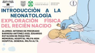
- 1. INTRODUCCIÒN A LA NEONATOLOGÌA EXPLORACIÒN FÌSICA DEL RECIÈN NACIDO ALUMNO: INTERNO DE PREGRADO BARRERA ANTÙNEZ AXEL EDUARDO ROTACIÒN DE PEDIATRÌA MEMORIAL HOSPITAL MILPA HIGH HOSPITAL GENERAL MILPA ALTA
- 2. DEFINICIÒN DE RECIEN NACIDO CLASIFICACIÒN DEL RECIÈN NACIDO ESCALAS USADAS EN EL RECIÈN NACIDO EXPLORACIÒN CLÌNICA DEL RECIÈN NACIDO 01 02 04 05 CONTENIDO TEMÀTICO 03 06 GRÀFICAS USADAS EN EL RECIÈN NACIDO ASPECTOS BÀSICOS DE REANIMACIÒN NEONATAL
- 3. INTRODUCCIÒN 01 RECIÈN NACIDO: Producto de la concepción desde el nacimiento hasta los 28 días de edad. VIVO: Después de concluir su separación del organismo materno manifiesta algún tipo de vida, tales como movimientos respiratorios, latidos cardiacos o movimientos definidos de músculos voluntarios. NORMA Oficial Mexicana NOM-007-SSA2-2016, Para la atención de la mujer durante el embarazo, parto y puerperio, y de la persona recién nacida. MUERTO: Después de concluir su separación del organismo materno no respira, ni manifiesta otro signo de vida tales como latidos cardiacos o funiculares o movimientos definidos de músculos voluntarios.
- 4. DEFINICIÒN DE RN PERIODO NEONATAL PRODUCTO VIVO DE LA CONCEPCIÒN DE ENTRE 37-42 SEMANAS, ESTÀ EN CONDICIONES ÒPTIMAS PARA ADAPTARSE AL AMBIENTE EXTRAUTERINO. TEMPRANO: 7 DÌAS TARDÌO: 8-28 DÌAS >MORBILETALIDAD HISTORIA MÈDICA DE LOS PADRES HISTORIA OBSTÈTRICA MATERA HISTORIA DEL EMBARAZO ACTUAL MICROAMBIENTE MATROAMBIENTE MACROAMBIENTE RODRÌGUEZ BONITO R, MANUAL DE NEONATOLOGÌA 2ª EDICIÒN, EDITORIAL MC GRAWHILL MÈXICO, 2012.
- 5. MICROAMBIENTE MACROAMBIENTE MATROAMBIENTE -CONTACTO ÌNTIMO CON EL PRODUCTO. -DINÀMICO. -L.A. -CORDÒN UMBILICAL -MEMBRANAS -PLACENTA -FÌSICAS. -CULTURALES. -SOCIALES. -ECONÒMICAS. -POLÌTICAS -ANATÒMICAS. -FISIOLÒGICAS. -BIOLÒGICAS. -PATOLÒGICAS. RODRÌGUEZ BONITO R, MANUAL DE NEONATOLOGÌA 2ª EDICIÒN, EDITORIAL MC GRAWHILL MÈXICO, 2012. DR. MONIE
- 6. ● FECHA ÙLTIMA MENSTRUACIÒN ● ECOGRAFÌA OBSTÈTRICA ● EXAMEN FÌSICO CLASIFICACIÒN EDAD GESTACIONAL PESO/EDAD GESTACIONAL PRETÈRMINO: 22-<32 SDG, PESO <2500 g HIPOTRÒFICO: PESO INFERIOR < PERCENTILA 10 TÈRMINO: 37-<42 SDG, >2500 g EUTRÒFICO: PESO ENTRE PERCENTILA 10-90 POSTÈRMINO: >= 42 SDG O >=294 DIAS HIPERTRÒFICO: PESO > PERCENTILA 90 INMADURO: 22-<28 SDG, PESO 501-1000 PESO BAJO: PESO <2500 g INDEPENDIENTE DE EG. • NORMA Oficial Mexicana NOM-007-SSA2-2016, Para la atención de la mujer durante el embarazo, parto y puerperio, y de la persona recién nacida. • NORMA Oficial Mexicana NOM-007-SSA2-1993, Atención de la mujer durante el embarazo, parto y puerperio y del recién nacido. Criterios y procedimientos para la prestación del servicio.
- 7. CLASIFICACIÒN DEL RECIÈN NACIDO 1. EDAD GESTACIONAL 2. PESO DEL NACIMIENTO 3. PESO PARA EDAD GESTACIONAL 4. ADAPTACIÒN A LA VIDA EXTRAUTERINA 1. EDAD GESTACIONAL FUM 1. CIERTA 2. SEGURA 3. CONFIABLE ECOGRAFÌA 1. PRECOZ 2. MARGEN DE ERROR CLÌNICA 1. SOMÀTICAS 2. >VARIABILIDAD (+-2 SEMANAS) • NEONATOLOGÌA: TEMAS PRÀCTICOS, BORBONET D, EDITORIAL EULAR.
- 8. CLASIFICACIÒN DEL RECIÈN NACIDO <32 SEMANAS PERIMETRO CEFÀLICO + 2 SEMANAS TÈRMINO 37-41 6 DÌAS PRETÈRMINO <37 SEMANAS POSTÈRMINO 42 O + SEMANAS PRETÈRMINO LEVE 35-36 SEMANAS PRETÈRMINO MODERADO 32-34 SEMANAS PRETÈRMINO SEVERO <=31 SEMANAS PRETÈRMINO EXTREMO <28 SEMANAS • NEONATOLOGÌA: TEMAS PRÀCTICOS, BORBONET D, EDITORIAL EULAR.
- 9. SEGÙN PESO AL NACER ● NORMOPESO: 2500-4000 g ● BAJO PESO AL NACER: <2500 g ● MUY BAJO PESO AL NACER: <1500 g ● MUY MUY BAJO PESO AL NACER: <1000 g ● MACROSÒMICO: >4000 g SEGÙN PESO AL NACER ● ADECUADO PARA EDAD GESTACIONAL (AEG): PERCENTILAS ENTRE 10-90. ● PEQUEÑOS PARA LA EDAD GESTACIONAL(PEG): <PERCENTILA 10. ● GRANDES PARA LA EDAD GESTACIONAL(GEG): >PERCENTILA 90. • NEONATOLOGÌA: TEMAS PRÀCTICOS, BORBONET D, EDITORIAL EULAR.
- 10. SEGÙN ADAPTACIÒN A LA VIDA EXTRAUTERINA ● 1953,VIRGINIA APGAR, ANESTESISTA OBSTÈTRICA. ● EVALUACIÒN DE LA VITALIDAD DEL RECIÈN NACIDO 1. FRECUENCIA CARDÌACA 2. TONO 3. COLORACIÒN DE PIEL 4. ESFUERZO RESPIRATORIO 5. RESPUESTA AL ESTÌMULO 1. VIGOROSOS: 7-10 2. DNM:4-6 3. DNS: 0-3 PRONÒSTICO NEUROLÒGICO MANIOBRAS DE REANIMACIÒN • NEONATOLOGÌA: TEMAS PRÀCTICOS, BORBONET D, EDITORIAL EULAR.
- 11. ULTRASONOGRAFÌA • DIÀMETRO BIPARIETAL • CIRCUNFERENCIA ABDOMINAL • CIRCUNFERENCIA CEFÀLICA • LONGITUD FEMORAL SEGURIDAD +- 1-2 SEMANAS, NO CAMBIAN MUCHO EN LAS PRIMERAS 24 HRS VIDA. CALCULO DE LA EDAD GESTACIONAL USHER FARR-DUBOWITZ MÈTODOS PRENATALES U OBSTÈTRICOS28 días de edad. MÈTODOS POSNATALES8 O PEDIÀTRICOS • Ceriani Cernadas J, Neonatologìa Pràctica 4ª Ediciòn, Buenos Aires, Editorial Panamericana 2009 • Nazer J, Neonatologìa, 1ª Ediciòn, Chile, Editorial Universitaria, Chile. CAPURRO BALLARD MODIFICADO • 1966: 10 CARACTERÌSTICAS SOMÀTICAS • 1970: 10 PARÀMETROS NEUROLÒGICOS • 21 ÌTEMS: PUNTAJE DE 0-2, 3, 4 O 5, SUMATORIA TOTAL 70 PUNTOS (35 C/GRUPO). PARKIN EVALÙA 4 PARÀMETROS: 1. NÒDULO MAMARIO 2. PABELLÒN AURICULAR 3. COLOR DE LA PIEL 4. TEXTURA DE LA PIEL PUNTAJE 0-3; TOTAL 12
- 12. CLASIFICACIÒN DEL RECIÈN NACIDO PESO INDEPENDIENTE DE EDAD GESTACIONAL MACROSÒMICO RN MBPN RN BPN PESO >= 4000 G PESO <1000 G PESO <2500 G PESO >= 4500 G PESO <1500 G RN DIMINUTO MICRONATO O NEONATO FETAL PESO 500-750 G • NEONATOLOGÌA DE NAZER,
- 13. TYPE A Venus is the second planet from the Sun Jupiter is the biggest planet of them all TYPE A TYPE A Despite being red, Mars is actually a cold place Saturn is a gas giant and has several rings TYPE A CÀLCULO DE EDAD GESTACIONAL
- 16. PUNTAJE DE PARKIN PUNTAJE SEMANAS 1 27 2 30 3 33 4 34.5 5 36 6 37 7 38.5 8 39.5 9 40 10 41 11 41.5 12 42
- 17. EG (SEMANAS)=PUNTAJE OBTENIDO/7 VARIABLES SOMÀTICAS 1. TEXTURA DE LA PIEL 2. FORMA DE LA OREJA 3. TAMAÑO DE LA GLÀNDULA MAMARIA 4. EXTENSIÒN Y CARACTERÌSTICAS DE LOS PLIEGUES PLANTARES MÈTODO DE CAPURRO (1978) VARIABLES NEUROLÒGICAS 1. MANIOBRA DE LA BUFANDA 2. SOSTÈN CEFÀLICO 200-309 DÌAS (28.4-44.1 SEMANAS) ERROR: +- 8,4 DÌAS VARIABLE SOMÀTICA ADICIONAL 5.- FORMACIÒN DEL PEZÒN (NO POSIBILIDAD DE PARÀMETROS NEUROLÒGICOS). • 204-298 DÌAS (29.1-42.4 SEMANAS) • ERROR: +-9,2 DÌAS
- 21. 1991: MODIFICACIONES PARA USO EN PREMATUROS MÀS PEQUEÑOS MÈTODO DE BALLARD
- 23. MÈTODO DE HITTNER RECIÈN NACIDOS <34 SDG
- 24. CURVAS DE CRECIMIENTO INTRAUTERINO
- 25. GRÀFICAS DE JURADO GARCÌA
- 27. SIGNOS VITALES SIGNOS VITALES TYPE 1 TYPE 3 TYPE 2 VENUS SATURN JUPITER MARS EARTH We all live on Earth Venus is really hot Mars is a cold place Saturn is a gas giant Neptune is far from us
- 28. SOMATOMETRÌA • El peso del RN en nuestro medio es de 3 kg ± 300 g, la talla 50 cm ± 1.5 cm. • El perímetro cefálico es 35-37 cm, y éste representa un valor igual a la mitad de la talla + 10. • La relación de segmentos resulta de la división del segmento superior (vértex/pubis) entre segmento inferior (pubis/talón); debe ser 1.7. • La longitud del pie tiene poca importancia clínica. Cárdenas-López, Cristina, Haua-Navarro, Karime, Suverza-Fernández, Araceli, & Perichart-Perera, Otilia. (2005). Mediciones antropométricas en el neonato. Boletín médico del Hospital Infantil de México, 62(3), 214-224. Recuperado en 16 de marzo de 2023, de http://www.scielo.org.mx/scielo.php?script=sci_arttext&pid=S1665-11462005000300009&lng=es&tlng=es.
- 31. ESCALA SILVERMAN Mercury is the closest planet to the Sun and the smallest one MERCURY Venus has a beautiful name and is the second planet from the Sun VENUS
- 32. EXPLORACIÒN FÌSICA DEL RN
- 33. REFLEJOS PRIMARIOS O ARCAICOS
- 35. MERCURY It’s the closest planet to the Sun and the smallest in the Solar System Venus has a beautiful name and is the second planet from the Sun Despite being red, Mars is actually a cold place. It’s full of iron oxide dust SYMPTOMS OF THE DISEASE VENUS MARS 01 02 03
- 36. CREDITS: This presentation template was created by Slidesgo, including icons by Flaticon and infographics & images by Freepik Do you have any questions? youremail@freepik.com +91 620 421 838 yourwebsite.com Please keep this slide for attribution THANKS!