
Recomendados
Más contenido relacionado
La actualidad más candente
La actualidad más candente (20)
Similar a membrana celular
Similar a membrana celular (20)
Último
Último (20)
membrana celular
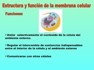
- 1. Aislar selectivamente el contenido de la célula del ambiente externo. Regular el intercambio de sustancias indispensables entre el interior de la célula y el ambiente externo Comunicarse con otras células
- 2. La clave del funcionamiento de las membranas radica en su estructura. Las membranas no son simplemente láminas uniformes: son estructuras complejas y heterogéneas cuyas diferentes partes desempeñan funciones perfectamente definidas y cambian de manera dinámica en respuesta al ambiente Las membranas son “mosaicos fluidos” en los que las proteinas se mueven dentro de capas de lípidos El modelo de mosaico fluido para las mambranas celulares fue desarrollado en 1972 por los biologos celulares S.J. Singer y G.L Nicolson
- 3. Según este modelo, una membrana, vista desde arriba, semeja un mosaico grumoso de azulejos en constante movimiento. Una doble capa de fosfolípidos forma una matriz de “cemento” fluida y viscosa, mientras que una variedad de proteínas son los “azulejos” que pueden desplazarse dentro de las capas fosfolipídicas.
- 4. fosfolípido hydrophilic heads fluido extracelular (ambiente acuoso) Cabezas hidrofílicas Colas hidrofóbicas citoplasma (ambiente acuoso) bicapa
- 6. glucoproteina fluido extracelular (exterior) cytoplasm (interior) cholesterol Bicapa fosfolipídica fosfolípido proteina de reconocimiento Proteina receptora Proteina de transporte protein filaments carbohidrato Sitio de unión
- 7. Hay tres categorías principales de proteínas de membrana, cada una de las cuales desempeña una función distinta: Las proteínas de transporte regulan el movimiento de moléculas hidrofílicas (solubles en agua) a través de la membrana plasmática. Algunas proteínas de transporte llamadas proteínas de canal, forman poros o canales que permiten a pequeñas moléculas solubles en agua atravesar la membrana. Las proteínas receptoras activan respuestas celulares cuando se unen a ellas moléculas específicas del fluido extracelular, como hormonas o nutrimentos. Las proteínas de reconocimiento, muchas de las cuales son glucoproteínas, sirven como etiquetas de identificación y como sitios de unión a la superficie celular. Las células del sistema inmunológico, por ejemplo, reconocen a una bacteria como invasor e inician su destrucción.
- 8. Transporte Pasivo Transporte que requiere energía Difusión simple Difusión facilitada Ósmosis Transporte activo Endocitosis Exocitosis
- 9. Transporte pasivo: movimiento de sustancias a través de una membrana, bajando por un gradiente de concentración, presión o carga eléctrica. No requiere que la célula gaste energía. Difusión simple: Difusión de agua, gases disueltos o moléculas solubles en lípidos a través de la bicapa fosfolipídica de una membrana. Difusión facilitada: difusión de moléculas (normalmente solubles en agua), a través de un canal o proteína portadora. Ósmosis: Difusión de agua a través de una membrana de permeabilidad diferencial; es decir, una que es más permeable al agua que a las moléculas disueltas.
- 10. Moleculas solubles en lípidos (O2, CO2, H2O) (fluido extracelular) (cytoplasm) difusión Simple
- 11. iones Proteinas De canal Difusión Facilitada a través de un canal
- 12. aminoacidos, azucares, Proteinas pequeñas Proteina portadora Difusión facilita a través de un portador (citoplasma) (fluido extracelular)
- 14. Transporte que requiere energía: Movimiento de sustancias a través de una membrana, casi siempre en contra de un gradiente de concentración, utilizando energía celular. Transporte activo: Movimiento de moléculas o iones a través de proteínas que llegan de un lado a otro de la membrana, utilizando energía celular, normalmente de ATP. Endocitosis: Movimiento de partículas grandes, o microorganismos enteros, hacia el interior de una célula que absorbe material extracelular, cuando la membrana plasmática forma bolsas delimitadas por membrana que se introducen en el citoplasma. Exocitosis: movimiento de materiales hacia el exterior de una célula envolviendo el material en una bolsa membranosa que se desplaza hacia la superficie de la célula, se funde con la membrana plasmática y se abre hacia el exterior, permitiendo que su contenido se difunda inmediatamente
- 15. ADP P La portadora suelta al Ion y a los residuos del ATP (ADP y P) y recupera Su configuración original. 3la proteina de transporte Se une a ATP y Ca2+ . 1 (cytoplasm) La energía del ATP altera La Forma de la proteina de Transporte y pasa el ion al otro lado de la membrana. 2 ATP Ca2+ Sitio de Unión a ATP Sitio de reconocimiento (extracellular fluid)
- 16. Pinocitosis 3 (fluido extracelular) (citoplasma) Vesicula que contiene fluido extracelular 2 1
- 17. (extracellular fluid) (citoplasma) 1 2 3 Fagocitosis Particula de alimento pseudopodos Vacuola alimentaria
- 18. (extracellular fluid) (citoplasma) 1 2 3 4 Endocitosis mediada por receptores nutrientes receptores Fosa recubierta Vesicula recubierta
- 19. plasma membrane plasma membrane (cytoplasm) vesícula 0.2 micrometer secreted material (extracellular fluid) Exocitosis
Notas del editor
- Figure: 4-UN2 Title: Phospholipid Bilayer
- Figure: 4-UN1 Title: Phospholipid
- Figure :4-1 Title: The plasma membrane is a fluid mosaic Caption: The plasma membrane is a bilayer of phospholipids in which various proteins are embedded. Many proteins have carbohydrates attached to them, forming glycoproteins.
- Figure :4-3 part a Title: Diffusion through the plasma membrane part a Simple diffusion Caption: (a) Simple diffusion: gases such as oxygen and carbon dioxide and lipid-soluble molecules can diffuse directly through the phospholipids.
- Figure :4-3 part b Title: Diffusion through the plasma membrane part b Facilitated diffusion through a channel Caption: (b) Facilitated diffusion through a channel: protein channels (pores) allow passage of some water-soluble molecules, principally ions, that cannot diffuse directly through the bilayer.
- Figure :4-3 part c Title: Diffusion through the plasma membrane part c Facilitated diffusion through a carrier Caption: (c) Facilitated diffusion through a carrier. Exercise Imagine an experiment that measures the initial rate of diffusion into cells placed in sucrose solutions of various different concentrations. Sketch a graph (initial diffusion rate versus solution concentration) that shows the result expected if diffusion is simple, and a graph that shows the result expected for facilitated diffusion.
- Figure :4-4 Title: Osmosis Caption: (a) Membrane pores allow “free” water molecules to pass through, but sugar molecules are too large. “Bound” water molecules, attracted to the sugars by hydrogen bonds, are also prevented from passing through the pore. (b) A bag is made of a membrane selectively permeable to free water molecules (white dots) but not to larger molecules, such as sugar (yellow hexagons) or water molecules held to the sugars by hydrogen bonds. If the bag is filled with a sugar solution and suspended in pure water, free water molecules will diffuse down their concentration gradient from the high concentration of water outside the bag to the lower concentration of water inside the bag. The bag will swell and may burst as water enters. Question Imagine a container of glucose solution, divided into two compartments (A and B) by a membrane that is permeable to water and glucose but not to sucrose. If some sucrose is added to compartment A, how will the contents of compartment B change?
- Figure :4-6 Title: Active transport Caption: Active transport uses cellular energy to move molecules across the plasma membrane, often against a concentration gradient. A transport protein (blue) has an ATP binding site and a recognition site for the molecules to be transported, in this case calcium ions (Ca2+).
- Figure :4-7 part a Title: Three types of endocytosis part a Pinocytosis Caption: Question Compare and contrast receptor-mediated endocytosis with active transport.
- Figure :4-7 part c Title: Three types of endocytosis part c Phagocytosis Caption: Question Compare and contrast receptor-mediated endocytosis with active transport.
- Figure :4-7 part b Title: Three types of endocytosis part b Receptor-mediated endocytosis Caption: Question Compare and contrast receptor-mediated endocytosis with active transport.
- Figure :4-9 Title: Exocytosis Caption: Exocytosis is functionally the reverse of endocytosis. Question How does exocytosis differ from diffusion of materials out of a cell?
- Figure :4-5 part a Title: The effects of osmosis part a Isotonic solution Caption: (a) If red blood cells are immersed in an isotonic salt solution, which has the same concentration of dissolved substances as the blood cells do, there is no net movement of water across the plasma membrane. The red blood cells keep their characteristic dimpled disk shape.
- Figure :4-5 part b Title: The effects of osmosis part b Hypertonic solution Caption: (b) A hypertonic solution, with too much salt, causes water to leave the cells, shriveling them up.
- Figure :4-5 part c Title: The effects of osmosis part c Hypotonic solution Caption: (c) A hypotonic solution, with less salt than is in the cells, causes water to enter, and the cells swell. Question All freshwater fish swim in a solution that is hypotonic to the fluid inside their bodies. Why don't freshwater fish swell up and burst?