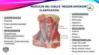
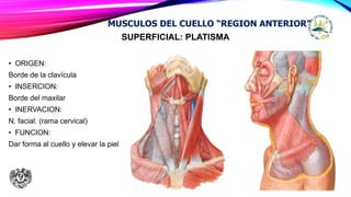
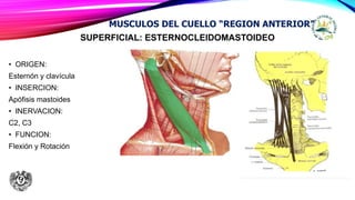
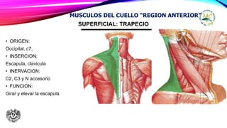
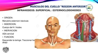
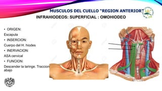
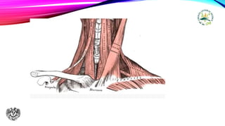
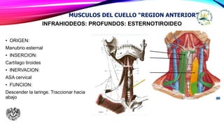
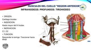
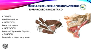
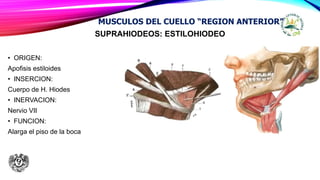
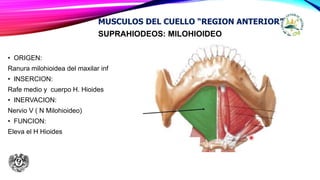
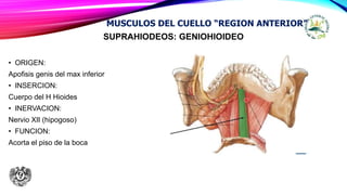
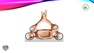
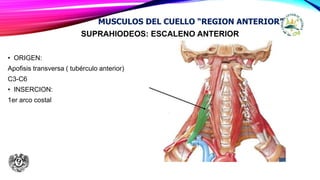
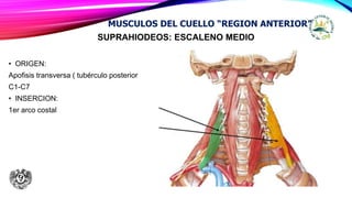
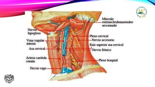
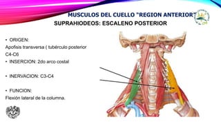
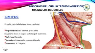
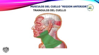
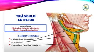
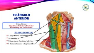
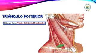
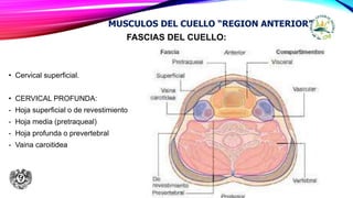
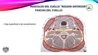
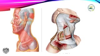
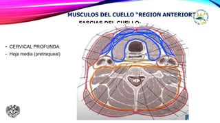
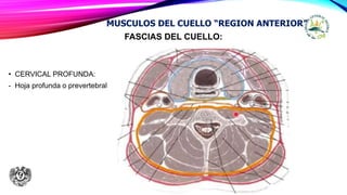
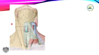
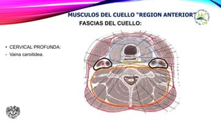
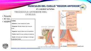
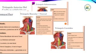
MUSCULOS DEL CUELLO
“REGION ANTERIOR”
• Ponente: HERNANDEZ SANTOS
• R1 CG
• HOSPITAL: Juan Ramon de la Fuente
CURSO DE POSGRADO EN CIRUGÍA GENERAL
CLASIFICACION:
• SUPERFICIALES
- Platisma
- Esternocleidomastoideo
- Trapecio
• INFRAHIODEOS
- Superficiales
(Esternocleidohiodeo,
Omohiodeo)
- Pofundos ( Esternotiroideo,
Tirohiodeo)
MUSCULOS DEL CUELLO “REGION ANTERIOR”
• SUPRAHIODEOS:
- Estilohiodeo
- Milohiodeo
- Digastrico
- Geniohiodeo
• ESCALENOS:
- Anterior
- Medio
- Posterior
• PREVERTEBRALES:
- Largo del cuello
- Largo de la cabeza
- Recto anterior menor
- Recto lateral de la cabeza
SUPERFICIAL: PLATISMA
• ORIGEN:
Borde de la clavícula
• INSERCION:
Borde del maxilar
• INERVACION:
N. facial. (rama cervical)
• FUNCION:
Dar forma al cuello y elevar la piel
MUSCULOS DEL CUELLO “REGION ANTERIOR”
SUPERFICIAL: ESTERNOCLEIDOMASTOIDEO
• ORIGEN:
Esternón y clavícula
• INSERCION:
Apófisis mastoides
• INERVACION:
C2, C3
• FUNCION:
Flexión y Rotación
MUSCULOS DEL CUELLO “REGION ANTERIOR”
SUPERFICIAL: TRAPECIO
• ORIGEN:
Occipital, c7,
• INSERCION:
Escapula, clavicula
• INERVACION:
C2, C3 y N accesorio
• FUNCION:
Girar y elevar la escapula
MUSCULOS DEL CUELLO “REGION ANTERIOR”
INFRAHIODEOS: SUPERFICIAL : ESTERNOCLEIDOHIODEO
• ORIGEN:
Manubrio esternon/ clavicula
• INSERCION:
Cuerpo del H. hiodes
• INERVACION:
ASA cervical
• FUNCION:
Descender la laringe. Traccionar hacia
abajo
MUSCULOS DEL CUELLO “REGION ANTERIOR”
INFRAHIODEOS: SUPERFICIAL : OMOHIODEO
• ORIGEN:
Escapula
• INSERCION:
Cuerpo del H. hiodes
• INERVACION:
ASA cervical
• FUNCION:
Descender la laringe. Traccionar hacia
abajo
MUSCULOS DEL CUELLO “REGION ANTERIOR”
INFRAHIODEOS: PROFUNDOS: ESTERNOTIROIDEO
• ORIGEN:
Manubrio esternal
• INSERCION:
Cartílago tiroides
• INERVACION:
ASA cervical
• FUNCION:
Descender la laringe. Traccionar hacia
abajo
MUSCULOS DEL CUELLO “REGION ANTERIOR”
INFRAHIODEOS: PROFUNDOS: TIROHIODEO
• ORIGEN:
Cartilago tiroides
• INSERCION:
Hasta mayor del H hiodes
• INERVACION:
C1, C2
• FUNCION:
Descender la laringe. Traccionar hacia
abajo
MUSCULOS DEL CUELLO “REGION ANTERIOR”
SUPRAHIODEOS: DIGASTRICO
• ORIGEN:
Apofisis mastoides
• INSERCION:
Borde post menton
• INERVACION:
Posterior VII y Anterior Trigenimo
• FUNCION:
Descender el mento hacia abajo
MUSCULOS DEL CUELLO “REGION ANTERIOR”
SUPRAHIODEOS: ESTILOHIODEO
• ORIGEN:
Apofisis estiloides
• INSERCION:
Cuerpo de H. Hiodes
• INERVACION:
Nervio VII
• FUNCION:
Alarga el piso de la boca
MUSCULOS DEL CUELLO “REGION ANTERIOR”
SUPRAHIODEOS: MILOHIOIDEO
• ORIGEN:
Ranura milohioidea del maxilar inf
• INSERCION:
Rafe medio y cuerpo H. Hioides
• INERVACION:
Nervio V ( N Milohioideo)
• FUNCION:
Eleva el H Hioides
MUSCULOS DEL CUELLO “REGION ANTERIOR”
SUPRAHIODEOS: GENIOHIOIDEO
• ORIGEN:
Apofisis genis del max inferior
• INSERCION:
Cuerpo del H Hioides
• INERVACION:
Nervio XII (hipogoso)
• FUNCION:
Acorta el piso de la boca
MUSCULOS DEL CUELLO “REGION ANTERIOR”
SUPRAHIODEOS: ESCALENO ANTERIOR
• ORIGEN:
Apofisis transversa ( tubérculo anterior)
C3-C6
• INSERCION:
1er arco costal
MUSCULOS DEL CUELLO “REGION ANTERIOR”
SUPRAHIODEOS: ESCALENO MEDIO
• ORIGEN:
Apofisis transversa ( tubérculo posterior
C1-C7
• INSERCION:
1er arco costal
MUSCULOS DEL CUELLO “REGION ANTERIOR”
SUPRAHIODEOS: ESCALENO POSTERIOR
• ORIGEN:
Apofisis transversa ( tubérculo posterior
C4-C6
• INSERCION: 2do arco costal
• INERVACION: C3-C4
• FUNCION:
Flexión lateral de la columna.
MUSCULOS DEL CUELLO “REGION ANTERIOR”
TRAINGULOS DEL CUELLO
MUSCULOS DEL CUELLO “REGION ANTERIOR”
TRAINGULOS DEL CUELLO
MUSCULOS DEL CUELLO “REGION ANTERIOR”
FASCIAS DEL CUELLO:
• Cervical superficial.
• CERVICAL PROFUNDA:
- Hoja superficial o de revestimiento
- Hoja media (pretraqueal)
- Hoja profunda o prevertebral
- Vaina caroitidea
MUSCULOS DEL CUELLO “REGION ANTERIOR”
FASCIAS DEL CUELLO:
- Hoja superficial o de revestimiento:
MUSCULOS DEL CUELLO “REGION ANTERIOR”
FASCIAS DEL CUELLO:
• CERVICAL PROFUNDA:
- Hoja media (pretraqueal)
MUSCULOS DEL CUELLO “REGION ANTERIOR”
FASCIAS DEL CUELLO:
• CERVICAL PROFUNDA:
- Hoja profunda o prevertebral
MUSCULOS DEL CUELLO “REGION ANTERIOR”
FASCIAS DEL CUELLO:
• CERVICAL PROFUNDA:
- Vaina caroitidea.
MUSCULOS DEL CUELLO “REGION ANTERIOR”
GRACIAS…….
CLASIFICACION:
• Ponente: HERNANDEZ SANTOS
• R1 CG
• HOSPITAL: Juan Ramon de la Fuente
MUSCULOS DEL CUELLO “REGION ANTERIOR”
ANA DE CUELLO.pptx

ANA DE CUELLO.pptx

  • 1.
    MUSCULOS DEL CUELLO “REGIONANTERIOR” • Ponente: HERNANDEZ SANTOS • R1 CG • HOSPITAL: Juan Ramon de la Fuente CURSO DE POSGRADO EN CIRUGÍA GENERAL
  • 2.
    CLASIFICACION: • SUPERFICIALES - Platisma -Esternocleidomastoideo - Trapecio • INFRAHIODEOS - Superficiales (Esternocleidohiodeo, Omohiodeo) - Pofundos ( Esternotiroideo, Tirohiodeo) MUSCULOS DEL CUELLO “REGION ANTERIOR” • SUPRAHIODEOS: - Estilohiodeo - Milohiodeo - Digastrico - Geniohiodeo • ESCALENOS: - Anterior - Medio - Posterior • PREVERTEBRALES: - Largo del cuello - Largo de la cabeza - Recto anterior menor - Recto lateral de la cabeza
  • 3.
    SUPERFICIAL: PLATISMA • ORIGEN: Bordede la clavícula • INSERCION: Borde del maxilar • INERVACION: N. facial. (rama cervical) • FUNCION: Dar forma al cuello y elevar la piel MUSCULOS DEL CUELLO “REGION ANTERIOR”
  • 4.
    SUPERFICIAL: ESTERNOCLEIDOMASTOIDEO • ORIGEN: Esternóny clavícula • INSERCION: Apófisis mastoides • INERVACION: C2, C3 • FUNCION: Flexión y Rotación MUSCULOS DEL CUELLO “REGION ANTERIOR”
  • 6.
    SUPERFICIAL: TRAPECIO • ORIGEN: Occipital,c7, • INSERCION: Escapula, clavicula • INERVACION: C2, C3 y N accesorio • FUNCION: Girar y elevar la escapula MUSCULOS DEL CUELLO “REGION ANTERIOR”
  • 7.
    INFRAHIODEOS: SUPERFICIAL :ESTERNOCLEIDOHIODEO • ORIGEN: Manubrio esternon/ clavicula • INSERCION: Cuerpo del H. hiodes • INERVACION: ASA cervical • FUNCION: Descender la laringe. Traccionar hacia abajo MUSCULOS DEL CUELLO “REGION ANTERIOR”
  • 8.
    INFRAHIODEOS: SUPERFICIAL :OMOHIODEO • ORIGEN: Escapula • INSERCION: Cuerpo del H. hiodes • INERVACION: ASA cervical • FUNCION: Descender la laringe. Traccionar hacia abajo MUSCULOS DEL CUELLO “REGION ANTERIOR”
  • 10.
    INFRAHIODEOS: PROFUNDOS: ESTERNOTIROIDEO •ORIGEN: Manubrio esternal • INSERCION: Cartílago tiroides • INERVACION: ASA cervical • FUNCION: Descender la laringe. Traccionar hacia abajo MUSCULOS DEL CUELLO “REGION ANTERIOR”
  • 11.
    INFRAHIODEOS: PROFUNDOS: TIROHIODEO •ORIGEN: Cartilago tiroides • INSERCION: Hasta mayor del H hiodes • INERVACION: C1, C2 • FUNCION: Descender la laringe. Traccionar hacia abajo MUSCULOS DEL CUELLO “REGION ANTERIOR”
  • 12.
    SUPRAHIODEOS: DIGASTRICO • ORIGEN: Apofisismastoides • INSERCION: Borde post menton • INERVACION: Posterior VII y Anterior Trigenimo • FUNCION: Descender el mento hacia abajo MUSCULOS DEL CUELLO “REGION ANTERIOR”
  • 13.
    SUPRAHIODEOS: ESTILOHIODEO • ORIGEN: Apofisisestiloides • INSERCION: Cuerpo de H. Hiodes • INERVACION: Nervio VII • FUNCION: Alarga el piso de la boca MUSCULOS DEL CUELLO “REGION ANTERIOR”
  • 14.
    SUPRAHIODEOS: MILOHIOIDEO • ORIGEN: Ranuramilohioidea del maxilar inf • INSERCION: Rafe medio y cuerpo H. Hioides • INERVACION: Nervio V ( N Milohioideo) • FUNCION: Eleva el H Hioides MUSCULOS DEL CUELLO “REGION ANTERIOR”
  • 15.
    SUPRAHIODEOS: GENIOHIOIDEO • ORIGEN: Apofisisgenis del max inferior • INSERCION: Cuerpo del H Hioides • INERVACION: Nervio XII (hipogoso) • FUNCION: Acorta el piso de la boca MUSCULOS DEL CUELLO “REGION ANTERIOR”
  • 17.
    SUPRAHIODEOS: ESCALENO ANTERIOR •ORIGEN: Apofisis transversa ( tubérculo anterior) C3-C6 • INSERCION: 1er arco costal MUSCULOS DEL CUELLO “REGION ANTERIOR”
  • 18.
    SUPRAHIODEOS: ESCALENO MEDIO •ORIGEN: Apofisis transversa ( tubérculo posterior C1-C7 • INSERCION: 1er arco costal MUSCULOS DEL CUELLO “REGION ANTERIOR”
  • 20.
    SUPRAHIODEOS: ESCALENO POSTERIOR •ORIGEN: Apofisis transversa ( tubérculo posterior C4-C6 • INSERCION: 2do arco costal • INERVACION: C3-C4 • FUNCION: Flexión lateral de la columna. MUSCULOS DEL CUELLO “REGION ANTERIOR”
  • 21.
    TRAINGULOS DEL CUELLO MUSCULOSDEL CUELLO “REGION ANTERIOR”
  • 22.
    TRAINGULOS DEL CUELLO MUSCULOSDEL CUELLO “REGION ANTERIOR”
  • 26.
    FASCIAS DEL CUELLO: •Cervical superficial. • CERVICAL PROFUNDA: - Hoja superficial o de revestimiento - Hoja media (pretraqueal) - Hoja profunda o prevertebral - Vaina caroitidea MUSCULOS DEL CUELLO “REGION ANTERIOR”
  • 27.
    FASCIAS DEL CUELLO: -Hoja superficial o de revestimiento: MUSCULOS DEL CUELLO “REGION ANTERIOR”
  • 29.
    FASCIAS DEL CUELLO: •CERVICAL PROFUNDA: - Hoja media (pretraqueal) MUSCULOS DEL CUELLO “REGION ANTERIOR”
  • 30.
    FASCIAS DEL CUELLO: •CERVICAL PROFUNDA: - Hoja profunda o prevertebral MUSCULOS DEL CUELLO “REGION ANTERIOR”
  • 32.
    FASCIAS DEL CUELLO: •CERVICAL PROFUNDA: - Vaina caroitidea. MUSCULOS DEL CUELLO “REGION ANTERIOR”
  • 34.
  • 40.
    CLASIFICACION: • Ponente: HERNANDEZSANTOS • R1 CG • HOSPITAL: Juan Ramon de la Fuente MUSCULOS DEL CUELLO “REGION ANTERIOR”

Notas del editor

  • #3 Dividirlo en 5 grupos:
  • #4 Tambien llamado MUSCULO CUTANEO DEL CUELLO, justo debajo del T ce Subcutaneo Fibras…. Deltoides y pectoral mayor
  • #5 Dos orígenes o ramas ( manubrio del esternon y tercio proximal de clavicula Linea curva del occipital Girar cuando solo un musculo se contrae
  • #6 Arriba de ella, V yugular ezterna Arriba de la fascia suerficla
  • #7 Origenes: línea curva occipital, protubencia occipital, Ligamento cervical posterior, C7, aposfisis espinosa de todas las dorsales, Inserccio. Borde supr escapula. Acromion, tercio externo de la clavicula
  • #8 Origenes:
  • #9 Dos vientres. Unidos por un temdon. Traccion hacia claviclua y externo (extensión aponeurótica)
  • #11 Dos vientres. Unidos por un temdon. Traccion hacia claviclua y externo (extensión aponeurótica) Asa cervical (c1-c3)
  • #12 Unico no por asa cervical
  • #13 Dos vientres. Posterior y anterior Tendon intermedio unido al h Hiodes por un retináculo
  • #14 Para diferenciar,el se une al hiodes
  • #16 Antagonista del estilohioideo…. Alarga Y este genio acorta
  • #17 Siguiente grupo escalenos Cuerpor vertebral cervical Anterior y posterior Apofisis Transversa : 2 (prolongaciones) tuberculos , anterior y posterior
  • #18 Tuberculo escalenico en 1ra costilla
  • #19 Cara sup de 1ra costilla Nervio frénico Plexo braquial Art Subclavio
  • #21 detras
  • #22 FORMANDO 2 TRIANGULOS: ANT Y POSTERIOR. LIMITES:
  • #23 TENER EN CUENTA. CUBIERTOS POR 2 ESTRUCTURAS. MUSCULO PLASTISMA HOJA SUPERFICAIL DE LA APONEUROSIS DEL CUELLO TECHO…..
  • #24 CARA LAT SOLO 3 TRIANGULOS SUBMAXILAR: glan parotida, sunmaxilar, arteria ay vena facial - borde inff deigastrico, borde posr omohiodieo y anter ECM (1/3 medio): carotida externa y ramas, V yugular interna, Nervios (vago, acces, hipoglo) Glandula tiroides, paratiroides, traquea , esofago
  • #25 EN MEDIO DE VIENTRE ANTERIOR DE DIGASTRICOS Ganglios .
  • #26 Occipital : bodr post ECM, Bord anter trapecio Y sup omohioideo Supraclavicular: Bord posr ECM, Clavicla, Boder inf omohioideo Plaxo braquila, ganglios linfáticos Art subclavia
  • #27 Justo debajo del t celur subcutaneo. Antes del platimsa, no se ve…. Revestimiento: fascia de COLLIS
  • #28 Dos hojas ant y posterior, envuelve a ECM, Trapecio Triangulo anterior (ant) Traingulo posterior (lado Techo. Hoja de revestimineto o hoja superficial.
  • #29 inserciones.: Protuberancia del occipucio, línea inf occipital, apófisis mastoides. Arco cigomáticos y maximalr inferior Infe, calvucual, esternos, espina de escapula. Envuelve por completo el cuello Perforada: plexo cervical y vena yugular externa
  • #30 Tiene 2 hojas. 1… para los muscuos infra hiodeos: omohioideo y esternocleidohioideo 2….. Glandula tiroide Apneurosis bucofaringeaa
  • #31 Fija, cuerpos vertebrales y espinas, apofosis Musculos posteriores de la columna.
  • #32 Amarilla: revestimientos, superficial Verde claro pretraqueal Morado prevertebral Azul vaina caroiteda
  • #33 Condensacion de las 3 fascias. Paquete neurovascular Carotida común interna Lat ve yugular interna Nervio vago. Com´patimientos