
Recomendados
Más contenido relacionado
La actualidad más candente
Similar a Trigliceridos (1)
Similar a Trigliceridos (1) (20)
Último
Último (20)
Trigliceridos (1)
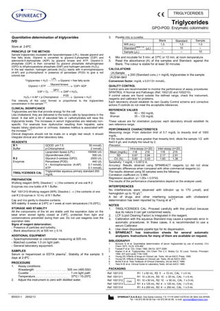
- 1. Triglycerides GPO-POD. Enzymatic colorimetric BSIS31-I 26/02/13 SPINREACT,S.A./S.A.U. Ctra.Santa Coloma, 7 E-17176 SANT ESTEVE DE BAS (GI) SPAIN Tel. +34 972 69 08 00 Fax +34 972 69 00 99. e-mail: spinreact@spinreact.com TRIGLYCERIDES Quantitative determination of triglycerides IVD Store at 2-8ºC PRINCIPLE OF THE METHOD Sample triglycerides incubated with lipoproteinlipase (LPL), liberate glycerol and free fatty acids. Glycerol is converted to glycerol-3-phosphate (G3P) and adenosine-5-diphosphate (ADP) by glycerol kinase and ATP. Glycerol-3- phosphate (G3P) is then converted by glycerol phosphate dehydrogenase (GPO) to dihydroxyacetone phosphate (DAP) and hydrogen peroxide (H2O2). In the last reaction, hydrogen peroxide (H2O2) reacts with 4-aminophenazone (4-AP) and p-chlorophenol in presence of peroxidase (POD) to give a red colored dye: Triglycerides + H2O LPL Glycerol + free fatty acids Glycerol + ATP kinaseGlycerol G3P+ ADP G3P + O2 GPO DAP + H2O2 H2O2 + 4-AP + p-Chlorophenol POD Quinone + H2O The intensity of the color formed is proportional to the triglycerides concentration in the sample 1,2,3 . CLINICAL SIGNIFICANCE Triglycerides are fats that provide energy for the cell. Like cholesterol, they are delivered to the body’s cells by lipoproteins in the blood. A diet with a lot of saturated fats or carbohydrates will raise the triglyceride levels. The increases in serum triglycerides are relatively non- specific. For example liver dysfunction resulting from hepatitis, extra hepatic biliary obstruction or cirrhosis, diabetes mellitus is associated with the increase 3,6,7 . Clinical diagnosis should not be made on a single test result; it should integrate clinical and other laboratory data. REAGENTS R 1 Buffer GOOD pH 7.5 p-Chlorophenol 50 mmol/L 2 mmol/L R 2 Enzymes Lipoprotein lipase (LPL) Glycerolkinase (GK) Glycerol-3-oxidasa (GPO) Peroxidase (POD) 4 – Aminophenazone (4-AP) ATP 150000 U/L 500 U/L 2500 U/L 440 U/L 0.1 mmol/L 0.1 mmol/L TRIGLYCERIDES CAL Triglycerides aqueous primary standard 200 mg/dL PREPARATION Working reagent (WR): Dissolve ( ) the contents of one vial R 2 Enzymes into one bottle of R 1 Buffer. Ref: 1001310 Working reagent (WR): Dissolve ( ) the contents of one vial R 2 Enzymes in 10 mL of R 1 Buffer. Cap and mix gently to dissolve contents. WR stability: 6 weeks at 2-8ºC or 1 week at room temperature (15-25ºC). STORAGE AND STABILITY All the components of the kit are stable until the expiration date on the label when stored tightly closed at 2-8ºC, protected from light and contaminations prevented during their use. Do not use reagents over the expiration date. Signs of reagent deterioration: - Presence of particles and turbidity. - Blank absorbance (A) at 505 nm > 0.14. ADDITIONAL EQUIPMENT - Spectrophotometer or colorimeter measuring at 505 nm. - Matched cuvettes 1.0 cm light path. - General laboratory equipment. SAMPLES Serum or heparinized or EDTA plasma 1 . Stability of the sample: 5 days at 2-8ºC . PROCEDURE 1. Assay conditions: Wavelength: . . . . . . . . . . . . . .. . . . . 505 nm (490-550) Cuvette: . . . . . . . . . . . . . . . . . . . . . . . . 1 cm light path Temperature . . . . . . . . . . . . . . . . . . . . 37ºC / 15-25ºC 2. Adjust the instrument to zero with distilled water. 3. Pipette into a cuvette: Blank Standard Sample WR (mL) 1.0 1.0 1.0 Standard (Note 1,2) ( L) -- 10 -- Sample ( L) -- -- 10 4. Mix and incubate for 5 min. at 37ºC or 10 min. at room temperature. 5. Read the absorbance (A) of the samples and Standard, against the Blank. The colour is stable for at least 30 minutes. CALCULATIONS dardtanS)A( Sample)A( x 200 (Standard conc.) = mg/dL triglycerides in the sample Conversion factor: mg/dL x 0.0113= mmol/L. QUALITY CONTROL Control sera are recommended to monitor the performance of assay procedures: SPINTROL H Normal and Pathologic (Ref. 1002120 and 1002210). If control values are found outside the defined range, check the instrument, reagents and calibrator for problems. Each laboratory should establish its own Quality Control scheme and corrective actions if controls do not meet the acceptable tolerances. REFERENCE VALUES Men 40 – 160 mg/dL Women 35 – 135 mg/dL These values are for orientation purpose; each laboratory should establish its own reference range. PERFORMANCE CHARACTERISTICS Measuring range: From detection limit of 0.7 mg/dL to linearity limit of 1000 mg/dL. If the results obtained were greater than linearity limit, dilute the sample 1/2 with NaCl 9 g/L and multiply the result by 2. Precision: Intra-assay (n=20) Inter-assay (n=20) Mean (mg/dL) 118 216 119 215 SD 0.67 0.94 2.17 2.91 CV (%) 0.60 0.43 1.83 1.36 Sensitivity: 1 mg/dL = 0.0012 A. Accuracy: Results obtained using SPINREACT reagents (y) did not show systematic differences when compared with other commercial reagents (x). The results obtained using 50 samples were the following: Correlation coefficient (r): 0.996. Regression equation: y= 1.00x + 0.0743. The results of the performance characteristics depend on the analyzer used. INTERFERENCES No interferences were observed with bilirubin up to 170 mol/L and hemoglobin up to 10 g/L 2 . A list of drugs and other interfering substances with cholesterol determination has been reported by Young et al 4, 5 . NOTES 1. TRIGLYCERIDES CAL: Proceed carefully with this product because due its nature it can get contamined easily. 2. LCF (Lipid Clearing Factor) is integrated in the reagent. 3. Calibration with the aqueous Standard may cause a systematic error in automatic procedures. In these cases, it is recommended to use a serum Calibrator. 4. Use clean disposable pipette tips for its dispensation. 5. SPINREACT has instruction sheets for several automatic analyzers. Instructions for many of them are available on request. BIBLIOGRAPHY 1. Buccolo G et al. Quantitative determination of serum triglycerides by use of enzimes. Clin Chem 1973; 19 (5): 476-482. 2. Fossati P et al. Clin. Chem 1982; 28(10): 2077-2080. 3. Kaplan A et al. Tryglycerides. Clin Chem The C.V. Mosby Co. St Louis. Toronto. Princeton 1984; 437 and Lipids 1194-1206. 4. Young DS. Effects of drugs on Clinical Lab. Tests, 4th ed AACC Press, 1995. 5. Young DS. Effects of disease on Clinical Lab. Tests, 4th ed AACC 2001. 6. Burtis A et al. Tietz Textbook of Clinical Chemistry, 3rd ed AACC 1999. 7. Tietz N W et al. Clinical Guide to Laboratory Tests, 3rd ed AACC 1995. PACKAGING Ref: 1001310 R1: 1 x 50 mL, R2: 5 10 mL, CAL: 1 x 5 mL Ref: 1001311 R1: 10 x 20 mL, R2: 10 20 mL, CAL: 1 x 5 mL Ref: 1001312 R1: 10 x 50 mL, R2: 10 50 mL, CAL: 1 x 5 mL Ref: 1001313 R1: 4 x 125 mL, R2: 4 125 mL, CAL: 1 x 5 mL Ref: 1001314 R1: 4 x 250 mL, R2: 4 250 mL, CAL: 1 x 5 mL Cont.
- 2. Triglicéridos GPO-POD. Enzimático colorimétrico BSIS31-E 26/02/13 SPINREACT,S.A./S.A.U. Ctra.Santa Coloma, 7 E-17176 SANT ESTEVE DE BAS (GI) SPAIN Tel. +34 972 69 08 00 Fax +34 972 69 00 99. e-mail: spinreact@spinreact.com TRIGLYCERIDES Determinación cuantitativa de triglicéridos IVD Conservar a 2-8ºC PRINCIPIO DEL METODO Los triglicéridos incubados con lipoproteinlipasa (LPL) liberan glicerol y ácidos grasos libres. El glicerol es fosforilado por glicerolfosfato deshidrogenasa (GPO) y ATP en presencia de glicerol quinasa (GK) para producir glicerol-3-fosfato (G3P) y adenosina-5-difosfato (ADP). El G3P es entonces convertido a dihidroxiacetona fosfato (DAP) y peróxido de hidrogeno (H2O2) por GPO. Al final, el peróxido de hidrogeno (H2O2) reacciona con 4-aminofenazona (4-AF) y p-clorofenol, reacción catalizada por la peroxidasa (POD) dando una coloración roja: Triglicéridos + H2O LPL Glicerol + Ácidos grasos libres Glicerol + ATP quinasaGlicerol G3P+ ADP G3P + O2 GPO DAP + H2O2 H2O2 + 4-AF + p-Clorofenol POD Quinona + H2O La intensidad del color formado es proporcional a la concentración de triglicéridos presentes en la muestra ensayada1,2,3 . SIGNIFICADO CLINICO Los triglicéridos son grasas que suministran energía a la célula. Al igual que el colesterol, son transportados a las células del organismo por las lipoproteínas en la sangre. Una dieta alta en grasas saturadas o carbohidratos puede elevar los niveles de triglicéridos. Su aumento es relativamente inespecífico. Diversas dolencias, como ciertas disfunciones hepáticas (cirrosis, hepatitis, obstrucción biliar) o diabetes mellitus, pueden estar asociadas con su elevación3,6,7, . El diagnostico clínico debe realizarse teniendo en cuenta todos los datos clínicos y de laboratorio. REACTIVOS R 1 Tampón GOOD pH 7,5 p-Clorofenol 50 mmol/L 2 mmol/L R 2 Enzimas Lipoprotein lipasa (LPL) Glicerol quinasa (GK) Glicerol-3-oxidasa (GPO) Peroxidasa (POD) 4 – Aminofenazona (4-AF) ATP 150000 U/L 500 U/L 2500 U/L 440 U/L 0,1 mmol/L 0,1 mmol/L TRIGLICERIDOS CAL Patrón primario acuoso de Triglicéridos 200 mg/dL PREPARACION Reactivo de trabajo (RT): Disolver ( ) el contenido de un vial de R 2 Enzimas en un frasco de R 1 Tampón. Ref: 1001310 Reactivo de trabajo (RT): Reconstituir ( ) el contenido de un vial de R 2 Enzimas en 10 mL de R 1 Tampón. Tapar y mezclar suavemente hasta disolver su contenido. RT Estabilidad: 6 semanas en nevera (2-8ºC) o una semana a 15-25ºC. CONSERVACION Y ESTABILIDAD Todos los componentes del kit son estables, hasta la fecha de caducidad indicada en la etiqueta, cuando se mantienen los frascos bien cerrados a 2-8ºC, protegidos de la luz y se evita su contaminación. No usar reactivos fuera de la fecha indicada. Indicadores de deterioro de los reactivos: - Presencia de partículas y turbidez. - Absorbancia (A) del Blanco a 505 nm > 0,14. MATERIAL ADICIONAL - Espectrofotómetro o analizador para lecturas a 505 nm. - Cubetas de 1,0 cm de paso de luz. - Equipamiento habitual de laboratorio. MUESTRAS Suero y plasma heparinizado o EDTA 1 . Estabilidad de la muestra: 5 días a 2-8ºC. PROCEDIMIENTO 1. Condiciones del ensayo: Longitud de onda: . . . . . . . . . . . . . . . . 505 (490-550) nm Cubeta:. . . . . . . . . . . . . . . . . . . . . . . .. .1 cm paso de luz Temperatura . . . . . . . . . . . . . . . . . . . . . . 37ºC / 15-25ºC 2. Ajustar el espectrofotómetro a cero frente a agua destilada. 3. Pipetear en una cubeta: Blanco Patrón Muestra RT (mL) 1,0 1,0 1,0 Patrón (Nota1,2) ( L) -- 10 -- Muestra ( L) -- -- 10 4. Mezclar e incubar 5 minutos a 37ºC o 10 min. a temperatura ambiente. 5. Leer la absorbancia (A) del Patrón y la muestra, frente al Blanco de reactivo. El color es estable como mínimo 30 minutos. CALCULOS Patrón)A( Muestra)A( x 200 (Conc. Patrón) = mg/dL de triglicéridos en la muestra Factor de conversión: mg/dL x 0,0113 = mmol/L. CONTROL DE CALIDAD Es conveniente analizar junto con las muestras sueros control valorados: SPINTROL H Normal y Patológico (Ref. 1002120 y 1002210). Si los valores hallados se encuentran fuera del rango de tolerancia, revisar el instrumento, los reactivos y el calibrador. Cada laboratorio debe disponer su propio Control de Calidad y establecer correcciones en el caso de que los controles no cumplan con las tolerancias. VALORES DE REFERENCIA Hombres: 40 – 160 mg/dL Mujeres: 35 – 135 mg/dL Estos valores son orientativos. Es recomendable que cada laboratorio establezca sus propios valores de referencia. CARACTERISTICAS DEL METODO Rango de medida: Desde el limite de detección de 0,7 mg/dL hasta el limite de linealidad de 1000 mg/dL. Si la concentración es superior al límite de linealidad, diluir la muestra 1/2 con ClNa 9 g/L y multiplicar el resultado final por 2. Precisión: Intraserie (n=20) Interserie (n=20) Media (mg/dL) 118 216 119 215 SD 0,67 0,94 2,17 2,91 CV (%) 0,60 0,43 1,83 1,36 Sensibilidad analítica: 1 mg/dL = 0,0012 A. Exactitud: Los reactivos SPINREACT (y) no muestran diferencias sistemáticas significativas cuando se comparan con otros reactivos comerciales (x). Los resultados obtenidos con 50 muestras fueron los siguientes: Coeficiente de correlación (r): 0,996. Ecuación de la recta de regresión: y= 1,00x + 0,0743. Las características del método pueden variar según el analizador utilizado. INTERFERENCIAS No se han observado interferencias con bilirrubina hasta 170 mol/L y hemoglobin hasta 10 g/L 2 . Se han descrito varias drogas y otras substancias que interfieren en la determinación de los triglicéridos 4,5 . NOTAS 1. TRIGLYCERIDES CAL: Debido a la naturaleza del producto, es aconsejable tratarlo con sumo cuidado ya que se puede contaminar con facilidad. 2. LCF(Lipid Clearing Factor) está integrado en el reactivo. 3. La calibración con el Patrón acuoso puede dar lugar a errores sistemáticos en métodos automáticos. En este caso, se recomienda utilizar calibradores séricos. 4. Usar puntas de pipeta desechables limpias para su dispensación. 5. SPINREACT dispone de instrucciones detalladas para la aplicación de este reactivo en distintos analizadores. BIBLIOGRAFIA 1. Buccolo G et al. Quantitative determination of serum triglycerides by use of enzimes. Clin Chem 1973; 19 (5): 476-482. 2. Fossati P et al. Clin. Chem 1982; 28(10): 2077-2080. 3. Kaplan A et al. Tryglycerides. Clin Chem The C.V. Mosby Co. St Louis. Toronto. Princeton 1984; 437 and Lipids 1194-1206. 4. Young DS. Effects of drugs on Clinical Lab. Tests, 4th ed AACC Press, 1995. 5. Young DS. Effects of disease on Clinical Lab. Tests, 4th ed AACC 2001. 6. Burtis A et al. Tietz Textbook of Clinical Chemistry, 3rd ed AACC 1999. 7. Tietz N W et al. Clinical Guide to Laboratory Tests, 3rd ed AACC 1995. PRESENTACION Ref: 1001310 R1: 1 x 50 mL, R2: 5 10 mL, CAL: 1 x 5 mL Ref: 1001311 R1: 10 x 20 mL, R2: 10 20 mL, CAL: 1 x 5 mL Ref: 1001312 R1: 10 x 50 mL, R2: 10 50 mL, CAL: 1 x 5 mL Ref: 1001313 R1: 4 x 125 mL, R2: 4 125 mL, CAL: 1 x 5 mL Ref: 1001314 R1: 4 x 250 mL, R2: 4 250 mL, CAL: 1 x 5 mL Cont.