Más contenido relacionado
La actualidad más candente (9)
Similar a New fundamentals in hemostasis (20)
New fundamentals in hemostasis
- 1. NEW FUNDAMENTALS IN HEMOSTASIS
Henri H. Versteeg, Johan W. M. Heemskerk, Marcel Levi, and Pieter H. Reitsma
Einthoven Laboratory for Experimental Vascular Medicine, Leiden University Medical Center, Leiden, The
Netherlands; Department of Biochemistry, Cardiovascular Research Institute Maastricht, Maastricht University,
Maastricht, The Netherlands; and Department of Medicine, Academic Medical Center, Amsterdam, The
Netherlands
L
Versteeg HH, Heemskerk JWM, Levi M, Reitsma PH. New Fundamentals in He-
mostasis. Physiol Rev 93: 327–358, 2013; doi:10.1152/physrev.00016.2011.—
Hemostasis encompasses the tightly regulated processes of blood clotting, platelet
activation, and vascular repair. After wounding, the hemostatic system engages a
plethora of vascular and extravascular receptors that act in concert with blood com-
ponents to seal off the damage inflicted to the vasculature and the surrounding tissue. The first
important component that contributes to hemostasis is the coagulation system, while the second
important component starts with platelet activation, which not only contributes to the hemostatic
plug, but also accelerates the coagulation system. Eventually, coagulation and platelet activation
are switched off by blood-borne inhibitors and proteolytic feedback loops. This review summarizes
new concepts of activation of proteases that regulate coagulation and anticoagulation, to give rise
to transient thrombin generation and fibrin clot formation. It further speculates on the (patho)phys-
iological roles of intra- and extravascular receptors that operate in response to these proteases.
Furthermore, this review provides a new framework for understanding how signaling and adhesive
interactions between endothelial cells, leukocytes, and platelets can regulate thrombus formation
and modulate the coagulation process. Now that the key molecular players of coagulation and
platelet activation have become clear, and their complex interactions with the vessel wall have been
mapped out, we can also better speculate on the causes of thrombosis-related angiopathies.
I. INTRODUCTION 327
II. NEW FUNDAMENTALS IN COAGULATION 328
III. NEW FUNDAMENTALS IN... 338
IV. NEW FUNDAMENTALS IN... 347
V. CONCLUSIONS 349
I. INTRODUCTION
Hemostasis enables an organism to 1) close off damaged
blood vessels, 2) keep the blood in a fluid state, and
3) remove blood clots after restoration of vascular integrity.
The hemostatic system is a highly conserved machinery,
from zebrafish to human, in which blood clotting, also re-
ferred to as coagulation, has a prominent role. Two millen-
nia ago, the Greek philosopher Plato already described that
the blood forms fibers once it leaves the heat of the body. He
was also the first one to coin the term fibrin, which nowa-
days refers to a key blood clotting protein composing those
fiber structures. Interestingly, Plato’s view on blood clot-
ting, which was shared by other early philosophers, such as
Aristotle and Galen, remained the leading concept until the
end of the 18th century. In the course of the 19th century,
groundbreaking discoveries were made on the biological
mechanism of coagulation. Around 1865, platelets were
discovered as well as their critical function in hemostasis
(30). It was proposed that a hypothetical protein termed
“thrombin” could induce the formation of fibrin. The ma-
jority of the key players in coagulation were discovered
during the course of the 20th century. In 1905, Morawitz
constructed the first coagulation model in which thromboplas-
tin, now known as tissue factor (TF), was released by damaged
vessels to convert prothrombin into thrombin in the presence
of calcium (246). Thrombin then converted fibrinogen into
fibrin resulting in the formation of a blood clot. However, this
four-clotting factor model could not fully explain the complex
process of coagulation. Around the 1950s, many of the re-
maining factors had been characterized, such as von Wille-
brand factor (VWF) and factors V, VII, VIII, IX, and XI (FV,
FVII, FVIII, FIX, FXI). Deficiency in some of these factors was
linked to bleeding diseases, such as FVIII deficiency in hemo-
philia A and FIX deficiency in hemophilia B (72). In the 1960s,
two independent groups constructed a model for coagulation
that resembled a waterfall or cascade. Therefore, this model
was aptly named the coagulation cascade model (67, 168).
Herein, each clotting factor consists of a proenzyme that is
converted to an active enzyme by the upstream activated clot-
ting factor (FIGURE 1). It was also suggested that two different
cascades exist that converge in FX activation. These are named
the intrinsic pathway, so called because all the components are
present in the blood, and the extrinsic pathway requiring an
external factor (TF from the extravascular tissue). The intrin-
sic pathway becomes activated in vitro once blood comes into
contact with hydrophilic surfaces. This is triggered by auto-
activated FXII cleaving prekallikrein into kallikrein, which
Physiol Rev 93: 327–358, 2013
doi:10.1152/physrev.00016.2011
3270031-9333/13 Copyright © 2013 the American Physiological Society
Downloaded from www.physiology.org/journal/physrev by ${individualUser.givenNames} ${individualUser.surname} (177.228.042.146) on February 11, 2018.
Copyright © 2013 American Physiological Society. All rights reserved.
- 2. leads to a subsequent activation pathway of FXI, FIX, FX, and
prothrombin. The extrinsic pathway starts with TF and acti-
vated FVII, which directly induces sequential activation of FX
and prothrombin.
The current concept of hemostasis, outlined in detail below, is
as follows. Upon vessel damage, platelets adhere to the dam-
aged site and aggregate through interactions of platelet recep-
tors with extracellular ligands and soluble proteins. Vascular
damage-induced exposure of subendothelial TF generates
trace amounts of thrombin with multiple effects on other co-
agulation factors and platelets. Via multiple enforcement
loops in the coagulation system and in platelet activation, large
amounts of fibrin are formed stabilizing earlier formed platelet
thrombi. In this review we will first discuss the latest insights in
function and regulation of the coagulation cascade, and then
discuss the complex interaction of platelets with the endothe-
lium and the extracellular matrix. As hemostasis is now con-
sidered to include wound healing and endothelial barrier pro-
tection, we will also discuss the role of coagulation factors in
these important processes. Of note, we will not extensively
discuss the fibrinolytic pathway.
II. NEW FUNDAMENTALS IN
COAGULATION
A. Various Phases of Coagulation
According to a widely used current model (179), coagulation
can be divided into three separate phases: 1) an initiation
phase, in which low amounts of active coagulant factors are
generated; 2) an amplification phase, in which the level of
active coagulation factors is boosted; and 3) a propagation
phase, in which coagulation factors bind to highly procoagu-
lant membranes of activated platelets and fibrin clots are
Subendothelium
Subendothelium
Endothelium
Platelets
VIIa
Xa
Xa
IXa
IXa
XIa
XIIa
VIIIa
Va
TF
Poly P
Poly P
XaX
VI
TF
I
F
a
y
XaIX
IIa
IIa
IIa
IXa
VIIIa
Fibrin
Xa
Va
brinninn
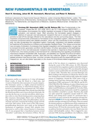
FIGURE 1. The coagulation cascade. Upon endothelial damage, tissue factor (TF) is exposed to the blood-
stream and binds factor VII, which is activated to factor VIIa. The TF:VIIa complex enables subsequent activation
of factor X and prothrombin, after which small amounts of thrombin activate the factor XI-IX feedback loop on
the platelet surface. Factor IXa will then activate additional factor X. Simultaneously, the trace amounts of
thrombin will then activate factors VIII (cofactor to factor IX) and V (cofactor to factor X), which dramatically
enhances catalytic activity of factors IX and X. Finally, thrombin (factor IIa) activation leads to fibrin deposition.
In parallel, local polyphosphate (polyP) release by activated platelets may additionally stimulate activation of
factor XII, factor V, and FXI and inhibit clot lysis.
VERSTEEG ET AL.
328 Physiol Rev • VOL 93 • JANUARY 2013 • www.prv.org
Downloaded from www.physiology.org/journal/physrev by ${individualUser.givenNames} ${individualUser.surname} (177.228.042.146) on February 11, 2018.
Copyright © 2013 American Physiological Society. All rights reserved.
- 3. formed. While this cell biological model of coagulation is gain-
ing attention, the more classical division between intrinsic and
extrinsic pathway is still widely used (170).
1. Initiation phase
The initiation phase, classically referred to as the extrinsic
pathway of coagulation, starts when the vasculature is dis-
rupted, and subendothelial cells like smooth muscle cells
and fibroblasts become exposed to the bloodstream (FIGURE
1) (179). These cells expose a key initiator of the coagula-
tion cascade, TF, which binds coagulation FVII. By acting
as a cofactor for FVII, TF promotes proteolysis and activa-
tion to FVIIa. TF largely resides on the cell surface in an
inactive (cryptic) configuration, but under certain condi-
tions it is readily decrypted as described below. It is not
precisely understood how FVII is cleaved into FVIIa, but a
proteolytic role is suggested for either the minute amounts
of FVIIa that circulate in the blood (100 pM) (198) or the
factor VII-activating protein (FSAP), but recent data argue
against the latter (276). At physiological concentrations,
FVIIa without TF shows little activity, because of a unique
sequence characteristic that retains the FVIIa in a zymogen-
like conformation (230). Thus FVIIa activity at physiolog-
ical levels is entirely TF dependent.
The TF/FVIIa complex proteolytically cleaves traces of FIX
and FX into FIXa and FXa, respectively. This allows FXa to
associate with cofactor FVa to form a prothrombinase com-
plex on TF-expressing cells (179), which serves to convert
prothrombin (FII) into thrombin. FXa may dissociate from
these TF-expressing cells to form prothrombinase com-
plexes on distant cell membranes. However, the presence of
protease inhibitors in plasma such as the Kunitz-type pro-
tease inhibitor tissue factor pathway inhibitor (TFPI), and
the serine protease inhibitor antithrombin (AT), will limit
such diffusion (40, 135). FIXa is not targeted by TFPI and
hence can diffuse more easily to other cell surfaces to par-
ticipate in the propagation phase.
2. Amplification phase
The slowly accumulating amounts of thrombin will further
activate platelets that have adhered to a site of injury, as
discussed in section IV. In parallel, thrombin will convert
(platelet-derived) FV into FVa, thus amplifying prothrom-
binase activity, and convert FVIII into FVIIIa, which acts as
a cofactor to FIXa on the surface of activated platelets to
support FXa generation. In addition, thrombin converts
FXI into FXIa (FIGURE 1).
3. Propagation phase
Whereas in current models the initiation phase takes place
on TF-expressing surfaces, the propagation phase occurs
away on surfaces containing procoagulant phospholipids,
such as activated platelets. Activated FXI converts FIX into
FIXa, which then associates with thrombin-cleaved FVIII
(FIGURE 1). Absence or near-absence of FVIII or FIX leads
to severe bleeding complications (hemophilia A and B, re-
spectively), thus underlining the importance of these coag-
ulation factors for normal hemostasis. On phosphatidylser-
ine-exposing cell membranes, the tenase complex of FIXa/
FVIIIa catalyzes the conversion of FX to FXa, after which
the FXa/FVa complex produces sufficient amounts of
thrombin to massively form fibrin fibers. As a final step, the
thrombin-activated plasma transglutaminase FXIIIa cata-
lyzes the formation of covalent crosslinks between adjacent
fibrin chains to yield an elastic, polymerized fibrin clot (8).
B. Reappraisal of the Intrinsic Coagulation
Pathway
According to the cell biological model of coagulation, the
intrinsic FXI-FXII pathway only serves as an amplification
loop initiated by the extrinsic TF pathway. Several pieces of
evidence indicate that this understates the role of the intrin-
sic pathway. Recent studies indicate that in mice, the intrin-
sic pathway is activated more or less in parallel with the
extrinsic pathway. Three physiological triggers of the in-
trinsic pathway have been discovered, namely, collagen
(302), linear phosphate polymers termed polyphosphates
(245), and neutrophil extracellular traps (NETs) (308).
Cell- and platelet-derived polyphosphates bind to and acti-
vate FXII, thereby leading to the subsequent activation of
plasma kallikrein, FIX, and further downstream coagula-
tion factors (184). In particular, a role has been proposed
for platelet-derived polyphosphates, as platelets are abun-
dantly present at the sites of vascular injury. Polyphos-
phate-dependent FXII activation does not appear to lead to
a faster clot formation, but rather to increased fibrin clot
stability (225, 240). This could also explain why high levels
of FXII associate with thrombosis, while FXII deficiency
may render the clot unstable and lead to embolization. Re-
cent findings further indicate that polyphosphates can act as
a cofactor for thrombin-mediated activation of FV and FXI
(50), and that it inhibits clot fibrinolysis, presumably
through activation of TAFI (275). Markedly, the shorter
chain polyphosphates secreted by platelets appear to be
more active in FV conversion, while longer chain polyphos-
phates more efficiently contribute to FXII activation, thus
suggesting that different polyphosphate pools act on these
distinct pathways (275). Another phosphate source, extra-
cellular RNA, was recently also shown to stimulate activa-
tion of the intrinsic pathway components such as FXII and
FXI, but this has been associated with thrombosis, rather
than with hemostasis (140). NETs also lead to the activa-
tion of FXII to FXIIa, and this influences clot formation.
Whether FXIa is formed remains unclear (308). Taken to-
gether, one can speculate that polyphosphate- and NET-
dependent activation of FV and FXI plays a role in hemo-
NEW FUNDAMENTALS IN HEMOSTASIS
329Physiol Rev • VOL 93 • JANUARY 2013 • www.prv.org
Downloaded from www.physiology.org/journal/physrev by ${individualUser.givenNames} ${individualUser.surname} (177.228.042.146) on February 11, 2018.
Copyright © 2013 American Physiological Society. All rights reserved.
- 4. stasis, while the polyphosphate- and RNA-dependent acti-
vation of FXII plays a role in thrombosis.
C. Coagulation Proteases
The coagulation factors are usually divided into two bio-
chemical classes: the serine proteases [i.e., (pro)thrombin,
FVII(a), FIX(a), FX(a), and FXI(a)] and serine protease co-
factors [i.e., thrombomodulin, TF, FV(a), and FVIII(a)].
The natural anticoagulant protein C also belongs to the
group of serine proteases. These proteolytic enzymes are the
active components of the clotting machinery and share
many structural features. Full-length FVII, FIX, FX, and
protein C consist of an NH2-terminal ␥-carboxylated glu-
tamic acid (Gla) domain, two epidermal growth factor-like
domains (EGF1 and EGF2), and a serine protease domain.
Prothrombin has a similar structure but contains two krin-
gle domains instead of the EGF domains, while FXI is a
homodimer with four apple domains in each subunit. Se-
quence analysis of these serine proteases in different organ-
isms including primates, rodents, and fish suggests that
these serine proteases all originate from a common ances-
tral gene and are the result of gene reduplications (66). The
prothrombin gene may originate from the same ancestral
gene, but appears to have undergone an EGF to kringle
domain substitution.
Posttranslational modification is of key importance to the
function of coagulation serine proteases. Glutamate resi-
dues in the NH2-terminal domain are converted into ␥-car-
boxylated glutamic acids by the enzyme ␥-glutamyl carbox-
ylase (GGCX), which is an endoplasmic reticulum resident
protein, mostly expressed in the liver. After hepatic secre-
tion into the circulation, two or three of the Gla residues of
the serine protease bind a Ca2ϩ
, which promotes a confor-
mational change (287). This conformational change con-
fers to the Gla domain-containing protease the ability to
bind to procoagulant phospholipid surfaces, which is a re-
quirement for efficient hemostasis, as discussed above.
D. Role and Activation of TF
TF, also known as thromboplastin or CD142, is a 47-kDa
transmembrane glycoprotein that is expressed in extravas-
cular tissue, particularly in fibroblasts and smooth muscle
cells, serving as a hemostatic “envelope,” poised to activate
coagulation upon vascular injury (78, 318). Generally, ac-
tive TF is not exposed to the bloodstream, but endothelial
cells and adhered leukocytes may express active TF as a
response to injury or to inflammatory stimuli such as endo-
toxin, chemokines, or cytokines (84, 311). As discussed
later, this may cause severe deregulation of hemostasis.
The mature TF protein comprises a 219-amino acid extra-
cellular region, a hydrophobic transmembrane region, and
a short intracellular tail of 21 amino acids. The external
part consists of two fibronectin-type III domains, each with
an extracellular disulfide bond (Cys49
-Cys57
and Cys186
-
Cys209
). Only breaking of the second bond distorts the co-
agulant function of TF. Intracellularly, TF may be anchored
in the cell membrane via acylation of palmitic and stearic
acids, likely serving to target TF to glycosphingolipid- and
cholesterol-rich microdomains (76). TF-like DNA se-
quences have also been identified in fish and in insects like
Anopheles gambiae and Drosophila. Structurally, TF
shares a high degree of homology with the class II interferon
receptors (20). On cells, TF can be regarded as a true recep-
tor for FVIIa, also given the fact that two cytoplasmic ser-
ines (Ser253
and Ser258
) become phosphorylated in the pres-
ence of FVIIa as a (co-)ligand (1).
E. TF Encryption/Decryption
Although extravascular cells amply express TF, TF proco-
agulant activity often remains surprisingly low. On the
other hand, stimulating these cells with agonists such as
Ca2ϩ
ionophore, hydrogen peroxide, or proteases, but also
disruption of cells, can lead to a dramatic increase, often up
to a 100-fold, in TF-dependent procoagulant activity (12,
176). Therefore, it has been suggested that procoagulant
activity is controlled by cellular mechanisms, that keep TF
in an inactive or “encrypted” state and regulate decryption
after an appropriate stimulus. Although substantial evi-
dence for this hypothesis is available, the exact nature of the
underlying mechanism remains controversial. Below we
will discuss earlier and recent models that have been put
forward to understand the regulation of TF decryption.
Classically, it has been assumed that TF encryption-decryp-
tion depends on the phospholipid environment (13), i.e.,
under cryptic conditions, TF is located in a noncoagulant
membrane. Upon cell activation and subsequent increased
cytosolic Ca2ϩ
, the inner plasma membrane leaflet-residing
phospholipid phosphatidylserine is transported to the outer
leaflet, a process modulated by flippase, floppase, and
scramblase-type lipid transporters (331) (FIGURE 2, A AND
A=). The identity of these proteins on the surface of TF-
exposing cells remains largely unknown, but ABC-class
transmembrane transporters and TMEM16F are candidate
floppases and scramblase, respectively (64, 282). The ex-
posed negatively charged phosphatidylserine accelerates co-
agulation reactions on TF-containing membrane surfaces
by stimulating tenase and prothrombinase activities (14,
150). The direct association of TF and phosphatidylserine
seems to restrict the orientation of the TF/FVIIa complex to
align the active site with the scissile peptide bonds in mem-
brane-bound FX/FIX (19). In support of this view, cells
typically show more TF-dependent procoagulant activity
upon phosphatidylserine exposure that occurs during apop-
tosis (108). Nevertheless, phosphatidylserine exposure does
not fully explain TF encryption/decryption. For instance,
VERSTEEG ET AL.
330 Physiol Rev • VOL 93 • JANUARY 2013 • www.prv.org
Downloaded from www.physiology.org/journal/physrev by ${individualUser.givenNames} ${individualUser.surname} (177.228.042.146) on February 11, 2018.
Copyright © 2013 American Physiological Society. All rights reserved.
- 5. PC/SphPS
Floppase ScramblaseFlippase
Unactivated
ATP
PS
Non-PS
ATP
PS
m
tiv
PS
Non-P
mblas
vated
se
PL
Activated
PL
va
Non P
L
ated
CaCa2+
CaCa2+
CaCa2+
A C
B D
A’
DD
asTF
Oxidoreductase function
?
Xa
VIIa
TF
Oxidized:
• High affinity for VII
• Coagulant active
Reduced:
• Low affinity for VII
• Coagulant inactivee
y for VIIy for VII
inactive
II
Reduced:
• Low affinity• Low affinity
C l t inacti• Coagulant
IIity for VVity for
t ti
V
ve
V
B Oxidized:
• High affini• High affini
C l tt activv• Coagulant
S-S S-S –SNO
CCC
aOxidoreductaPDI
PDI
Chaperone
function
FIGURE 2. Regulation of TF activity. A: induced exposure of phosphatidylserine (PS). Under resting condi-
tions, TF-exposing cells maintain a nonsymmetrical membrane composition, resulting in low PS contents in the
outer membrane leaflet and high contents in the inner leaflet. This asymmetry is maintained through ATP-
dependent inward transport of PS by flippases and outward transport of non-PS by floppases. Upon stimulation,
calcium transients will inhibit flippase and stimulate the nonselective lipid transporter scramblase, resulting in
PS exposure and the creation of a negatively charged surface that functions to bind coagulation factors. B: TF
disulfide regulation. TF is kept on the cell surface in an inactive state through PDI- and/or nitric oxide
(NO)-dependent reduction of the TF allosteric disulfide. Oxidation of the allosteric disulfide restores TF coagulant
function. C: intravascular cells, such as activated monocytes, PDI-dependently shed TF-positive MPs. D: asTF
on cellular surfaces or MPs may synergize with normally spliced TF to enhance TF procoagulant activity.
NEW FUNDAMENTALS IN HEMOSTASIS
331Physiol Rev • VOL 93 • JANUARY 2013 • www.prv.org
Downloaded from www.physiology.org/journal/physrev by ${individualUser.givenNames} ${individualUser.surname} (177.228.042.146) on February 11, 2018.
Copyright © 2013 American Physiological Society. All rights reserved.
- 6. annexin A5, which binds to phosphatidylserine and thus
competes with TF/FVIIa and FX for binding sites, does not
appear to inhibit the TF/FVIIa-induced FX to FXa conver-
sion (155), e.g., on ovarian carcinoma cells.
Another model explains TF-dependent procoagulant activ-
ity by oxidation and reduction of the TF COOH-terminal
Cys186
-Cys209
bond. This disulfide bond between two an-
tiparallel -strands is less stable because of a strained bond
geometry. Breaking of this disulfide bond may cause con-
formational changes that alter the affinity of TF for FVIIa.
TF mutants lacking this disulfide bond show a much lower
affinity for FVIIa and loss of coagulant function (242, 301),
and pools of TF showing either high or low affinity for
FVIIa may be found on the surface of cells that endoge-
nously express TF (2, 155).
The Cys186
-Cys209
bond shows a typical right-handed sta-
ple allosteric disulfide conformation and may thus be prone
to modulation by protein disulfide isomerases (PDI) (263)
(FIGURE 2B). Indeed, these isomerases have been proposed
to break the allosteric disulfide bond to produce coagulant
inactive TF, while PDI-dependent oxidation of this bond
restores coagulant activity. PDI is a 64-kDa protein that is
critically involved in protein folding and quality control in
the endoplasmic reticulum, by using catalytical thiols that
are used to enzymatically break or form disulfides, so-called
oxidoreductase function, or reshuffle disulfides between
cysteine residues, which is known as “isomerization” (319).
However, PDI is also present on the surface of various cells,
through electrostatic interaction with the membrane (3, 87,
328). In keratinocytes, PDI covalently complexes with TF
and keeps TF in an inactivated state, but strong oxidants
induce a PDI-dependent oxidative activation of TF function
(2). Moreover, PDI activates TF on the surface of micropar-
ticles and stimulates thiol-dependent shedding of active TF
on monocyte-derived microparticles (98). Finally, in mu-
rine models, PDI enhances coagulation by converting inac-
tive TF into active disulfide bond-containing TF, a feature
that is sensitive to TF free thiol-blockade (243), and pro-
motes fibrin generation (49, 243). PDI-dependent covalent
modification of TF cysteines with nitric oxide and/or gluta-
thione, which also produces coagulant-inactive TF, has also
been postulated and may form an additional level of regu-
lation of TF. In this context, it is noteworthy that vascular-
protective NO synthesis is frequently perturbed in athero-
sclerosis, diabetes, or inflammation (80), all conditions as-
sociated with thrombotic risk. Therefore, uncoupling of
NO synthesis may shift cell-surface TF activity towards
coagulation.
Although allosteric disulfide modulation forms an attrac-
tive explanation for TF decryption, this model has been
extensively debated in the field. Some studies find that TF
disulfide mutants are completely inactive and cannot be
decrypted, whereas other studies attributed significant
function and decryption potential to these mutants. Finally,
TF redox switching does not appear to take place in path-
ological settings such as cancer, and this may account for
the relatively high TF activity often observed on the surface
of cancer cells (227).
A third model assumes that decryption relies on TF dimer
formation. Like other members of the class II interferon
receptors, TF indeed has the capability to dimerize in a
manner determined by the redox environment and the ex-
posure of phosphatidylserine (254). However, both mono-
meric and dimeric forms of TF appear to possess procoagu-
lant activity, depending on the experimental conditions (13,
301).
A last model predicts that TF becomes encrypted after lo-
calization to lipid rafts, as lipid rafts are known to be poor
in phosphatidylserine (38). In endothelial cells, assembly of
the ternary TF/FVIIa/FXa complex indeed results in TF
translocation to caveolae, where TF is then rendered inac-
tive (269). In agreement with this, microparticles derived
from cholesterol-rich monocyte rafts contain inactive TF.
TF is activated upon microparticle fusion with platelets
(70), presumably by platelet-dependent phosphatidylserine
enrichment. In contrast, disruption of lipid rafts by choles-
terol depletion appeared to decrease TF activity in fibro-
blasts, and it was suggested that raft-localized cholesterol
functions as a positive regulator of TF function by main-
taining TF receptors in a high-affinity state for FVIIa bind-
ing (171). Thus the importance of lipid rafts for TF function
is still unclear. Based on these inconsistencies, one might
speculate that cryptic TF is not a single entity and that
encryption is differentially regulated in different cells. Al-
ternatively, it is possible that TF needs to proceed through
sequential steps, e.g., relocalization from rafts to phosphati-
dylserine-rich microdomains, followed by oxidation, to be-
come active.
F. Blood-Borne TF
The traditional assumption is that expression of active TF is
confined to extravascular cells. However, especially under
pathological conditions, detectable amounts of TF are
found in circulating blood. Recent insights predict that the
coagulant activity of these TF pools may be tightly regu-
lated by the environment and may also contribute to normal
hemostasis under physiological conditions.
1. TF on intravascular cells
During sepsis, cell wall components from Gram-negative
bacteria like lipopolysaccharide Toll receptor-dependently
induce the expression of active TF on intravascular cells,
such as circulating monocytes and endothelial cells in the
microvascular system (77, 226). The consequence is wide-
spread activation of coagulation, formation of fibrin, and
VERSTEEG ET AL.
332 Physiol Rev • VOL 93 • JANUARY 2013 • www.prv.org
Downloaded from www.physiology.org/journal/physrev by ${individualUser.givenNames} ${individualUser.surname} (177.228.042.146) on February 11, 2018.
Copyright © 2013 American Physiological Society. All rights reserved.
- 7. consumption of clotting factors. Whether intravascular
cells other than monocytes and endothelial cells express TF
has been the subject of debate for over 10 years. Expression
of TF on neutrophils has been reported by some authors
(102, 125), but denied by others (221). This controversy
may be explained by the possibility that neutrophils can
take up TF from other blood cells (113).
Expression of TF on platelets forms another controversial
topic. Platelets appear to acquire TF through interaction
with TF-bearing microparticles from monocytes (70, 88,
127), but platelets also contain TF pre-mRNA, which can
be spliced into mature mRNA upon platelet activation, cul-
minating in limited TF protein expression and procoagulant
activity (267). Because platelets secrete large quantities of
the TF antagonist tissue factor pathway inhibitor (TFPI),
the physiological relevance of this low expression of TF to
hemostasis is unclear.
2. TF on microparticles
Microparticles are 50- to 1,000-nm membrane vesicles that
are released from various cells and may mediate intercellu-
lar signaling and regulate hemostasis (95). Especially dis-
ease states such as sepsis, inflammatory responses, and can-
cer are accompanied by shedding of TF-positive micropar-
ticles (FIGURE 2C), and microparticles may also contribute
to thrombotic risk. Thus enhanced TF activity on tumor
cell-derived microparticles correlates with disease progres-
sion, and also associates with an increased risk of venous
thrombosis (291). Here, we will only discuss the role of
microparticle-associated TF pools in the regulation of nor-
mal hemostasis.
The available evidence suggests a prohemostatic effect of
TF-positive microparticles. Although numbers in blood
from healthy individuals may vary, microparticles can ac-
celerate or increase formation of fibrin-rich thrombi (185).
TF-positive vesicular structures are detected in thrombi
formed under flow (102), and they appear to home to de-
veloping thrombi in vivo by interaction of microparticle
P-selectin with PSGL-1-expressing platelets (89). This sug-
gests that this pool of blood-borne TF stimulates fibrin for-
mation after a thrombus has buried the extravascular
source of TF. This view in which microparticle TF replen-
ishes the TF pool at the site of clot formation has been
criticized. First, in healthy subjects, the blood concentration
and activity of microparticle-associated TF is relatively low
(136, 291). Second, in static models of coagulation, this TF
pool does not appear to mediate clot growth (215, 219). In
a physiological context, however, blood flow and therefore
continuous supply of microparticles may lead to local TF
concentrations that can drive thrombus growth in vivo.
Cellular sources of coagulant active TF on microparticles
include monocytes, platelets and endothelial cells (70, 185),
but in vivo models predict that especially hematopoietic
cells contribute to hemostatic TF-MP generation (224). In-
terestingly, TF on platelet and monocyte microparticles
likely requires activation by certain changes in the intravas-
cular environment. Notably, TF microparticles generated
from platelets become procoagulant upon association with
activated neutrophils (185), and TF on monocyte-derived
microparticles shows activity after fusion with phosphati-
dylserine-expressing platelets, but not resting platelets (70).
As discussed above, PDI-dependent decryption of micro-
particle TF may play a role here (307).
3. Alternatively spliced TF
The primary transcript encoding full-length TF (flTF) con-
tains six exons, but an alternatively spliced form of TF
(asTF) exists in which exon 5 is spliced out. Because of a 3=
frame shift mutation, the flTF transmembrane and cyto-
plasmic tail are replaced with a hydrophobic COOH-termi-
nal domain, which renders asTF soluble. asTF is expressed
in lung, pancreas, placenta, heart, endothelium, and mono-
cytes (31, 285, 286). Although the level of asTF in human
plasma may be substantial, amounting to 10–30% of total
TF (107), the question of whether it contributes to coagu-
lation is a matter of debate. Several observations suggest
that asTF has a role in hemostasis: 1) asTF localizes on
platelet surfaces in experimental blood clots; 2) the 165–
166 lysine doublet involved in FVIIa binding is maintained
in asTF, and theoretically, activity of the TF/FVIIa complex
is maintained; 3) when added to plasma in vitro, asTF
shortens the clotting time (31); and 4) while cell-expressed
asTF alone does not appear to have procoagulant properties
(32, 47), endothelial asTF expression in the presence of TF
may be procoagulant, as depletion of asTF reduced TF-
dependent coagulant activity (285). Thus asTF and flTF
may cooperate in a synergistic fashion (FIGURE 2D), but
such a mechanism, as well as how asTF would enhance
function of normally spliced TF, remains speculative.
In conclusion, on the basis of the above-described studies on
blood-borne TF, a model is likely in which damaged vascu-
lar cells shed MP-TF that fuse with platelets and leukocytes
to form a “second-generation” procoagulant platform that
is in part dependent on asTF for optimal activity.
G. Inhibition of the Coagulation Process
Considerable efforts have been made in recent years to un-
ravel the suppressor mechanisms of the coagulation pro-
cess. Studies with patients showing deficiency in specific
coagulation inhibitors and genetically modified mice have
clearly shown that extensive negative control of coagula-
tion is essential, to prevent uncontrolled, widespread clot
formation. First, circulating protease inhibitors, such as an-
tithrombin, heparin cofactor II, TFPI, and C1 inhibitor,
eliminate activated coagulation factors by attacking their
active sites. The second anticoagulant modality is provided
NEW FUNDAMENTALS IN HEMOSTASIS
333Physiol Rev • VOL 93 • JANUARY 2013 • www.prv.org
Downloaded from www.physiology.org/journal/physrev by ${individualUser.givenNames} ${individualUser.surname} (177.228.042.146) on February 11, 2018.
Copyright © 2013 American Physiological Society. All rights reserved.
- 8. by the enzyme-based protein C/protein S pathway. Interest-
ingly, the latter is implicated in endothelial-based pathways
of coagulation inactivation.
1. The protein C/protein S pathway
Coagulant activity of tenase and prothrombinase com-
plexes is dependent on the cofactors FVIIIa and FVa, re-
spectively. Since the 1980s, it is known that activated pro-
tein C (APC) in complex with protein S establishes proteo-
lytic inactivation of FVIIIa and FVa, thus suppressing
tenase and prothrombinase actions (FIGURE 3).
Protein C is a 419-amino acid anticoagulant factor with
high homology to the vitamin K-dependent procoagulant
factors (see above). For full anticoagulant control, it needs
to be cleaved into APC and bind to its cofactor, protein S,
also a vitamin K-dependent protein (635 amino acids). Pro-
tein C, aside from an NH2-terminal phospholipid-binding
Gla domain, contains a thrombin-sensitive region, four
EGF-like domains that are required for protein S interac-
tion, and two laminin G-type domains which synergistically
with FV target FVIIIa (see below) (61).
As its concentrations gradually rise during coagulation,
thrombin binds to thrombomodulin, a 60-kDa trans-
membrane protein that is expressed on endothelial cells
(283). The extracellular domain of thrombomodulin
consists of an NH2-terminal lectin-like domain, six EGF-
like repeats, and a short serine/threonine-rich domain, of
which the EGF-like repeats 5 and 6 bind to exosite I of
thrombin. Modification of the serine/threonine-rich re-
gion by chondroitin sulfate can induce binding to exosite
II of thrombin (298).
Once bound to thrombomodulin, thrombin proteolytically
cleaves and activates protein C that is bound to nearby
endothelial protein C receptor (EPCR), an activity that is
dependent on thrombomodulin’s EGF-like repeats 4–6
(146, 190). Activation of protein C occurs after cleavage at
Arg169
, thereby removing the activation peptide. Other
thrombomodulin domains do not appear to play a role in
anticoagulant activity but are important in inflammation, a
link that will not be discussed here.
There is evidence that binding of thrombin to thrombo-
modulin is not strictly required for protein C activation,
although its cleavage is extremely slow in the absence of
thrombomodulin (86). Due to the large endothelial surface
area in capillary beds, activation of protein C in these small
vessels is relatively efficient. In larger vessels, where the
endothelial surface area-blood volume ratio is low, addi-
Subendothelium
Endothelium
Xa
Xa
Xa Va
Prot S
TFPI
APC
Prot C
TM
EPCR
TFPITFPI
Xa
T
X
TFPI
IIa
IXa VIIIa
AT
AT
AT
Prot S
IIa
TFPIα
FIGURE 3. Negative regulation of the coagulation cascade. TFPI binds to FXa or the TF-FVIIa-FXa complex to
restrict coagulation function. Protein S (prot S) may additionally bind to TFPIa to further inhibit FXa activity.
Generated thrombin at sufficient amounts binds to thrombomodulin and is presented to protein C in complex
with EPCR, after which protein C (prot C) is activated (APC). APC in complex with its cofactor protein S then
inactivates FVa and FVIIIa. Antithrombin (AT) forms another level of control as it inhibits function of thrombin,
FIXa, and FX.
VERSTEEG ET AL.
334 Physiol Rev • VOL 93 • JANUARY 2013 • www.prv.org
Downloaded from www.physiology.org/journal/physrev by ${individualUser.givenNames} ${individualUser.surname} (177.228.042.146) on February 11, 2018.
Copyright © 2013 American Physiological Society. All rights reserved.
- 9. tional presence of EPCR is required for protein C binding
and presenting it to the thrombomodulin-thrombin com-
plex (152).
Structurally, EPCR is a class I transmembrane glycoprotein,
which shares homology with the histocompatibility class 1
family of receptors (MHC1) involved in immune and in-
flammation responses (96). Strikingly, both MHCI and
EPCR contain a binding groove that tightly binds phospho-
lipids and glycolipids, respectively. However, whereas gly-
colipid binding by MHC1 functions to stimulate the im-
mune response to bacteria, the phospholipid binding by
EPCR is necessary for tight protein C binding. (213). Stud-
ies with mice lacking EPCR indicate that EPCR not only
suppresses thrombosis, but is also essential for normal em-
bryonic development (110). Embryos deficient in EPCR die
around day 10 due to a dramatic increase in TF-dependent
fibrin formation around the trophoblast giant cells and in
the primitive placenta.
Depending on the experimental system, the presence of
EPCR enhances APC formation by 6-fold (cell cultures) to
20-fold (in vivo) (277, 289). This agrees with the presump-
tion that the binding and activation of protein C are endo-
thelial cell membrane-restricted processes. On the mem-
brane, APC associates with its cofactor protein S to form a
complex that proteolytically attacks FVa and FVIIIa, which
are mostly membrane-bound as well. The structural re-
quirements for this cofactor inactivation have been studied
in detail (62). Both FV and FVIII are characterized by an
A1-A2-B-A3-C1-C2 domain structure. The A domains
shape into a globular structure with the B domain protrud-
ing from it. The latter is removed during activation of the
cofactors by thrombin or FXa (61). While the A domains
interact with FXa and FIXa, respectively, the C domains
establish binding to the phospholipid membrane surface
(92, 138, 220, 228).
The inactivation complex of APC with protein S cleaves
FVa at Arg306
, Arg506
and Arg679
leading to complete
downregulation of prothrombinase activity (139, 172). In-
terestingly, in the presence of protein S, intact FV functions
as an additional cofactor to APC in the cleavage of FVa and
FVIIIa (271). For FV to function as an APC cofactor, integ-
rity of the FV B domain as well as APC-dependent FV
cleavage at Arg506
are required (200).
The majority of protein S in plasma circulates in complex
with C4b-binding protein (C4BP). Recent evidence indi-
cates that C4BP-bound protein S also facilitates APC-
dependent FVa cleavage, but at a reduced rate (174).
Protein S also displays anticoagulant activity in the ab-
sence of APC by various mechanisms, i.e., by competing
with prothrombin for direct binding to FVa, by inhibiting
FXa, or by promoting the FXa-TFPI interaction (111,
119, 120).
Until recently, the APC pathway was seen as a means to
terminate coagulation by cleaving FVIIIa and FVa. New
insights have shed a different light on the role of APC. A key
observation was that APC only inactivates FVa when the
thrombin-generating surface is provided by endothelial
cells, but not if it comes from platelets (214). In agreement
with this, other authors report that platelets offer protec-
tion against FVa cleavage by APC (43). Taking into consid-
eration the claim that activation of protein C is restricted to
endothelium of vascular beds where thrombomodulin ex-
pression is high, it is now assumed that APC does not so
much function to switch-off coagulation, but rather to pre-
vent clotting reactions on healthy, uninjured vessels and in
capillary beds.
Factor VLeiden
is a common gene defect that is detected in
about one-third of the (Caucasian) patients suffering from
venous thromboembolism (253). Because of an Arg to Gln
mutation at Arg506
, this form of FV cannot be cleaved by
APC and cannot support the APC-driven inactivation of
FVIIIa (28). As a result, individuals who are heterozygous
or homozygous for FVLeiden
have a 5- or 50-fold increased
risk of venous thrombosis, respectively (61). Several other
mutations or polymorphisms of coagulation factor genes
are known to associate with an altered thrombotic risk
(253), but due to space limitations, these cannot be men-
tioned.
Apart from controlling the protein C pathway, thrombo-
modulin and EPCR can also affect coagulation in other
ways. Thrombomodulin appears to impair clot lysis by sup-
pressing fibrinolytic activity (18). Via its EGF-like domains
3–6, it binds the plasma zymogen thrombin-activable fibri-
nolysis inhibitor (TAFϩI) (144). The latter is activated by
thrombin into TAFIa. TAFIa removes COOH-terminal
lysine residues from partially degraded fibrin, and because
these residues are important for further stimulation of the
fibrinolytic pathway, their removal leads to impaired fibri-
nolysis. However, TAFIa has other functions. It inhibits
binding of plasminogen to a fibrin clot, decreases the tissue-
type plasminogen activator (tPA) cofactor activity of par-
tially degraded fibrin, and reduces the capacity of fibrinogen
to protect plasmin from inactivation by ␣2-antiplasmin
(258, 265, 310). The activation of TAFI by thrombin ap-
pears to be much more efficient in the presence of throm-
bomodulin (18). Therefore, the thrombin-thrombomodulin
complex is considered to be the main physiological activa-
tor of TAFI.
Recent findings provide evidence that EPCR also functions
as a scavenging receptor for FVII/FVIIa. Structurally, the
Gla domains of protein C and FVIIa are highly homolo-
gous, and both proteins hence bind to EPCR with similar
affinity (100). The relevance of this interaction in terms of
coagulation remains a matter of speculation, but a damp-
ening effect is likely. Binding to EPCR also results in inter-
NEW FUNDAMENTALS IN HEMOSTASIS
335Physiol Rev • VOL 93 • JANUARY 2013 • www.prv.org
Downloaded from www.physiology.org/journal/physrev by ${individualUser.givenNames} ${individualUser.surname} (177.228.042.146) on February 11, 2018.
Copyright © 2013 American Physiological Society. All rights reserved.
- 10. nalization of FVIIa by endothelial cells, thereby clearing
FVIIa from the circulation. In addition, EPCR binding re-
duces TF/FVIIa-dependent FX activation, which provides
an alternative explanation for the inhibitory role of EPCR
on coagulation initiation (165). Another proposed mecha-
nism is that EPCR inhibits FVII activation by FXa on endo-
thelial cells, which may suggest that EPCR inhibits coagu-
lation by sequestering FVII(a) from the procoagulant phos-
pholipid environment (236). Some controversy exists with
regard to the exact mechanism underlying EPCR-depen-
dent inhibition of FXa generation. While some researchers
primarily attribute this effect to inhibitory effects of EPCR
on the FVIIa Gla domain, others find that the FX/FXa Gla
domain is the primary EPCR target (74). Despite the lack of
experiments demonstrating the relevance of these findings
in vivo, EPCR appears to be a bona fide inhibitor of coag-
ulation initiation.
Similar to other receptors involved in hemostasis, both
thrombomodulin and EPCR can be cleaved from the cell
surface. Thrombomodulin cleavage is induced by neutro-
phil-associated proteases, metalloproteinases, and rhom-
boids, whereas EPCR cleavage is accomplished by metallo-
proteinases and thrombin (164, 323). In healthy persons,
the cleaved soluble thrombomodulin and EPCR fragments
circulate in plasma at appreciable concentrations of 50 and
100 ng/ml, respectively, leaving open the possibility that
these soluble proteins have a biological function. This is
confirmed by the observation that soluble EPCR retains its
affinity for protein C and APC. Interestingly, soluble throm-
bomodulin concentrations in plasma correlate with a de-
creased risk of coronary heart disease (322), whereas solu-
ble EPCR levels may correlate with an increased risk of deep
venous thrombosis (192, 260), implying that soluble EPCR
but not soluble thrombomodulin, functions as a negative
regulator of protein C activation. However, in mice ex-
pressing increased levels of soluble EPCR, a role for this
fragment in APC formation could not be established (329).
Hence, the main function of the cleavage products of these
endothelial receptors still remains unclear.
2. Coagulation protease inhibitors
Out of the many plasma proteins that exert negative regu-
latory control on the coagulation process, TFPI and anti-
thrombin are the most studied and best understood inhibi-
tors. For this reason, we will primarily focus on these two
proteins, each representing different classes of protease in-
hibitors: TFPI is a Kunitz-type protease inhibitor that limits
coagulation initiation, while antithrombin is a serpin (acro-
nym for serine protease inhibitor) type of inhibitor.
The Kunitz-type domains in TFPI act on coagulation pro-
teases by mimicking their substrates. Upon binding of the
enzyme, cleavage of the pseudosubstrate occurs at slow
rates, or not at all. TFPI is present in platelets and on the
microvascular endothelium, where it remains associated
with the cell surface in a yet poorly characterized manner
(39). Eighty percent of the circulating pool of TFPI is asso-
ciated with lipoproteins, while the remaining 20% is lipid-
free (114). In human plasma, three TFPI isoforms, resulting
from alternative splicing, have been identified. Best charac-
terized is TFPI␣, which contains all three Kunitz domains. It
circulates at a concentration of ϳ0.2 nM (39), but levels
increase after heparin infusion (206), or by secretion from
platelets at sites of vascular injury (207). In the second
isoform, TFPI, the third Kunitz domain and COOH ter-
minus are replaced by a neo COOH terminus containing a
glycophosphatidylinositol anchor. This isoform is ex-
pressed in endothelial cells, albeit at lower levels than
TFPI␣, and was identified as the major endothelial cell sur-
face-associated form of TFPI (104). Given that it is easily
cleaved from the phospholipid anchor, TFPI may well be
the predominant TFPI pool available in the body (104). A
third TFPI isoform containing only Kunitz domains 1 and 2,
TFPI␦, has only been identified at the mRNA level.
TFPI inhibits coagulation in two distinct manners, namely,
by direct inhibition of free FXa and by interaction with the
transient TF/FVIIa/FXa complex (FIGURE 3) (105). Kunitz
domain 2 in TFPI serves to block FXa activity, while inac-
tivation of TF/FVIIa is mediated via the Kunitz domain 1
(105). Recently, protein S has been identified as an impor-
tant cofactor for TFPI-dependent inhibition of FXa, but not
TF/FVIIa, at low procoagulant stimuli (FIGURE 3) (111,
194). Protein S potently increases the affinity of TFPI for
FXa in a procoagulant phospholipid-dependent manner,
bringing TFPI concentrations needed to block FXa function
well within the range of free TFPI␣ concentrations. TFPI-
protein S complexes exist in plasma, and protein S defi-
ciency results in lower TFPI levels as well, suggesting that
protein S improves TFPI stability in plasma (45). Although
the function of TFPI Kunitz domain 3 remains to be firmly
established, it may be that this domain and the COOH
terminus serve to dock TFPI onto phospholipid-bound pro-
tein S that is in close proximity to phospholipid-bound FXa
(195), and it is then likely that protein S only serves as a
cofactor for full-length TFPI␣.
It is currently unclear why different pools of TFPI (trun-
cated TFPI and TFPI␣) exist. Nonetheless, it should be
noted that the TFPI␣/protein S does not inhibit thrombin
generation in the presence of high TF concentrations, unless
sufficient APC at later points in coagulation is generated to
slow down thrombin generation (229). Thus it may be that
TFPI␣ and carboxy-truncated TFPI fulfill different antico-
agulant actions during different phases of coagulation.
In conclusion, given the fact that 1) TFPI function is par-
tially dependent on protein S, 2) both are contained in plate-
lets at high concentration, and 3) protein S deficiency lowers
TFPI levels, the field may witness a dramatic reevaluation of
TFPI function in hemostasis.
VERSTEEG ET AL.
336 Physiol Rev • VOL 93 • JANUARY 2013 • www.prv.org
Downloaded from www.physiology.org/journal/physrev by ${individualUser.givenNames} ${individualUser.surname} (177.228.042.146) on February 11, 2018.
Copyright © 2013 American Physiological Society. All rights reserved.
- 11. Antithrombin (previously antithrombin III) is considered
one of the most important inhibitors of thrombin genera-
tion and function. This is exemplified by its high affinity
towards three key coagulation proteases, i.e., FIXa, FXa,
and thrombin (FIGURE 3). Clinically, heterozygous defi-
ciency in antithrombin confers a 10-fold higher risk of ve-
nous thrombosis, while true homozygous deficiency has
never been observed in humans, probably because it is lethal
(71). Antithrombin-deficient mice also die in utero, sup-
porting the notion that complete AT deficiency is incompat-
ible with life (131).
Serpins (serine protease inhibitor) like antithrombin belong
to a class of protease inhibitors distinct from Kunitz-type
inhibitors. They display a typical protruding reactive center
loop (RCL), which is subject to protease attack. After cleav-
age, the protease covalently links to the cleaved RCL, and
the RCL incorporates into the main body of the serpin
(154). The direct effects of these events are threefold: 1) the
serpin adopts a hyperstable conformation, 2) the protease
catalytic domain is distorted, and 3) the protease structure
is disordered, adopting a zymogen-like state (129).
The natural inactivation of coagulation proteases by anti-
thrombin is strongly enhanced by heparin. Long-chain
heparins function to bind antithrombin as well as protease
FIXa, FXa, or thrombin, thereby bringing these compo-
nents together in close proximity (217, 218). Additionally,
heparin pentasaccharide binding to antithrombin results in
a conformational change that promotes antithrombin rec-
ognition by FIXa and FXa, but not thrombin (160, 216).
H. Roles of Protease-Activated Receptors in
Coagulation, Wound Repair, and
Endothelial Barrier Function
Key proteases of the coagulation cascade cleave and acti-
vate a particular class of intravascular receptors, the family
of protease-activated receptors (PARs). PARs are seven-
transmembrane domain G protein-coupled receptors that
are activated by proteolytic removal of their NH2-terminal
exodomains (59). Receptor cleavage results in the liberation
of a neo-NH2 terminus, which serves as a tethered ligand
that folds back into the ligand-binding pocket of the recep-
tor. This results in the activation of a set of signal-transmit-
ting GTP-binding proteins, namely, Gi, G12/G13, and Gq
(212). There are four isoforms of PAR, PAR1–4, each with
different specificities towards coagulation proteases. PAR1
is primarily activated by thrombin, but is also subject to
cleavage by other proteases such as APC, FXa, the ternary
TF/FVIIa/FXa complex, high concentrations of plasmin,
and matrix metalloproteinase-1 (33, 149, 247–249, 309).
Thrombin most effectively activates PAR1, since its exosite
binds a hirudin-like sequence in PAR1 thus promoting effi-
cient thrombin-PAR1 assembly (163). PAR2 is not acti-
vated by thrombin, but rather by FXa, TF/FVIIa, trypsin,
and mast cell tryptase (42, 209, 249). On endothelial cells,
FXa is a main trigger of PAR2-induced signaling (65).
PAR3 and PAR4 are mainly thrombin receptors, but
marked interspecies differences in activation of these recep-
tors exist. The isoform PAR3, when present on mouse plate-
lets but not on human platelets, is not cleaved by thrombin,
but serves as a coreceptor for PAR4 (191). Thrombin can
bind to a hirudin-like sequence of PAR3 while directly tar-
geting the PAR4 NH2 terminus. On human vascular
smooth muscle cells, PAR3 appears to be directly targeted
by thrombin (37, 222). PAR4 is cleaved and activated by
thrombin in a similar way as PAR1. Members of the PAR
family have a variety of functions in hemostasis. The roles
of PAR1/PAR4 in human platelets and PAR3/PAR4 in
mouse platelets are described below in section IV.
PAR1, PAR2, PAR4, and possibly also PAR3 are expressed
on the endothelium (210, 264). In endothelial cells, throm-
bin stimulates the release of procoagulant factors, such as
VWF and platelet-activating factor, and mediates the sur-
face exposure of TF and P-selectin, especially via PAR1 (58,
151, 280). Endothelial cell activation and secretion induced
via PAR2 have been demonstrated as well (65, 143).
PAR2 is expressed on endothelial cells, smooth muscle cells
and fibroblasts, but is absent from platelets. Activation of
PAR2 by TF/FVIIa or FXa is not necessarily directly rele-
vant for coagulation, but it is currently speculated that this
event is more important for wound healing, angiogenesis
and tissue remodeling, now regarded as a downstream ef-
fects of coagulation (126). Especially PAR2-dependent an-
giogenesis appears to be critically regulated by TF, as phos-
phorylation of the TF cytoplasmic tail enhances PAR2 ac-
tivation (23). PAR2 signaling via TF/FVIIa also promotes
the proliferation and migration of epithelial cells into a
wound (126). As squamous epithelial cells express high
levels of PAR2, it is likely that TF/FVIIa signaling via
PAR2 also contributes to cutaneous wound healing
(259). In support of this notion, TF/FVIIa signaling in
keratinocytes upregulates expression of “wound-heal-
ing” genes such as hbEGF, CTGF, FGF-5, IL-8, the PGE2
receptor, as well as MMP-1 and -13 (42). In keratino-
cytes, TF phosphorylation as a consequence of PAR2
signaling also regulates ␣31 integrin function and cell
migration on extracellular matrix components (75).
Other involvement of the coagulation system in wound
healing occurs via thrombin-induced PAR1 signaling reg-
ulating angiogenesis, and via a scaffold function of fibrin
networks for reepithelialization (153). As these processes
are key in embryonic development and cancer, it is tempt-
ing to speculate that the roles of coagulation factors in
wound healing mimic those in development and disease
states. Indeed, overexpression of many coagulation fac-
tors is observed in tumors, leading to an exaggerated
“wound-healing” and angiogenic response (300).
NEW FUNDAMENTALS IN HEMOSTASIS
337Physiol Rev • VOL 93 • JANUARY 2013 • www.prv.org
Downloaded from www.physiology.org/journal/physrev by ${individualUser.givenNames} ${individualUser.surname} (177.228.042.146) on February 11, 2018.
Copyright © 2013 American Physiological Society. All rights reserved.
- 12. Vascular integrity is maintained by endothelial cells, pro-
viding a well-controlled barrier for blood components. Loss
of the barrier function is accompanied by exposure of the
subendothelial matrix, which as indicated triggers hemo-
static responses. In a pathological context, thrombin-in-
duced endothelial activation, e.g., upon sepsis, may impair
vascular integrity. Recent findings indicate that thrombin at
high (Ͼ100 pM), but not at low (Ͻ50 pM) concentrations
can disrupt the endothelial barrier via activation of PAR1
(15, 93). Several downstream signaling events may be im-
plicated, among which caveolin-1-dependent weakening of
endothelial tight junctions (147), and G12/G13-dependent
RhoA activation and concomitant actin stress fiber forma-
tion (303).
In contrast to thrombin, the anticoagulant protease APC
appears to promote endothelial barrier function via signal-
ing pathways dependent on Rac activation and cortical ac-
tin rearrangements (94). Intriguingly, also the APC-depen-
dent barrier effects are elicited via PAR1 activation. The
proposed mechanism is that APC-activated PAR1 generates
the bioactive lipid sphingosine 1-phosphate, which in turn
activates G protein-coupled EDG receptors to improve bar-
rier function (93). This raises the question how PAR1 stim-
ulation by thrombin or APC can have opposing effects on
cellular responses, especially considering that thrombin is a
more potent activator of PAR1 than APC (167). A reason-
able explanation is that PAR1 cleaved and activated by high
thrombin concentrations is rapidly internalized and broken
down, whereas PAR1 activated by APC remains on the
endothelial cell surface, even in the presence of thrombin
(266). Another explanation is that binding of APC to EPCR
results in relocalization of PAR1 out of raft domains, ren-
dering PAR1 more sensitive to APC-mediated cleavage
(16). Thus EPCR may be implicated in the activation of
PAR1 by thrombin or APC. Jointly, this points to a complex
interplay between PAR1 and EPCR in regulating endothe-
lial activation, secretion, and barrier function. Given the
multifunctionality of these receptors with, in part, different
physiological consequences, we speculate that still un-
known adjunct receptors or receptor-associated proteins
are involved in the fine-tuning of the effects of thrombin and
APC.
At this point, it is not clear why thrombin would disturb
vascular integrity. However, it is known that coagulation
further amplifies the inflammatory response, and it may be
speculated that sepsis-induced coagulation, both as a result
of TF upregulation and increased exposure of extracellular
matrix, functions as a positive feedback loop to help clear
invading pathogens.
III. NEW FUNDAMENTALS IN PLATELET-
VESSEL WALL INTERACTION
In the adult human body, 1 ϫ 1012
blood platelets contin-
uously flow over 1,000 m2
of vascular surface with nor-
mally minimal adhesion or aggregation. Upon disruption of
the vessel wall or at sites of activated or damaged endothe-
lium, swift and complex interactions occur between vascu-
lar cells, extracellular matrix components, platelets, and the
coagulation system. The traditional concept of “sealing” a
damaged vessel wall assumes that platelets first aggregate to
form a primary plug, after which a fibrin clot forms as a
consequence of activation of the coagulation system, phases
that are termed primary and secondary hemostasis. As will
be pointed out, current insights point to a more dynamic
and intricate interplay between platelet responses, coagula-
tion proteins, and components of the vessel wall where the
relative contribution of many of the numerous molecules in
thrombus formation still is a matter of debate.
Resting platelets are kept in a discoid and nonadherent state
by the activity of endothelial cells, which on the one hand
produce substances that strongly inhibit platelets like pros-
taglandin I2 and nitric oxide, and on the other hand metab-
olize platelet agonists like ADP and thrombin to inactive
products (54). Platelets become activated upon endothelial
dysfunction or disruption: they change in shape, increase in
adhesiveness, and acquire a prohemostatic surface. Studies
with genetically modified mice and with patients showing
gene defects (208) have convincingly demonstrated that
several classes of surface glycoproteins, which are expressed
in large numbers, are essential for these primary platelet
responses. Increased platelet adhesiveness is achieved by a
variety of mechanisms: assembly and clustering of recep-
tors, formation of neoepitopes on receptors due to confor-
mational changes, increased expression of receptors by
pseudopod formation and secretion, and sudden availabil-
ity of receptor agonists. Interestingly, the subsequent acti-
vation changes in platelets can be well compared with the
fast intercellular communication pathways in nerve termi-
nal synapses, in terms of regulation by ion channel activities
and release of soluble autocrine and paracrine agents. In the
following, we discuss current insights into the relative im-
portance of platelet glycoproteins (leucine-rich repeat, im-
munoglobulin, and integrin receptors), their adhesive li-
gands, and platelet receptors for soluble (autocrine) ago-
nists. It is aimed to link the consequences of receptor
ligandation to common signaling pathways and physiolog-
ical responses of platelets, with emphasis on hemostasis and
thrombosis.
A. Platelet Leucine-Rich Repeat and
Immunoglobulin Family Receptors
Both in vivo studies with animals (usually mice), investigat-
ing platelet adhesion upon vascular damage (79), and flow
chamber studies, monitoring platelet adhesion during per-
fusion of isolated blood over a vessel wall substrate (261),
have yielded important new information about the platelet
activation processes, the various sets of receptors, and the
complex intracellular signaling pathways that are involved
VERSTEEG ET AL.
338 Physiol Rev • VOL 93 • JANUARY 2013 • www.prv.org
Downloaded from www.physiology.org/journal/physrev by ${individualUser.givenNames} ${individualUser.surname} (177.228.042.146) on February 11, 2018.
Copyright © 2013 American Physiological Society. All rights reserved.
- 13. in the buildup of a multiplatelet thrombus under physiolog-
ical flow conditions. Although details may differ, various
researchers have described quite similar models of throm-
bus formation (36, 101, 186, 261, 279, 313). This is exem-
plified in FIGURE 4, which also visualizes key interactions
between platelet and coagulation processes. In brief, discoid
platelets interact via adhesive receptors with the extracellu-
lar matrix components VWF and collagen, resulting in un-
stable and later stable adhesion. The adhered platelets be-
come activated, change their shape to become rounded and
form pseudopods, express activated integrins, and secrete
autocrine agents. Multiple flowing platelets then aggregate
via fibrinogen bridges, produce fibrin clots through the ac-
tion of thrombin, and finally contract to form a tightly
packed thrombus. Patches of procoagulant platelets gener-
ate a phosphatidylserine-exposing membrane surface (188),
which dramatically increases the formation of tenase and
prothrombinase complexes of activated coagulation fac-
tors, leading to massive thrombin generation. Phosphati-
dylserine exposure thus firmly links the processes of platelet
activation and thrombin generation (332). Thrombin in
turns plays a central role in activating coagulation factors as
well as platelets (284). Recent studies point to a gradient in
platelet activation, with discoid, loosely aggregated plate-
lets at the surface of a thrombus (197), and a gradually
increased activation level deeper in the thrombus where
tight platelet-platelet contacts are formed (11, 36). Patho-
logical thrombus formation, which we will not discuss in
detail, additionally involves other factors like high shear
stress and dysfunctions of endothelial cells (255). The func-
tions and activation mechanisms of major platelet adhesive
receptors are described below.
Interaction of the GPIb-V-IX receptor with VWF is one of
the first steps in platelet adhesion and tethering. VWF is
stored inside Weibel-Palade bodies and ␣-granules in endo-
thelial cells and platelets, respectively, but it also circulates
in blood plasma. Once released from the endothelial cells, it
VWF
Collagen
GpIb-V-IX Integrins Fibrinogen GPVI GPCR PS
Resting
(Un)stable adhesion Aggregation Thrombin generation Contraction/clotting
Autacoids FIX, FII
FV, FVIII,
FIX, FX
Thrombin
FXII, FXI
Subendothelium
Shape
change
Integrin
activation
Secretion Procoagulant activityInt
acti
ogeoge
tegrin
ivation
SecretionS ti Procoaggugula tivityant act
ddotodotddotodot
VFF
IXFF
ddotdodot
nnenen
mmmheliumtht e uheliummmmmmheliumhtthelium
I,IVIIFVV,
XFXX, FXX
mmmheliumtht mmmmhhehelium
GppIb-V-IX IntegrIntegr
X, FIIFIXFIXF
rins Fibrinorins Fibrino
Auuttacoooids
bendSubeSSSubendbendSubeSSSubenddbenddSubeS bS bS eeSSSSubend
PCR PS
hrohT
GPPVVI GGG
oommmbinnnbinnnnnnnhrohT oomm
XFX FFFXIII,XX FFXI
PCR PSG
Fibrin
FIGURE 4. Stages of platelet activation and thrombus formation. Platelets adhere to a von Willebrand factor
(VWF)/collagen matrix, get activated, secrete granular contents, aggregate via integrins, produce thrombin
after developing a procoagulant surface, and form a contracted thrombus with fibrin. Heat map with color
codes from green (low Ca2ϩ
signal) to red (high Ca2ϩ
signal). Interactions of platelets with coagulation factor
are indicated, as described. Note that procoagulant platelets provide a phosphatidylserine (PS)-exposing
surface for the tenase complex (activated FVIII and FIX) and the prothrombinase complex (activated FV and FX).
Formed thrombin provides positive-feedback reactions to activate platelets via GPCR, to activate coagulation
factors, and to convert fibrinogen into fibrin.
NEW FUNDAMENTALS IN HEMOSTASIS
339Physiol Rev • VOL 93 • JANUARY 2013 • www.prv.org
Downloaded from www.physiology.org/journal/physrev by ${individualUser.givenNames} ${individualUser.surname} (177.228.042.146) on February 11, 2018.
Copyright © 2013 American Physiological Society. All rights reserved.
- 14. adheres to the cell surface membrane as ultra-large multim-
ers that form characteristic strings before cleavage by AD-
AMTS-13. In addition, VWF binds to subendothelial ma-
trix proteins, in particular collagen types I and III. Platelet
adhesion to immobilized VWF via GPIb-V-IX is consider-
ably accelerated by the high shear forces present in the
arterial circulation, as a consequence of conformational
changes in the immobilized VWF (133).
The transmembrane complex GPIb-V-IX is generally con-
sidered to consist of two pairs of the subunits GPIb␣, GPIb
and GPIX, which are all glycoproteins with leucine-rich
repeats, next to one GPV subunit (6). About 25,000 copies
of the complex are present on the cell membrane of a plate-
let. Tight connections between GPIb-V-IX and the mem-
brane cytoskeleton, via the actin-binding protein 14–3-3,
are important in platelet production. Mutations in the var-
ious leucine-rich subunits give rise to a bleeding disorder,
Bernard-Soulier syndrome, which is characterized by the
presence of low numbers of giant platelets (macrothrombo-
cytopenia) (208). A similar phenotype is seen in mice lack-
ing the GPIb␣  chains. Other dysfunctional mutations are
present in subtypes of von Willebrand disease (208). Plate-
lets also contain other classes of leucine-rich repeat motif
receptors, such as Toll-like receptors, but the role of these is
still under investigation (53).
A direct effect of engagement of GPIb-V-IX by VWF is
restructuring of the actin cytoskeleton (132). The ligand-
bound GPIb␣ also transmits weak intracellular signals by
activating Src-related protein kinases, phosphoinositide
3-kinases (PI3K) and small GTPases, which cause Ca2ϩ
fluxes and mediate integrin ␣IIb3 activation and platelet
spreading (44, 133). GPIb-dependent platelet activation en-
gages further lipid signaling, in that phospholipase D1 ac-
tivation produces phosphatidic acid, which may contribute
to integrin activation (82).
The current understanding is that GPIb-V-IX has a much
broader role than only serving as a VWF receptor, as the
complex also interacts with several plasma proteins and
with counterreceptors on other cells. For instance, it con-
tributes to platelet rolling on the endothelium via P-selectin
(252), and it binds to the neutrophil receptor integrin ␣M2
(Mac-1), thus controlling platelet-neutrophil interactions
(274). Furthermore, GPIb-V-IX binds with high affinity to
thrombin, which can support thrombin-induced activation
of the receptors PAR1 and PAR4, although the physiologi-
cal importance of the GPIb pathway is debated (68). In
addition, GPIb␣ has been reported to bind the coagulation
factors FXI, FXII, and high-molecular-weight kininogen (7)
and may also function as a coreceptor for FVIIa (314) and
FXI(a) (17). On the other hand, recent findings suggest that
FXI binds to platelets via the receptor LRP8 (ApoeER2)
(317). LRP8 may also serve as a binding site for APC (326).
Interestingly, 2 glycoprotein-1, a plasma protein impli-
cated in lupus anticoagulant, has been shown to interfere
with the binding of VWF to GPIb␣ (156). One can speculate
that it is because of this combination of properties that
GPIb-V-IX is so important in normal hemostasis, such as
apparent from the severe bleeding phenotype in patients
with type 3 von Willebrand disease, lacking this glycopro-
tein complex (162).
By a mechanism that is not fully understood, but is consid-
ered to involve binding of VWF and coagulation factors, the
GPIb-V-IX complex enhances platelet procoagulant activ-
ity (surface exposure of phosphatidylserine) and thereby the
formation of thrombin (22, 316) and fibrin (56). Several
glycoprotein chains of the complex can be inactivated by
ectodomain cleavage. Under conditions that are not well
clarified, the extracellular protease ADAM-17 cleaves both
GPIb␣ and GPV in activated platelets (7).
GPVI, the major signaling collagen receptor on platelets, is
a member of the immunoglobulin superfamily, which also
includes Fc receptors and the T- and B-cell receptors (52,
203). Deficiency in GPVI leads to impaired collagen-in-
duced platelet adhesion and aggregation and is associated
with a mild bleeding disorder (182). GPVI expression is
dependent on association with the FcR ␥-chain, which ex-
presses as a homodimer containing two so-called ITAM
motifs (two adjacent YxxL sequences), requiring tyrosine
phosphorylation for signal transmission (313). Collagen
binding and dimerization of GPVI mediates activation of
Lyn tyrosine kinase and other Src-family kinases (SFK)
leading to phosphorylation of the ITAM motifs (237,
262). This in turn leads to binding of protein tyrosine
kinase Syk and generation of a large signaling complex
(signalosome). A major effector protein in this complex is
phospholipase C␥2 (PLC␥2), which becomes activated
with support of the scaffold membrane proteins LAT and
SLP-76, the GTP-exchange factors Vav1 and Vav3, the
small GTPase Rac1, several isoforms of PI3K and Tec
family tyrosine kinases, and other proteins (312). Initial
phosphorylation events in GPVI-induced activation are
under control of the membrane tyrosine phosphatase
CD148 (268).
Both in vitro and in vivo thrombosis studies have estab-
lished that GPVI is a crucial receptor in collagen-dependent
thrombus formation (118, 148, 201). GPVI is a low-affinity
receptor for collagen, and the interaction is enhanced by
three mechanisms: 1) coadhesion via integrin ␣21 which
complements and extends GPVI-dependent signaling (uni-
fying two-site two-step model) (10, 48); 2) GPIb-V-IX bind-
ing to collagen-bound VWF, which also enhances GPVI
activation (130, 273); and 3) receptor dimerization. GPVI is
furthermore implicated in platelet adhesion to laminin,
likely with integrin ␣61 as coreceptor and dependent on
GPIb-V-IX under flow conditions (130).
VERSTEEG ET AL.
340 Physiol Rev • VOL 93 • JANUARY 2013 • www.prv.org
Downloaded from www.physiology.org/journal/physrev by ${individualUser.givenNames} ${individualUser.surname} (177.228.042.146) on February 11, 2018.
Copyright © 2013 American Physiological Society. All rights reserved.
- 15. Recent findings point to a subtle regulation of GPVI expres-
sion on platelets. Once activated, GPVI is easily shed from the
platelet surface by the extracellular proteases ADAM-10 and
-17, which abolishes GPVI-induced responses (24). Inter-
estingly, under clotting conditions this proteolytic cleavage
appears to be regulated by FXa, but not by thrombin (4).
This finding demonstrates a novel negative-feedback mech-
anism of the coagulation process on a platelet activation
pathway. Unfortunately, patients with GPVI deficiency are
extremely rare (208) so that the physiological role of this
receptor in human hemostasis is still unclear.
The C-type lectin receptor CLEC-2, highly expressed on
platelets relative to other hematopoietic cells, is character-
ized by the intracellular presence of a hem-ITAM motif (one
YxxL sequence), which needs to become tyrosine-phospho-
rylated to transmit intracellular signals via the tyrosine ki-
nase Syk. Clustering of CLEC-2 evokes a similar set of
signaling events as seen with GPVI, including tyrosine phos-
phorylation by SFK, Syk, and Tec-family kinases and signal
complex assembly around scaffold proteins (313). Platelet
stimulation via CLEC-2 relies on autocrine effects via re-
lease of secondary mediators like ADP and thromboxane
A2 (233). There is still discussion on the possible existence
of a physiological ligand for CLEC-2 receptors on platelets,
which could potentially be CLEC-2 itself (281). An estab-
lished ligand is the transmembrane protein podoplanin,
which is widely expressed outside of the vasculature on
tissues such as lymphatic endothelial cells and certain can-
cer cells, but is absent on platelets and endothelial cells.
Depletion of CLEC-2 in mouse platelets impairs thrombus
formation consistent with the presence of a physiological
ligand in platelets or in plasma (175, 281).
The low-affinity IgG receptor Fc␥RIIa, which contains one
ITAM, also evokes Syk-dependent signaling pathways in
human platelets that are enforced by autocrine stimulation
(159). Signaling via Fc␥RIIa is induced by clustering of two
or more receptor chains, e.g., by autoimmune antibodies
against the complex of platelet factor 4 and heparin, leading
to heparin-induced thrombocytopenia.
B. Platelet Integrins and Other Adhesive
Receptors
Integrins are required for the stable adhesion of platelets to
the vessel wall or a growing thrombus. Integrins exist as
noncovalent heterodimeric complexes of a transmembrane
␣ and  chain, both of which are largely extracellular and
contain short cytoplasmic tails (124). It is currently ac-
cepted that integrins need to be in an active conformation,
achieved by declasping of the ␣ and  membrane domains
and bending out of the extracellular domains, to develop
high-affinity ligand binding (231, 270). Integrins on plate-
lets, once activated, interact with several adhesive proteins
of the extracellular matrix (collagens, elastin, laminin, vi-
tronectin) or in the blood plasma (VWF, fibrinogen, fi-
bronectin) (116).
Integrin ␣IIb3 (GPIIb/IIIa) is the most abundant glycopro-
tein with more than 80,000 copies expressed per platelet
and additional pools of up to 40,000 copies held in the open
canicular system and in ␣-granules (231). In the active,
“open” conformation, ␣IIb3 binds to several bivalent li-
gands, particularly fibrinogen, VWF, fibronectin, vitronec-
tin, and CD40L. Integrin antagonists and knockout mice
have established that ␣IIb3 is the most important adhesive
receptor for platelet aggregation. Activation of ␣IIb3 is
evoked by the vast majority of platelet agonists and is the
result of a complex chain of intracellular signal transduc-
tion events, referred to as inside-out signaling. Integrin-
activating agents include the soluble agonists ADP, epi-
nephrine, thromboxane A2, and thrombin, as well as the
ligands of adhesive receptors like GPVI (collagen) and
CLEC-2 (podoplanin). At least in response to ADP and low
concentrations of most agonists, integrin activation is a re-
versible process, requiring continuous signaling to maintain
its active conformation (55). Congenital absence of ␣IIb3,
as in Glanzmann’s thrombasthenia, results in a severe
bleeding phenotype (208). In agreement with its crucial
role, integrin ␣IIb3 is a widely used target of antiplatelet
medication (drugs: abciximab, eptifibatide, and tirofiban).
Activated integrin ␣IIb3 also contributes to the platelet
activation process (41). This so-called outside-in signaling
by the integrin relies on the clustering of ligand-occupied
integrins. In spite of the short cytoplasmic integrin domains,
they appear to assemble large signaling complexes consist-
ing of adaptor proteins, cytoskeletal proteins (talin-1, skel-
emin), and multiple protein phosphatases and kinases (231,
270). The integrin-associated tyrosine kinases Src and Syk
in particular regulate further downstream signaling. Integ-
rin outside-in signaling regulates platelet spreading over a
fibrinogen surface and platelet-dependent clot retraction,
suggesting that this pathway is particularly relevant for
platelets adhered to a surface.
Integrin ␣21 (GPIa/IIa) is a less abundant platelet integrin,
expressed at 1,500–4,000 copies per platelet (53). In the
active, extended conformation, it binds with increased af-
finity to collagens (83, 183). One way to achieve the con-
formational change of ␣21 is by ␣IIb3-dependent signal-
ing (299). At least in mouse blood, both the ␣2 and 1
chains are redundant for collagen-dependent platelet adhe-
sion and thrombus formation under flow (148, 201). How-
ever, ␣21 integrin does sustain and enforce human platelet
adhesion via GPIb-V-IX and GPVI (273). As platelets on
immobilized ␣21 substrates show limited signaling, it ap-
pears that this integrin mainly serves to support platelet
interaction via the other collagen receptor, GPVI (187,
203).
NEW FUNDAMENTALS IN HEMOSTASIS
341Physiol Rev • VOL 93 • JANUARY 2013 • www.prv.org
Downloaded from www.physiology.org/journal/physrev by ${individualUser.givenNames} ${individualUser.surname} (177.228.042.146) on February 11, 2018.
Copyright © 2013 American Physiological Society. All rights reserved.
- 16. Integrin ␣v3 (binding vitronectin and fibrinogen), integrin
␣51 (binding fibronectin), and integrin ␣61 (binding
laminin) are moderately expressed on platelets at ϳ1,000
copies per cell (53). The relative importance of these adhe-
sive integrins likely is also moderate, as their contribution in
thrombus formation is only demonstrated in the absence of
other functional integrins (␣21 and ␣IIb3) (109).
The glycoprotein CD36 (GPIV, GPIIIb) is abundantly ex-
pressed on platelets (10,000–25,000 copies) as well as on
mononuclear cells, macrophages, and endothelial cells.
While recognized as a fatty acid binding/transporting pro-
tein in some cells, on platelets and other cells of the vascular
system it acts as a receptor for thrombospondin-1, oxidized
low-density lipoprotein, and oxidized lipids (scavenger re-
ceptor function) (196, 232). Platelet adhesion to (immobi-
lized) thrombospondin causes significant signaling events,
enforced by autocrine agents. These involve several protein
kinases, including SFK and Syk, and result in Ca2ϩ
rises.
Another reported pathway is inhibition of the cAMP-de-
pendent protein kinase A (PKA) (250). In the past, CD36
has been proposed as a collagen receptor. However, it later
appeared that CD36 deficiency did not alter platelet inter-
action with collagen (141). Its role in the thrombus may be
confined to conditions of dyslipidemia and oxidant stress
(232).
In addition to the key suppressors of platelet activation,
prostaglandin I2 and nitric oxide, platelet inhibition may
also be achieved via adhesive receptors. Platelet-endothelial
cell adhesion molecule-1 (PECAM-1), present at 5,000–
9,000 copies per cell, acts as a negative regulator of platelet
activation (101, 205). Homophilic PECAM-1-PECAM-1
interactions can occur, but may not be involved in platelet
PECAM-1 function. However, it is shown that cross-link-
ing of two PECAM-1 molecules results in phosphorylation
of their inhibitory ITIM motifs, and causes recruitment of
the tyrosine phosphatases SHP-1 and SHP-2 (51, 205). In
vivo experiments with mice have demonstrated that platelet
PECAM-1 is a negative regulator of collagen-dependent
thrombus formation (89). Two other ITIM-containing
membrane proteins, also recruiting SHP-1 and also nega-
tively regulating platelet signaling by collagen, are the
weakly effective glycoprotein CEACAM-1 (320) and the
more active immunoglobulin receptor G6b-B (181).
C. Platelet Receptors for Soluble Agonists
Platelet activation by extracellular matrix proteins results in
the production of a range of autacoids or soluble secondary
mediators, which can activate and trap nearby platelets.
Most or all of these autacoids are only transiently present,
and most become degraded or inactivated within minutes
(133). Examples are the nucleotides ADP and ATP released
from platelet-dense granules (degraded by the endothelial
exonucleotidase, CD39), serotonin from the same granules
(taken up by platelets), thromboxane A2 produced by the
platelet cyclooxygenase/thromboxane synthase complex
(unstable eicosanoid, spontaneously decaying), and throm-
bin formed at the platelet surface (inactivated by antithrom-
bin and other plasma components). Below we discuss cur-
rent insights in platelet activation via receptors for these
soluble agonists.
Platelets respond to soluble agonists mostly via G protein-
coupled receptors (GPCR) (FIGURE 6). Once ligand-bound,
these receptors regulate various heterotrimeric G proteins,
the activities of which depend on whether the G protein
␣-subunits have bound GTP instead of GDP. Regulator of
G protein signaling (RGS) proteins help to hydrolyze GTP,
and thereby shorten the time period of G protein activation
and thus of signaling (35). Various prominent GPCR on
platelets signal to inhibit cAMP formation via adenylate
cyclase, i.e., the adrenergic ␣2A-receptor for epinephrine
(coupled to Gz, also to Gi), the EP3 receptor for PGE2 (cou-
pled to Gi), and the P2Y12 receptor for ADP (also coupled to
Gi) (211). Current insight is that PAR isoforms couple but
indirectly to Gi via PAR-mediated ADP secretion and P2Y12
signaling. The classical concept is that Gi activation and
adenylate cyclase inhibition lead to diminished activation of
PKA, which in turn functions as an overall inhibitor of
platelet responses. However, current perception is that
there is a more important role for Gi in stimulating PI3K
activation (46). On the other hand, adenylate cyclase and
hence PKA are stimulated by endothelial-derived prosta-
glandin I2 (prostacyclin) binding to the platelet IP receptor
(coupled to the Gs protein). The consequence of treatment
with prostaglandin I2 and PKA activity is overall platelet
inhibition. The alpha2 type of adenosine receptors also sup-
press platelet aggregation by coupling to Gs (325).
The ADP receptor P2Y12 is a well-established target to sup-
press platelet function in cardiovascular disease and to pre-
vent secondary thrombosis, e.g., after stent placement (46).
The successful clinical use of (pro)drugs directed against
this receptor, i.e., clopidogrel, prasugrel, and ticagrelor,
demonstrate that autocrine secretion of ADP and ensuing
integrin activation via the P2Y12 is a main driving force of
thrombus formation. It is speculated but not completely
sure that the clinical efficacy of P2Y12 inhibition relies on
signal transmission to Gi and PI3K (regulating integrin ac-
tivation), rather than on P2Y12-mediated suppression of
adenylate cyclase.
Platelets contain two GPCR for thrombin, PAR1 and PAR4
in human and PAR3 and PAR4 in mouse. Stimulation of
these thrombin receptors results in robust platelet activa-
tion, which is predominantly mediated by the coupling of
PAR1/4 to the Gq protein and activation of PLC- isoforms.
The high potency of thrombin to activate human platelets is
primarily dependent on PAR1 but enforced by PAR4,
which is believed to be cleaved at higher thrombin concen-
VERSTEEG ET AL.
342 Physiol Rev • VOL 93 • JANUARY 2013 • www.prv.org
Downloaded from www.physiology.org/journal/physrev by ${individualUser.givenNames} ${individualUser.surname} (177.228.042.146) on February 11, 2018.
Copyright © 2013 American Physiological Society. All rights reserved.
- 17. trations (5). Markedly, recent clinical trials aiming to re-
duce platelet function with PAR1 antagonists are of miscel-
laneous success and even show increased (intercranial)
bleeding (294). Whether platelet inhibition is the reason for
this undesired side effect needs to be established.
As indicated above, there is evidence for PAR1 cleavage and
activation by other proteases (295). The physiological im-
portance of cleavage by non-thrombin proteases however is
unclear. The PAR isoforms couple not only to Gq, but also
to G13 family proteins, which mediate activation of the
small G protein RhoA (178). RhoA contributes to platelet
secretion but, in particular, controls the activity of RhoA
kinase (ROCK). The latter protein kinase inhibits myosin
light-chain phosphatase and, hence, augments phosphory-
lation of the myosin light chain (MLC), which is a require-
ment for platelet shape change.
The second platelet ADP receptor, P2Y1, coupling to Gq,
has a prominent role in initial platelet responses, in contrast
to P2Y12 which is more active in later stages (117). The TP
receptor for autocrine produced thromboxane A2 (sup-
pressed by the drug aspirin) and the 5-HT2A receptor for
serotonin also couple to Gq, the latter of which has limited
signaling capacity (211). Platelets contain several cation
and anion channels, but their function in hemostasis is
rather unexplored. Only in the case of the P2X1 Ca2ϩ
chan-
nel has a significant enhancing role been established in col-
lagen-induced platelet activation and thrombus formation
(97, 223). Jointly, for the receptors of soluble platelet ago-
nists, only the P2Y12 (ADP) and TP (thromboxane A2) re-
ceptors have been shown to be successful targets for platelet
inhibition, suggesting that especially these two play major
roles in pathological platelet activation.
D. Platelet Common Signaling Responses
Stimulation of most platelet receptors leads to integrin
␣IIb3 activation and platelet aggregation. By implication,
integrin activation is an easily inducible platelet response
that even occurs at low cytosolic Ca2ϩ
levels. On the other
hand, only combinations of strong agonists (collagen,
thrombin) induce the platelet procoagulant response, which
is highly dependent on elevated cytosolic Ca2ϩ
. Important
recent progress has been made in deciphering the signaling
pathways leading to both integrin activation and Ca2ϩ
sig-
nal generation.
Studies using knockdown strategies in mice have helped to
unravel the molecular pathways of integrin ␣IIb3 activation.
Key signaling pathways involve activation of PI3K (through
3-phosphorylated phosphoinositides and possibly the Akt
protein kinases) and, in parallel, activation of the small G
protein regulator CalDAG-GEF1. Both pathways establish
stimulation of the G protein Rap1b, which binds to and
switches on its molecular effector RIAM (26, 321). The latter,
in a still unsolved way, prompts cytoskeleton-bound talin-1
together with kindlin-3 to unclasp the integrin ␣ and  chains
(202, 270). Patients with a deleterious kindlin-3 mutation (leu-
kocyte adhesion deficiency 3) thus have platelets that are de-
fective in ␣IIb3 activation (26, 270). The precise mechanism
of kindlin-3 action is still discussed (327). In agreement with
the early evidence that direct stimulation of protein kinase C
(PKC) results in platelet aggregation (272), the PLC/PKC
pathway also triggers integrin activation, possibly also involv-
ing Rap1b, and secretion of ADP. Full-size thrombus forma-
tion most likely requires the participation of the various sig-
naling proteins, including CalDAGGEF1 and Rap1b as well as
isoforms of PI3K and PKC (134).
Elevation of cytosolic Ca2ϩ
is a key signaling event, in that
short-term or prolonged Ca2ϩ
rises differentially control
the type of platelet responses (FIGURES 5 AND 6). Several
new proteins regulating Ca2ϩ
signal generation have recently
been discovered. The common stimulus for Ca2ϩ
elevation is
activation of PLC isoforms, either via the tyrosine kinase Syk
(PLC-␥2) or via the Gq protein (PLC-). Both PLC isoforms
produce the second messengers inositol 1,4,5-trisphosphase
(InsP3) and 1,2-diacylglycerol (121). The former releases Ca2ϩ
through InsP3 receptors in the endoplasmic reticulum mem-
brane, while the latter activates conventional and novel iso-
forms of PKC (see below). Initial Ca2ϩ
release from endoplas-
mic reticulum storage sites acts as a trigger for further Ca2ϩ
rises through the process of store-regulated Ca2ϩ
entry.
Herein, the reticular membrane protein STIM1 plays a crucial
role in sensing Ca2ϩ
store depletion and interacting with the
plasma membrane Ca2ϩ
channel Orai1 (304). Thus mice de-
ficient in either platelet STIM1 or Orai1 are greatly impaired
in thrombus formation and platelet procoagulant activity
(103, 304). The precise role of other proposed Ca2ϩ
channels
in platelets is still unclear.
Platelet responses induced by short-term rises in Ca2ϩ
are
shape change and granular secretion, the latter of which
also requires PKC-dependent phosphorylation events.
Weak platelet agonists (epinephrine, PGE2, serotonin, ATP)
induce minimal Ca2ϩ
responses without secretion, unless in
combination with other agonists (272). Moderate agonists
like ADP and thromboxane A2 cause repetitive spiking
Ca2ϩ
transients, and limited secretion (123). Dependent on
a prolonged Ca2ϩ
rise is the platelet procoagulant response
(phosphatidylserine exposure), which is only induced by
combinations of strong agonists, like thrombin and colla-
gen (121). For a long time, investigators have been search-
ing for the Ca2ϩ
-dependent phospholipid scramblase,
which is responsible for phosphatidylserine externalization
and platelet-dependent thrombin generation (332). On the
basis of genetic analysis of patients with Scott syndrome,
whose platelets are deficient in phosphatidylserine expo-
sure, the membrane protein TMEM16F has now been rec-
ognized as a good candidate protein implicated in phospho-
lipid scrambling (282). In secretory cells like platelets, Ca2ϩ
NEW FUNDAMENTALS IN HEMOSTASIS
343Physiol Rev • VOL 93 • JANUARY 2013 • www.prv.org
Downloaded from www.physiology.org/journal/physrev by ${individualUser.givenNames} ${individualUser.surname} (177.228.042.146) on February 11, 2018.
Copyright © 2013 American Physiological Society. All rights reserved.
- 18. acts to trigger many effector molecules, among which actin
cytoskeleton proteins, PKC, Ca2ϩ
-calmodulin kinase (Ca-
CAMK), and the Ca2ϩ
-regulated protease calpain. The role
of PKC, however, is more complex than earlier anticipated.
The two conventional isoforms, PKC␣ and -, stimulated
by diacylglycerol, not only evoke integrin activation and
secretion, but also support Ca2ϩ
rises in part by promoting
Ca2ϩ
extrusion pumping (122). A novel downstream medi-
ator of PKC is the protein kinase PKD2 (145). The precise
phosphorylation targets of these protein kinases are for the
most part unclear. On the other hand, the novel PKC iso-
forms, PKC-␦ and -, seem to have different functions. In-
terestingly, the novel isoform PKC- even suppresses colla-
gen-dependent platelet activation and procoagulant activity
in a not fully clarified way (115). Downregulation of plate-
let Ca2ϩ
signaling is provided by back-pumping via Ca2ϩ
-
ATPases in the endoplasmic reticulum (SERCA) and
plasma membrane (PMCA) (241).
An increasing number of soluble and membrane-bound
proteins, stored in the platelet ␣-granules, are found to play
a role in platelet aggregation and thrombus formation.
These secreted proteins trigger additional feed-forward
loops that appear to be required for stable and tight plate-
let-platelet contacts. For instance, activated platelets ex-
press on their surface members of the TAM family of Gas6
receptors which, by interacting with plasma Gas6, support
integrin activation and thrombus formation (57). Tight in-
tercellular contacts of platelets in a thrombus are estab-
lished by a number of signaling interactions: 1) the EphA4
receptor kinase with ephrinB1, 2) the receptor semaphorin
4D (CD100) with plexin (36), 3) P-selectin (CD62P, in
␣-granules) with its ligand PSGL-1 (292), and 4) the TNF-
family receptor CD40 with CD40L (161). Thrombus for-
mation is further supported by other secretion products
from the platelet ␣-granules (thrombospondin-1, VWF, fi-
brinogen, FV) and from the dense granules (ATP, ADP,
polyphosphates) (36). On the other hand, some receptor-
ligands pairs provide a negative feedback in platelet activa-
tion and thrombus formation. These include the CTX fam-
ily receptor members, ESAM (in ␣-granules) and JAM-A,
likely functioning by forming homophilic interactions (36);
VWF
GpIb-V-IX
Collagen
α2β1 αIIbβ3
Podoplanin VWF
Fibronectin
Fibrinogen
Fibronectin
Vitronectin
Fibronectin LamininThrombospondin
14-3-3ζ
Src
PI3K
FcRγ
SFK
Syk
Akt1/2
Rap1b
RIAM
PLD1
CalDAG-GEF1
PKD2
PLCγ2
PKCα/β
CaCAMK
IP3-R
STIM1/Orai1
TMEM16F
LAT
PI3K
SFK
Syk
SFK
Syk
Src
Syk
? ??
GPVI CLEC-2 CD36αvβ3 α5β1 α6β1
Shape changeIntegrin activation Secretion Procoagulant activity
FIGURE 5. Ligands and key signaling events of major adhesive receptors of platelets. Signaling proteins that
are associated with the various receptors are placed close to the receptor symbols. Gray box shows networks
of signal transmitting proteins that act independently of the type of receptor. Only key network interactions are
indicated. Heat map is shown with color codes from green (low Ca2ϩ
signal) to red (high Ca2ϩ
signal). Platelet
activation pathways may also be graded on other axes than the amount of Ca2ϩ
signal.
VERSTEEG ET AL.
344 Physiol Rev • VOL 93 • JANUARY 2013 • www.prv.org
Downloaded from www.physiology.org/journal/physrev by ${individualUser.givenNames} ${individualUser.surname} (177.228.042.146) on February 11, 2018.
Copyright © 2013 American Physiological Society. All rights reserved.
- 19. and Wnt3a interaction with the Wnt frizzled-6 receptor
(278). Some of the above-mentioned receptors are known
to be shed from the membrane of activated platelets, yield-
ing soluble cleavage products, for example, semaphorin 4D,
P-selectin, and CD40L (7). This cleavage can provide an-
other negative feedback mechanism limiting thrombus for-
mation.
Given the many signaling proteins that play a role in platelet
adhesion, activation, and thrombus formation and given
the multiple interactions that exist between these signaling
proteins, it is more and more difficult to construct a simple,
arrow-based signaling scheme explaining cellular re-
sponses. Furthermore, platelets in an “activating” environ-
ment will be exposed to multiple agonists at the same time,
implying that signaling pathways triggered by receptors for
various agonists will operate in synergy. Efforts are now
directed at the development of multiedge networks to incor-
porate all known weak and strong interactions between
signaling proteins. FIGURES 5 AND 6 provide a framework
for the linking of platelet receptors, the signaling pathways,
and networks controlled by these receptors with the ulti-
mate platelet responses, assuming the extent of intracellular
Ca2ϩ
rise as a main parameter of platelet activation. Similar
frameworks can clearly be constructed for other platelet
activation parameters. The current challenge is to extend
such frameworks for (physiologically relevant combina-
tions of) all ligands, receptors, intracellular signaling pro-
teins, and secretory mediators, to get a complete under-
standing of how the interaction with a specific vessel wall
segment determines platelet activation. A public accessible
database, in which the molecular events of platelet signaling
are linked together, as well as the biochemical reactions of
coagulation and clot dissolution, is provided by the Reac-
tome (http://www.reactome.org). The reaction lists of such
databases may be integrated into frameworks as described
above, e.g., by providing a weighted contribution of each of
the reactions to the overall process of platelet activation.
Such systems biology approaches may finally explain why
dysfunction of some platelet proteins, but not of others,
associate with a bleeding phenotype not only in mouse
models, but also in patients with certain gene defects (208,
315). In addition, this may give insight into the conditions
and reactions of platelets that give rise to hyperfunction and
thrombosis.
E. Adhesive Proteins and Platelet Function
Thrombus formation to stop bleeding or in thrombosis of-
ten is started by platelet interaction with a vulnerable or
Akt1/2
Rap1b
RIAM
MLC
CalDAG-GEF1
PKD2
PLCγ2
PKCα/β
CaCAMK
IP3-R
STIM1/Orai1
TMEM16F
IP
PGI2
Gs
α2A
Epinephrin
Gz
EP3
PGE2
Gi
P2Y12
ADP
Gi
P2Y1
ADP
Gq
PAR1/4
Thrombin
G12/13 Gq
TP
TxA2
Gq
5HT-2A
Serotonin
Gq
P2X1
ATP
Ca2+
AC AC AC AC PI3K RhoA
PKA –
Shape changeIntegrin activation Secretion Procoagulant activity
FIGURE 6. Ligands and key signaling events of major G protein-coupled receptors of platelets. Signaling
proteins that are associated with the various receptors are placed close to the receptor symbols. Gray box
shows networks of signal transmitting proteins that act independently of the type of receptor. Only key network
interactions are indicated. Heat map is shown with color codes from green (low Ca2ϩ
signal) to red (high Ca2ϩ
signal). Platelet activation pathways may also be graded on other axes than the amount of Ca2ϩ
signal.
NEW FUNDAMENTALS IN HEMOSTASIS
345Physiol Rev • VOL 93 • JANUARY 2013 • www.prv.org
Downloaded from www.physiology.org/journal/physrev by ${individualUser.givenNames} ${individualUser.surname} (177.228.042.146) on February 11, 2018.
Copyright © 2013 American Physiological Society. All rights reserved.