
Recomendados
Más contenido relacionado
Similar a anatomia del cuello.pptx
Similar a anatomia del cuello.pptx (20)
Último
Último (20)
anatomia del cuello.pptx
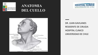
- 1. DR. JUAN GAVILANES RESIDENTE DE CIRUGIA HOSPITAL CLINICO UNIVERSIDAD DE CHILE ANATOMIA DEL CUELLO
- 2. GENERALIDADES • Entre la mandíbula y las clavículas • Estructuras vitales • Separa cabeza del tórax • Divisiones del cuello y su contenido • Capas del cuello y su implicancia quirúrgica
- 3. DIVISION • Límites • ECM y omohioideo y digástrico • Triángulo anterior • Muscular • Carotídeo • Submandibular • Submentoniano • Triángulo posterior • Occipital • Subclavio
- 4. TRIANGULO ANTERIOR • Bordes: • Borde anterior del ECM • Borde inferior de la mandíbula • Línea media del cuello • Hioides: omohioideo y músculo digástrico • Submandibular, carotídeo, muscular y submentoniano • Mentoniano impar • Suprahioides: • Milohoideo • Digástrico • Estilohioideo • Geniohioideo • Infrahioides: • Omohioideo • Esternohioideo • Esternotiroideo • Tirohioideo
- 5. TRIANGULO SUBMENTONIANO • Bordes: • Cuerpo de hioides • Digástrico anterior derecho e izquierdo • Piso: • Milohioideo • Techo: • Peso • Fascia cervical superficial c/ platisma • Fascia cervical profunda • Impar • Yugular anterior • Nódulos submentonianos
- 6. TRIANGULO SUBMANDIBULAR • Triángulo digástrico • Bordes: • Borde inferior de mandíbula • Vientre anterior y posterior del digástrico • Piso: • Hiogloso • Milohioideo • Contrictor medio • Techo: • Piel • Fascia superficial c/platisma • Fascia profunda
- 7. TRIANGULO SUBMANDIBULAR • Tres triángulos clínicamente significativos : • Lesser • Pirogov • Beclard • N. Hipogloso (superficial al hiogloso) y a. lingual (profundo al hiogloso) • Lesser: nervio hipogloso,digástrico (anterior y posterior) • Pirogov: n.hipogloso, tendón intermedio del digástrico y milohioideo • Beclard: Cuernos mayores del hioides, digástrico posterior, hiogloso
- 8. TRIANGULO SUBMANDIBULAR • Arterias y venas: • Facial • Submentoniana • Lingual • Nervios: • Milohioieo • Hipogloso • Lingual • Facial (ramas de la marginal y cervical) • Estructuras: • Glándula submandibular • Linfonodos submandibulares • Porción inferior de parótida
- 9. TRIANGULO CAROTIDEO • Tres carótidas dentro • Bordes: • Borde anterior de ECM • Digastrico posterior • Omohioideo Superior • Piso del triángulo: • Hiogloso • Tirohioideo • Constrictor medio • Constrictor inferior • Techo • Piel • Fascia superficial con platisma • Fascia profunda • Pareado
- 10. TRIANGULO CAROTIDEO • Contenido: • Arterias: • Carótida común (cuerpo carotídeo) • Carótida interna (seno carotídeo) • Carótida externa: • Tiroidea superior (n.laríngeo superior) • Lingual • Facial • Faringea ascendente • Occipital
- 11. TRIANGULO CAROTIDEO • Contenido: • Venas: • Yugular interna • Facial común • Tiroidea superior • Tiroidea media • Nervios: • Vago • Laringeo externo • Laringeo Superior • Espinal • Hipogloso • Asa cervical (asa superior) • Tronco simpático • Estructuras • Laringe y tiroides
- 12. TRIANGULO MUSCULAR • Bordes: • Borde anterior del ECM • Omohioideo superior • Línea media • Piso: • Esternohioideo • Esternotiroideo • Techo: • Piel • Fascia superficial con platisma • Fascia cervical profunda • Pareado
- 13. TRIANGULO MUSCULAR • Arteria: • Tiroidea superior • Vena: • Tiroidea superior • Tiroidea inferior • Yugular anterior • Nervios: • Asa cervical • Estructuras: • Músculo: • Esternohioideo • Esternotiroideo • Tirohioideo • Glándula tiroidea • Paratiroides • Laringe • Tráquea • Esófago • Liinfonodos
- 14. TRIANGULO POSTERIOR • Anterior: Borde posterior ECM • Posterior: Borde anterior de trapecio • Inferior: tercio medio de Clavícula • Techo: • Piel • Fascia superficial c/ platisma • Capa superficial de fascia cervical profunda • Piso: músculo semiespinoso de la cabeza, esplenio de la cabeza, escaleno posterior, medio y anterior
- 15. TRIANGULO POSTERIOR • Contenido: • Arterias: • 1/3 subclavia • Occipital • Suprascapular • Cervical transversa • Dorsal de la escápula • Venas • Yugular externa y tributarias • Occipital • Suprascapular • Cervical Transversa • Nervios: • Plexo cervical (sensoriales) • Occipitalmenor • Cervical transversa • Auricular mayor • Supraclavicular • Espinal • Escapular dorsal • Torácico largo • Supraescapular • Ramas del plexo braquial • Frénico
- 16. TRIANGULO POSTERIOR • OCCIPITAL: • Piso: esplenio de la cabeza, elevador de la escápula, escaleno medio • Techo: piel, fascia superficial, platisma • XI detrás del ECM hacia el trapecio • Nervios cutáneos en fascia profunda que lo cubre • SUPRACLAVICULAR: • Porción terminal de arteria subclavia • Plexo braquial • Ramas de tronco tirocervical • Tributarias cutáneas de yugular externa • Cúpula pleural
- 17. FASCIA • Fascia superficial • Fascia profunda -3 capas • Fascia superficial • Debajo de la piel • Tejido conectivo laxo, grasa, platisma, nervios y vasos sanguíneos • Nervios cutáneos y yugulares externas entre el platisma y fascia cervical profunda (no se retraen)
- 18. FASCIA • Fascia profunda: • Capa superficial o anterior • Envuelve dos músculos: trapecio y ECM y dos glándulas (parótidas y submaxilar) • Dos espacios: supraclavicular y supraesternal
- 19. FASCIA • Fascia profunda: • Capa media o pretraqueal: • Anterior: envuelve los músculos pretiroideos • Posterior: envuelve la glándula tiroides-cápsula falsa de la tiroides • Capa profunda o prevertebral: • Anterior a músculos prevertebrales • Cubre los músculos de la columna cervical • Se divide en frente de los cuerpos vertebrales: fascia alar (anterior) y fascia prevertebral (posterior)
- 20. FASCIA • Vaina carotídea • Fascia bucofaríngea • Posterior y lateral de faringe con capa alar de fascia prevertebral • Fascia axilar –originada en fascia prevertebral
- 23. APORTE VASCULAR SUBCLAVIO DEL CUELLO
- 24. APORTE VASCULAR SUBCLAVIO DEL CUELLO
- 25. APORTE VASCULAR SUBCLAVIO DEL CUELLO
- 26. APORTE VASCULAR SUBCLAVIO DEL CUELLO
- 27. APORTE VASCULAR CAROTIDEO DEL CUELLO
- 28. APORTE VASCULAR CAROTIDEO DEL CUELLO
- 29. APORTE VASCULAR CAROTIDEO DEL CUELLO
- 30. APORTE VASCULAR CAROTIDEO DEL CUELLO
- 31. APORTE VASCULAR CAROTIDEO DEL CUELLO
- 32. APORTE VASCULAR YUGULAR DEL CUELLO • Yugular interna • Occipital • Facial común • Facial • Lingual • Faríngea • Tiroidea superior • Tiroidea media • Subclavia • Vertebral • Yugular externa • Cervical transversa • Supraescapular • Yugular anterior • Braquiocefálico • Tiroidea inferior
- 33. APORTE VASCULAR YUGULAR DEL CUELLO
- 34. NIVELES CERVICALES • NIVEL I • SUPERIOR: MANDIBULA • INFERIOR: HIOIDES Y MUSCLO DIGASTRICO • IA: (Grupo submentoniano): entre los vientres anteriores de los dos músculos digástricos • IB (submandibular ) entre vientre anterior y posterior del digástrico: glándula submaxilar)
- 35. NIVELES CERVICALES • NIVEL II • Parte superior de VYI • SUPERIOR: Base del cráneo • INFERIOR: Bifurcación carotídea y linea horizontal a la altura de hueso hioides • ANTERIOR: estilohoideo • POSTERIOR: Borde anterosuperior del músculo trapecio • IIA: ganglios por delante del n. espinal • IIB: ganglios por detrás del n. espinal
- 36. NIVELES CERVICALES • NIVEL III • Parte media de VYI • SUPERIOR: Línea horizontal a la altura del hioides. • INFERIOR: Línea horizontal a la altura de cartílago cricoides • ANTERIOR: borde lateral del esternohioideo • POSTERIOR: Borde posterior de músculo ECM
- 37. NIVELES CERVICALES • NIVEL IV • Terco inferior de VYI • SUPERIOR: Línea horizontal a la altura de cartílago cricoides • INFERIOR: la clavícula • ANTERIOR: borde lateral del esternohioideo • POSTERIOR: Borde posterior de músculo ECM • IVa: nodos hacia la cabeza de la clavícula, la fascia de v. yugular y a. carótida. • IVb: nodos hacia cuerpo de clavícula por detrás de la yugular, a la altura del conducto torácico.
- 38. NIVELES CERVICALES • NIVEL V • Triángulo posterior del cuello • ANTERIOR: Borde posterior de músculo ECM • POSTERIOR:Borde anterior del trapecio • INFERIOR: la clavícula • Va: apex del triángulo cervical posterior; por encima de una línea horizontal a la altura del cricoides • Superior e inferior (hueso hioides) • Vb: debajo del plano horizontal, del borde inferior del cricoides. Desde el vientre posterior omohioideo hasta clavícula.
- 39. NIVELES CERVICALES • NIVEL VI • Zona central del cuello • Hipofaringe, laringe, traquea, esófago, NLRs, tiroides y paratiroides. • SUPERIOR: hioides • INFERIOR: tronco braquiocefálico o a. innominada • LATERALES: carótida • Nodos pretraqueales, paratraqueales, prelaringeos, peritiroideos y perirrecurrenciales
Notas del editor
- The superior laryngeal nerve has two branches: the internal laryngeal nerve pierces the thyrohyoid membrane and is sensory to the mucosa of pharynx and larynx from the level of epiglottis to vocal folds; the external laryngeal nerve is motor to cricothyroid.
- Diagrammatic cross section through the neck below the hyoid bone showing the layers of the deep cervical fascia and the structures that they envelop
- Diagrammatic cross section of the neck through the thyroid gland at the level of the sixth cervical vertebra showing the fascial planes, muscles, and vessels that may be encountered in an incision for thyroidectomy
- Diagrammatic cross section of the neck through the thyroid gland at the level of the sixth cervical vertebra showing the fascial planes, muscles, and vessels that may be encountered in an incision for thyroidectomy
- Diagrammatic cross section of the neck through the thyroid gland at the level of the sixth cervical vertebra showing the fascial planes, muscles, and vessels that may be encountered in an incision for thyroidectomy
- Diagrammatic cross section of the neck through the thyroid gland at the level of the sixth cervical vertebra showing the fascial planes, muscles, and vessels that may be encountered in an incision for thyroidectomy
- Diagrammatic cross section of the neck through the thyroid gland at the level of the sixth cervical vertebra showing the fascial planes, muscles, and vessels that may be encountered in an incision for thyroidectomy
- Diagrammatic cross section of the neck through the thyroid gland at the level of the sixth cervical vertebra showing the fascial planes, muscles, and vessels that may be encountered in an incision for thyroidectomy
- Diagrammatic cross section of the neck through the thyroid gland at the level of the sixth cervical vertebra showing the fascial planes, muscles, and vessels that may be encountered in an incision for thyroidectomy