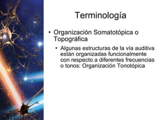
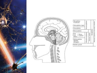
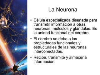
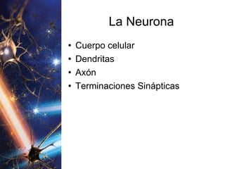
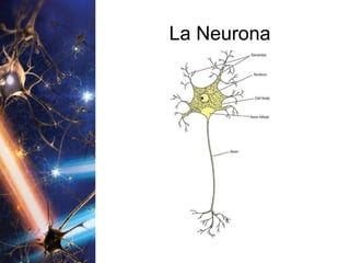
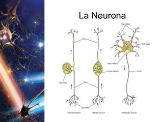
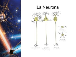
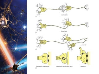
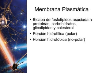
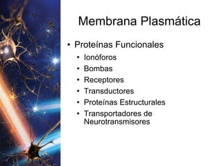
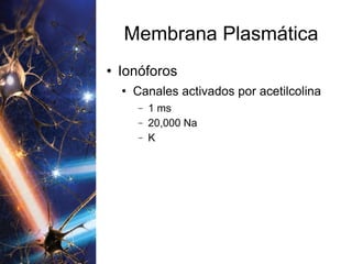
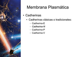
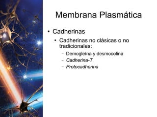
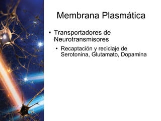
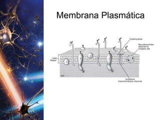
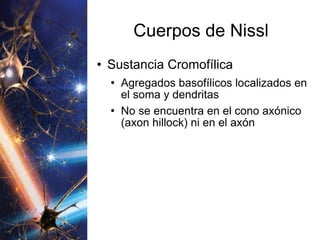
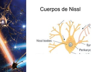
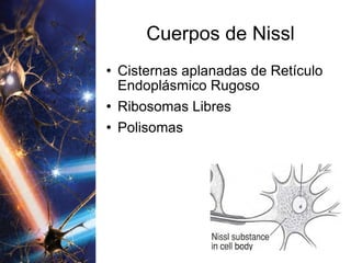
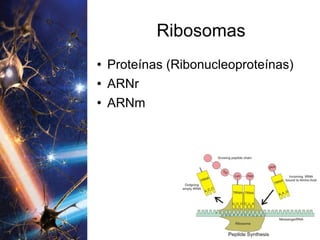
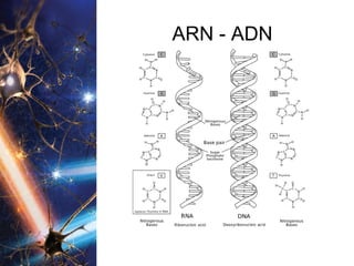
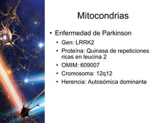
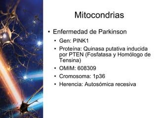
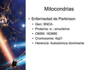
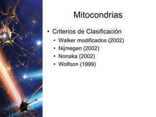
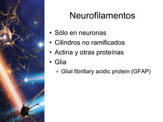
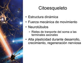
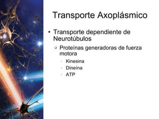
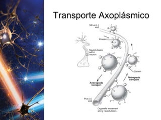
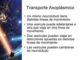
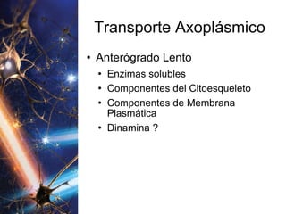
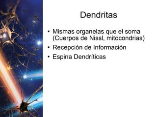
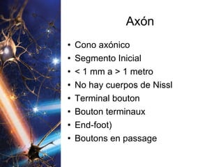
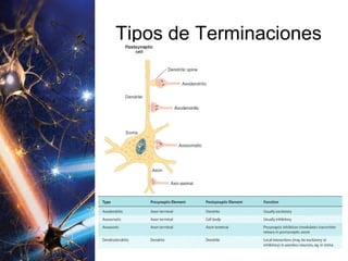
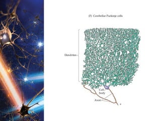
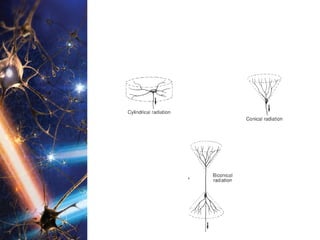
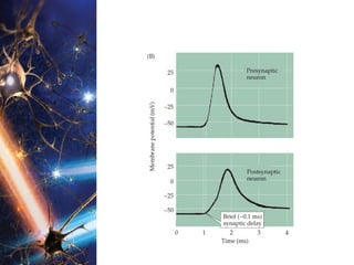
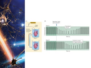
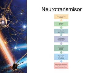
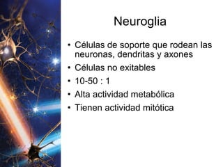
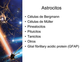
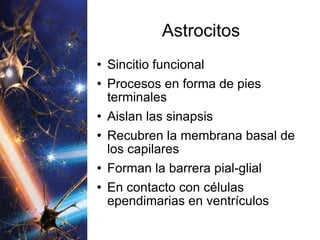
Introducción a la Neurología Dr. Edgar Avalos Herrera Neurólogo Internista
El Cerebro Humano Es lo que separa a los humanos del resto de las especies, permitiéndonos lograr maravillas como caminar sobre la luna y crear obras maestras de literatura, arte y música. (SfN)
El Cerebro Humano Ha sido comparado con una super-computadora, pero el cerebro es mucho más complicado, un hecho que los científicos confirma diariamente con cada nuevo descubrimiento.(SfN)
El Cerebro Humano La extensión de las capacidades del cerebro es desconocida, pero es la estructura viviente más compleja en todo el universo. (SfN)
El Cerebro Humano Cómo es que 100 billones de células nerviosas son producidas, crecen, se organizan y forman sistemas activos y funcionales.  De forma breve, el cerebro es lo que nos hace humanos. (SfN)
Divisiones Anatómica SN Central SN Periférico Funcional SN Somático SN Autonómico
Divisiones SN Periférico Nervios Craneales (Cerebro) Nervios Espinales (Médula Espinal)
Divisiones Señales SN Periférico Órganos de los Sentidos y Receptores Sensitivos hacia el SNC (AFERENTE) Del SNC hacia músculos y glándulas del cuerpo (EFERENTE)
Divisiones Señales SN Somático (SNP - SNC) Procesamiento y transmisión de informacion consciente e inconsciente. Visión, dolor, tacto, aferencias musculares inconscientes desde músculos de la cabeza, tronco y extremidades hacia el SNC
Divisiones Señales SN Somático (SNP - SNC) Eferencias para el control motor voluntario del músculo estriado
Divisiones Señales SN Autonómico (SNC - SNP) Procesamiento y transmisión de información sensitiva desde órganos viscerales (sistema digestivo, sistema cardiovascular, otros)
Divisiones Señales SN Autonómico (SNC - SNP) Control motor del músculo liso, músculo cardíaco y glándulas
Información La información sensitiva de receptores periféricos es transmitida a través del sistema nervioso por medio de  vías sensitivas Vías de dolor Vías de temperatura Vías visuales Otras
Información La información que llega al sistema nervioso central puede ser utilizada De forma consciente De forma inconsciente
Información La información nerviosa para la actividad motora es transmitida a través del sistema nervioso central hacia los músculos y glándulas por medio de  vías motoras
Información Cada vía sensitiva (aferente, ascendente) y motora (eferente, descendete) tiene centros de procesamiento (núcleos, ganglios, cortezas) a distintos niveles en la médula espinal y en el cerebro
Información Los centros de procesamiento de la información constituyen las computadoras de estas vías de alta velocidad
Información Desde los receptores periféricos hacia los centros más altos del cerebro las señales eléctricas siguen una misma secuencia básica:
Información Receptores Sensitivos  Fibras Nerviosas  Centros de Procesamiento en Médula Espinal y Cerebro  Fibras Nerviosas que ascienden de forma ipsilateral o se decusan  Centros Superiores de Procesamiento  Fibras ipsilaterales que ascienden y terminan en  Los Centros más altos en la Corteza Cerebral
Orientación El eje largo trazado a través de la médula espinal y el cerebro se denomina neuroeje, en forma de letra “T”
Orientación
Orientación
Terminología
Organización Sistema Nervioso Central Sustancia Gris Cuerpos Neuronales, Dendritas, Terminaciones Axónales, Sinapsis, Células Gliales, muy vascularizada Sustancia Blanca Axones, Células Gliales, sin neuronas excepto en los núcleos basales, menos vascularizada. Su color más blanco se debe a la mielina.
Terminología Comisuras Haces de fibras nerviosas que cruzan la línea media y que interconectan estructuras pares de ambos hemisferios cerebrales
Terminología Modalidad Cualidad de un estímulo, tipo de sensación transmitida (tacto, dolor, sonido, visión)
Terminología Organización Somatotópica o Topográfica Porciones específicas de ciertos tractos, núcleos, o áreas de la corteza cerebral asociadas con regiones restringidas y específicas del cuerpo
Terminología Organización Somatotópica o Topográfica Algunas estructuras de la vía visual están topográficamente relacionadas con regiones específicas de la retina: Organización Retinotópica
Terminología Organización Somatotópica o Topográfica Algunas estructuras de la vía auditiva están organizadas funcionalmente con respecto a diferentes frecuencias o tonos: Organización Tonotópica
 
La Neurona Célula especializada diseñada para transmitir información a otras neuronas, múlculos y glándulas. Es la unidad funcional del cerebro. El cerebro se debe a las propiedades funcionales y estructurales de las neuronas interconectadas. Recibe, transmite y almacena información
La Neurona Cuerpo celular Dendritas Axón Terminaciones Sinápticas
La Neurona
La Neurona
La Neurona


Núcleo Separado del citoplasma por una doble capa, la cubierta nuclear La membrana nuclear esta perforada por los poros nucleares La parte externa de la membrana se continúa con el retículo endoplásmico liso
Núcleo La parte interna de la membrana se une a la cromatina nuclear y con estructuras que controlan el diámetro de los poros nucleares
Núcleo Se sintetizan 3 clases de proteínas Sintetizadas en el citosol por ribosomas y polisomas, que permanecen en el citosol Distribuidas por transporte axoplásmico lento, incluidas enzimas esenciales para catalizar procesos metabólicos del citoesqueleto
Núcleo Se sintetizan 3 clases de proteínas Sintetizadas en el citosol por ribosomas y polisomas, que son incorporadas al núcleo, mitocondrias, peroxisomas Enzimas incluidas en la síntesis de ADN, ARN, factores de transcripción que regulan expresión genética
Núcleo Se sintetizan 3 clases de proteínas Sintetizadas en asociación con el retículo endoplásmico y el aparato de Golgi Distribuidas por transporte axonal rápido hacia organelas, hacia vesículas secretoras (neurotransmisores) y hacia la membrana plasmática para mantenimiento estructural
Nucléolo Maquinaria productora de ribosomas Constituido principalmente por ARN y menos ADN Sitio de ensamblaje inicial y producción de ARNr Bastante desarrollado en neuronas activamente productoras de péptidos y proteínas
Nucléolo El cerebro utiliza más genes que cualquier otro órgano del cuerpo 15,000 genes son únicos al tejido neural Neuronas femeninas Cuerpos de Barr
Núcleo
Membrana Plasmática Bicapa de fosfolípidos asociada a proteínas, carbohidratos, glicolípidos y colesterol Porción hidrofílica (polar) Porción hidrofóbica (no-polar)
Membrana Plasmática Proteínas intrínsecas o integrales Proteínas periféricas
Membrana Plasmática Reconocimiento Molecular Reconocimiento Celular Adhesión Celular
Membrana Plasmática Difusión de moléculas hidrosolubles Permeabilidad selectiva a ciertas moléculas Transducción
Membrana Plasmática Sodio, Potasio, Calcio, Cloro Oxígeno Nutrientes Desechos moleculares
Membrana Plasmática Transporte Pasivo Difusión Facilitada Transporte Activo Endocitosis Exocitosis
Membrana Plasmática Bomba Na-K ATPasa Integral ? Periférica ? Intrínseca ?
Membrana Plasmática Proteínas Funcionales Ionóforos Bombas Receptores Transductores Proteínas Estructurales Transportadores de Neurotransmisores
Membrana Plasmática Ionóforos Canales activados por acetilcolina 1 ms 20,000 Na K
Membrana Plasmática Bombas Iónicas A favor de gradiente ? Contra gradiente ?
Membrana Plasmática Receptores protéicos Hormonas Neurotransmisores
Membrana Plasmática Transductores Ligando Receptor Enzimas Segundos Mensajeros (AMPc)
Membrana Plasmática Proteínas Estructurales Moléculas de adhesión celular (MAC) MAC-N: Moléculas de adhesión celular neuronal Reconocimiento interneuronal Redes neuronales funcionales Migración neuronal Afinidad de terminaciones nerviosas Interacciones neurona-neurona neurona-glia
Membrana Plasmática MAC-N: Moléculas de adhesión celular neuronal Adherencia neuronal Interactúan con integrinas y cadherinas
Membrana Plasmática Integrinas Crecimiento celular. División celular. Supervivencia celular. Diferenciación celular. Apoptosis.
Membrana Plasmática Cadherinas Cadherinas clásicas o tradicionales:  Cadherina-E  Cadherina-N Cadherina-P Cadherina-V
Membrana Plasmática Cadherinas Cadherinas no clásicas o no tradicionales:  Demogleína y desmocolina Cadherina-T Protocadherina
Membrana Plasmática Transportadores de Neurotransmisores Recaptación y reciclaje de Serotonina, Glutamato, Dopamina
Membrana Plasmática
Cuerpos de Nissl Sustancia Cromofílica Agregados basofílicos localizados en el soma y dendritas No se encuentra en el cono axónico (axon hillock) ni en el axón
Cuerpos de Nissl
Cuerpos de Nissl Cisternas aplanadas de Retículo Endoplásmico Rugoso Ribosomas Libres Polisomas
Ribosomas Proteínas (Ribonucleoproteínas) ARNr ARNm
ARN - ADN
Tipos de ARN ARNr ARNm ARNt ARNi ARNmt ARN antisentido ARNnc largo Riboswitch
Codones UAG, UAA, UGA AUG
RER Proteínas Neurosecretoras Proteínas Integrales de la Membrana Plasmática Proteínas Lisosomales Ribosomas libres y polisomas Proteínas citosólicas Proteínas no integrales de la membrana plasmática
REL Triglicéridos Colesterol Esteroides
Mitocondrias Plantas de Energía de las neuronas Energía (ATP), Agua, CO2
Mitocondrias Fosforilación Oxidativa - Complejos I a V ?
Mitocondrias Deleciones y Duplicaciones esporádicas de ADNmt Síndrome de Kearns-Sayre Síndrome de Pearson Oftalmoplejía Externa Crónica Progresiva Esporádica Diabetes y Sordera Oftalmoplejía Externa Crónica Progresiva de herencia materna
Mitocondrias Mutaciones Puntuales de ADNmt Polipéptidos Encefalomiopatía mitocondrial con acidosis láctica y episodios parecidos a eventos cerebrovasculares ( Mitochondrial encephalomyopathy with lactic acidosis and stroke-like episodes - MELAS )
Mitocondrias Mutaciones Puntuales de ADNmt Polipéptidos Intolerancia al ejercicio Miopatía aislada Neuropatía óptica hereditaria de Leber Síndrome de Neuropatía Ataxia y Retinitis Pigmentosa Síndrome de Leigh
Mitocondrias Mutaciones Puntuales de ADNmt ARNt mitocondrial MELAS Epilepsia Mioclónica con Fibras Rojas Rasgadas ( Myoclonic epilepsy with ragged-red fibers MERRF )  Cardiomiopatía-Miopatía Oftalmoplejía Externa Crónica Progresiva Miopatía aislada
Mitocondrias Mutaciones Puntuales de ADNmt ARNt mitocondrial Diabetes y Sordera Sordera Neurosensorial Cardiomiopatía Hipertrófica Tubulopatía
Mitocondrias Mutaciones Puntuales de ADNmt ARNr mitocondrial Sordera no sindrómica inducida por aminoglucósidos Cardiomiopatía Hipertrófica
Mitocondrias Enfermedad de Alzheimer Gen: APP Proteína: Precursor Proteína Amiloide beta A4 OMIM: 104760 Cromosoma: 21q21 Herencia: Autosómica dominante
Mitocondrias Enfermedad de Huntington Gen: IT-15 Proteína: Proteína Huntingtina OMIM: 143100 Cromosoma: 4p16.3 Herencia: Autosómica dominante
Mitocondrias Enfermedad de Parkinson Gen: HTRA2 Proteína: PRSS25 OMIM: 606441 Cromosoma: 2p12 Herencia: Esporádica
Mitocondrias Enfermedad de Parkinson Gen: LRRK2 Proteína: Quinasa de repeticiones ricas en leucina 2 OMIM: 609007 Cromosoma: 12q12 Herencia: Autosómica dominante
Mitocondrias Enfermedad de Parkinson Gen: PARK2 Proteína: Parkina OMIM: 602544 Cromosoma: 6q25.2-q27 Herencia: Autosómica recesiva
Mitocondrias Enfermedad de Parkinson Gen: PARK7 Proteína: DJ-1 OMIM: 606324 Cromosoma: 1p36 Herencia: Autosómica recesiva
Mitocondrias Enfermedad de Parkinson Gen: PINK1 Proteína: Quinasa putativa inducida por PTEN (Fosfatasa y Homólogo de Tensina) OMIM: 608309 Cromosoma: 1p36 Herencia: Autosómica recesiva
Mitocondrias Enfermedad de Parkinson Gen: SNCA Proteína:  α  - sinucleína OMIM: 163890 Cromosoma: 4q21 Herencia: Autosómica dominante
Mitocondrias Criterios de Clasificación Walker modificados (2002) Nijmegen (2002) Nonaka (2002) Wolfson (1999)
Peroxisomas Detoxificación Neuroprotección del H2O2
Peroxisomas Síndrome de Zellweger Adrenoleucodistrofia  Neonatal Enfermedad de Refsum Condrodisplasia punctata rizomélica
Lisosomas Sistema digestivo intracelular Enzimas hidrolíticas Gránulos amarillos de lipofuscina
Lisosomas Mucopolisacaridosis Síndrome de Hurler-Scheie Síndrome de Hunter Síndrome de Sanfilippo (A-D) Síndrome de Morquio (A-B) Síndrome de Maroteaux-Lamy Síndrome de Sly Síndrome de Natowicz Síndrome de Austin
Lisosomas Esfingolipidosis Enfermedad de Fabry Lipogranulomatosis de Farber Enfermedad de Gaucher Leucodistrofia de Células Globoides (Krabbe) Leucodistrofia  Metacromática Niemann–Pick (A-B) Gangliosidosis GM1
Lisosomas Esfingolipidosis Gangliosidosis GM1 Gangliosidosis GM2 (Tay–Sachs) Gangliosidosis GM3 (Sandhoff)
Lisosomas Oligosacaridosis y glucoproteinosis Aspartilglucosaminuria Fucosidosis α- Mannosidosis β- Mannosidosis Sialidosis Enfermedad de Schindler
Lisosomas Glucogenosis Enfermedad de Pompe
Aparato de Golgi Cisternas Vesículas Túbulos membranosos
Aparato de Golgi Proteínas integrales de membrana plasmática Proteínas empacadas en vesículas secretoras liberadas en respuesta a señales Enzimas lisosomales
Citoesqueleto Neurotúbulos Neurofilamentos Microfilamentos de actina
Neurotúbulos Cilindros no ramificados Polímeros de tubulina Transportan organelas por toda la neurona
Neurofilamentos Sólo en neuronas Cilindros no ramificados Actina y otras proteínas Glia Glial fibrillary acidic protein (GFAP)
Citoesqueleto Estructura dinámica Fuerza mecánica de movimiento Neurotúbulos Rieles de tranporte del soma a las terminales axonales Alta plasticidad durante desarrollo, crecimiento, regeneración nerviosa
Transporte Axoplásmico Transporte Axonal Anterógrado u Ortógrado Del soma hacia las dendritas o el axón Transporte Axonal Retrógrado De las dendritas y el axón hacia el cuerpo neuronal
Transporte Axoplásmico Rápido 200 – 400 mm al día Anterógrado y retrógrado Lento 1 – 5 mm al día Anterógrado
Transporte Axoplásmico Anterógrado Rápido Mitocondrias Precursores de REL Precursores de Vesículas Sinápticas Precursores de Membrana Plasmática
Transporte Axoplásmico Retrógrado Rápido Mitocondrias Cuerpos multivesiculados (degradación) Vesículas que contienen ligandos (factores de crecimiento neural) captados por endocitosis
Transporte Axoplásmico Transporte dependiente de Neurotúbulos Proteínas generadoras de fuerza motora Kinesina Dineína ATP
Transporte Axoplásmico
Transporte Axoplásmico Un mismo neurotúbulo  tiene distintas líneas de movimiento Una vesícula puede adelantarse a otra que viaja en otra línea de movimiento Dos vesículas pueden viajar en direcciones opuestas en distintas líneas de movimiento Las vesículas pueden cambiarse de neurotúbulo
Transporte Axoplásmico Anterógrado Lento Enzimas solubles Componentes del Citoesqueleto Componentes de Membrana Plasmática Dinamina ?
Dendritas Mismas organelas que el soma (Cuerpos de Nissl, mitocondrias) Recepción de Información Espina Dendríticas
Axón Cono axónico Segmento Inicial < 1 mm a > 1 metro No hay cuerpos de Nissl Terminal bouton Bouton terminaux End-foot) Boutons en passage
Tipos de Terminaciones
Neurópilo Axones Dendritas Sinapsis Neuroglia Sustancia gris


Sinapsis Sherrington “ Doctrina de la Neurona” Santiago Ramón y Cajal “ Teoría Reticular” Golgi
Sinapsis Conexión interneuronal Hendidura sináptica (200 A) De pocas a miles de sinapsis (axón) Hasta 15,000 sinapsis (dendritas, neurona)
Sinapsis Membrana Presináptica Membrana Postsináptica Membrana Subsináptica
Sinapsis
Sinapsis Mecanismo de Transmisión Químicas Canal iónico y Receptor Canal iónico receptor Eléctricas


Sinapsis Químicas Neurotransmisores Neuromoduladores
Sinapsis Químicas Una Neurona = Un Neurotransmisor Neurotransmisor + Co-transmisor
Neurotransmisor Presente en la terminal presináptica Liberado en respuesta a la despolarización presináptica, liberación calcio-dependiente Receptores específicos en la membrana postsináptica
Neurotransmisor
Neuroglia Células de soporte que rodean las neuronas, dendritas y axones Células no exitables 10-50 : 1 Alta actividad metabólica Tienen actividad mitótica
Neuroglia
Astrocitos Funcional y morfológicamente heterogéneos Rodean a todas la neuronas Protoplásmicos (Sustancia Gris) Fibrosos (Sustancia Blanca)
Astrocitos Células de Bergmann Células de Müller Pinealocitos Pituicitos Tanicitos Otros Glial fibrillary acidic protein (GFAP)
Astrocitos Sincitio funcional Procesos en forma de pies terminales Aislan las sinapsis Recubren la membrana basal de los capilares Forman la barrera pial-glial En contacto con células ependimarias en ventrículos
Astrocitos Almacenan y transfieren metabolitos como glucosa que llevan de los capilares a las neuronas Eliminan es exceso de K del espacio extracelular Eliminan el exceso de glutamato Eliminan neurotoxinas
Astrocitos Spatial Buffering Spreading Depression Neurotrofinas
Astrocitos
Oligodendrocitos Producción de mielina en SNC Mantenimiento estructural de mielina
Oligodendrocitos Células satélite perineuronales Células interfasciculares
Oligodendrocitos
Oligodendrocitos Mielinización de internodos en hasta 70 axones Mayoría de vías centrales no se mielinizan hasta los 2 años Remielinización espontánea
Microglia Células microgliales en reposo Microglia activada o reactiva no fagocítica Microglia fagocítica
Microglia Activada por Cuerpos extraños Lesión cerebral Productos de degradación Inflamación
Microglia Sintetizan Factores de crecimiento Moléculas de adhesión Citoquinas
Microglia Interacción inmune SNC-SNP Vigilancia inmunológica de cerebro y médula espinal
Epéndimo Canal central de la médula espinal Paredes ventriculares Junto a los astrocitos forman la barrera LCR-Cerebro Tanicitos Transporte entre ventrículos y vasos sanguíneos

Introducción a la Neurología

  • 1.
    Introducción a laNeurología Dr. Edgar Avalos Herrera Neurólogo Internista
  • 2.
    El Cerebro HumanoEs lo que separa a los humanos del resto de las especies, permitiéndonos lograr maravillas como caminar sobre la luna y crear obras maestras de literatura, arte y música. (SfN)
  • 3.
    El Cerebro HumanoHa sido comparado con una super-computadora, pero el cerebro es mucho más complicado, un hecho que los científicos confirma diariamente con cada nuevo descubrimiento.(SfN)
  • 4.
    El Cerebro HumanoLa extensión de las capacidades del cerebro es desconocida, pero es la estructura viviente más compleja en todo el universo. (SfN)
  • 5.
    El Cerebro HumanoCómo es que 100 billones de células nerviosas son producidas, crecen, se organizan y forman sistemas activos y funcionales. De forma breve, el cerebro es lo que nos hace humanos. (SfN)
  • 6.
    Divisiones Anatómica SNCentral SN Periférico Funcional SN Somático SN Autonómico
  • 7.
    Divisiones SN PeriféricoNervios Craneales (Cerebro) Nervios Espinales (Médula Espinal)
  • 8.
    Divisiones Señales SNPeriférico Órganos de los Sentidos y Receptores Sensitivos hacia el SNC (AFERENTE) Del SNC hacia músculos y glándulas del cuerpo (EFERENTE)
  • 9.
    Divisiones Señales SNSomático (SNP - SNC) Procesamiento y transmisión de informacion consciente e inconsciente. Visión, dolor, tacto, aferencias musculares inconscientes desde músculos de la cabeza, tronco y extremidades hacia el SNC
  • 10.
    Divisiones Señales SNSomático (SNP - SNC) Eferencias para el control motor voluntario del músculo estriado
  • 11.
    Divisiones Señales SNAutonómico (SNC - SNP) Procesamiento y transmisión de información sensitiva desde órganos viscerales (sistema digestivo, sistema cardiovascular, otros)
  • 12.
    Divisiones Señales SNAutonómico (SNC - SNP) Control motor del músculo liso, músculo cardíaco y glándulas
  • 13.
    Información La informaciónsensitiva de receptores periféricos es transmitida a través del sistema nervioso por medio de vías sensitivas Vías de dolor Vías de temperatura Vías visuales Otras
  • 14.
    Información La informaciónque llega al sistema nervioso central puede ser utilizada De forma consciente De forma inconsciente
  • 15.
    Información La informaciónnerviosa para la actividad motora es transmitida a través del sistema nervioso central hacia los músculos y glándulas por medio de vías motoras
  • 16.
    Información Cada víasensitiva (aferente, ascendente) y motora (eferente, descendete) tiene centros de procesamiento (núcleos, ganglios, cortezas) a distintos niveles en la médula espinal y en el cerebro
  • 17.
    Información Los centrosde procesamiento de la información constituyen las computadoras de estas vías de alta velocidad
  • 18.
    Información Desde losreceptores periféricos hacia los centros más altos del cerebro las señales eléctricas siguen una misma secuencia básica:
  • 19.
    Información Receptores Sensitivos Fibras Nerviosas Centros de Procesamiento en Médula Espinal y Cerebro Fibras Nerviosas que ascienden de forma ipsilateral o se decusan Centros Superiores de Procesamiento Fibras ipsilaterales que ascienden y terminan en Los Centros más altos en la Corteza Cerebral
  • 20.
    Orientación El ejelargo trazado a través de la médula espinal y el cerebro se denomina neuroeje, en forma de letra “T”
  • 21.
  • 22.
  • 23.
  • 24.
    Organización Sistema NerviosoCentral Sustancia Gris Cuerpos Neuronales, Dendritas, Terminaciones Axónales, Sinapsis, Células Gliales, muy vascularizada Sustancia Blanca Axones, Células Gliales, sin neuronas excepto en los núcleos basales, menos vascularizada. Su color más blanco se debe a la mielina.
  • 25.
    Terminología Comisuras Hacesde fibras nerviosas que cruzan la línea media y que interconectan estructuras pares de ambos hemisferios cerebrales
  • 26.
    Terminología Modalidad Cualidadde un estímulo, tipo de sensación transmitida (tacto, dolor, sonido, visión)
  • 27.
    Terminología Organización Somatotópicao Topográfica Porciones específicas de ciertos tractos, núcleos, o áreas de la corteza cerebral asociadas con regiones restringidas y específicas del cuerpo
  • 28.
    Terminología Organización Somatotópicao Topográfica Algunas estructuras de la vía visual están topográficamente relacionadas con regiones específicas de la retina: Organización Retinotópica
  • 29.
    Terminología Organización Somatotópicao Topográfica Algunas estructuras de la vía auditiva están organizadas funcionalmente con respecto a diferentes frecuencias o tonos: Organización Tonotópica
  • 30.
  • 31.
    La Neurona Célulaespecializada diseñada para transmitir información a otras neuronas, múlculos y glándulas. Es la unidad funcional del cerebro. El cerebro se debe a las propiedades funcionales y estructurales de las neuronas interconectadas. Recibe, transmite y almacena información
  • 32.
    La Neurona Cuerpocelular Dendritas Axón Terminaciones Sinápticas
  • 33.
  • 34.
  • 35.
  • 36.
  • 37.
  • 38.
  • 39.
  • 40.
  • 41.
  • 42.
    Núcleo Separado delcitoplasma por una doble capa, la cubierta nuclear La membrana nuclear esta perforada por los poros nucleares La parte externa de la membrana se continúa con el retículo endoplásmico liso
  • 43.
    Núcleo La parteinterna de la membrana se une a la cromatina nuclear y con estructuras que controlan el diámetro de los poros nucleares
  • 44.
    Núcleo Se sintetizan3 clases de proteínas Sintetizadas en el citosol por ribosomas y polisomas, que permanecen en el citosol Distribuidas por transporte axoplásmico lento, incluidas enzimas esenciales para catalizar procesos metabólicos del citoesqueleto
  • 45.
    Núcleo Se sintetizan3 clases de proteínas Sintetizadas en el citosol por ribosomas y polisomas, que son incorporadas al núcleo, mitocondrias, peroxisomas Enzimas incluidas en la síntesis de ADN, ARN, factores de transcripción que regulan expresión genética
  • 46.
    Núcleo Se sintetizan3 clases de proteínas Sintetizadas en asociación con el retículo endoplásmico y el aparato de Golgi Distribuidas por transporte axonal rápido hacia organelas, hacia vesículas secretoras (neurotransmisores) y hacia la membrana plasmática para mantenimiento estructural
  • 47.
    Nucléolo Maquinaria productorade ribosomas Constituido principalmente por ARN y menos ADN Sitio de ensamblaje inicial y producción de ARNr Bastante desarrollado en neuronas activamente productoras de péptidos y proteínas
  • 48.
    Nucléolo El cerebroutiliza más genes que cualquier otro órgano del cuerpo 15,000 genes son únicos al tejido neural Neuronas femeninas Cuerpos de Barr
  • 49.
  • 50.
    Membrana Plasmática Bicapade fosfolípidos asociada a proteínas, carbohidratos, glicolípidos y colesterol Porción hidrofílica (polar) Porción hidrofóbica (no-polar)
  • 51.
    Membrana Plasmática Proteínasintrínsecas o integrales Proteínas periféricas
  • 52.
    Membrana Plasmática ReconocimientoMolecular Reconocimiento Celular Adhesión Celular
  • 53.
    Membrana Plasmática Difusiónde moléculas hidrosolubles Permeabilidad selectiva a ciertas moléculas Transducción
  • 54.
    Membrana Plasmática Sodio,Potasio, Calcio, Cloro Oxígeno Nutrientes Desechos moleculares
  • 55.
    Membrana Plasmática TransportePasivo Difusión Facilitada Transporte Activo Endocitosis Exocitosis
  • 56.
    Membrana Plasmática BombaNa-K ATPasa Integral ? Periférica ? Intrínseca ?
  • 57.
    Membrana Plasmática BombaNa-K ATPasa Integral ? Periférica ? Intrínseca ?
  • 58.
    Membrana Plasmática ProteínasFuncionales Ionóforos Bombas Receptores Transductores Proteínas Estructurales Transportadores de Neurotransmisores
  • 59.
    Membrana Plasmática IonóforosCanales activados por acetilcolina 1 ms 20,000 Na K
  • 60.
    Membrana Plasmática BombasIónicas A favor de gradiente ? Contra gradiente ?
  • 61.
    Membrana Plasmática Receptoresprotéicos Hormonas Neurotransmisores
  • 62.
    Membrana Plasmática TransductoresLigando Receptor Enzimas Segundos Mensajeros (AMPc)
  • 63.
    Membrana Plasmática ProteínasEstructurales Moléculas de adhesión celular (MAC) MAC-N: Moléculas de adhesión celular neuronal Reconocimiento interneuronal Redes neuronales funcionales Migración neuronal Afinidad de terminaciones nerviosas Interacciones neurona-neurona neurona-glia
  • 64.
    Membrana Plasmática MAC-N:Moléculas de adhesión celular neuronal Adherencia neuronal Interactúan con integrinas y cadherinas
  • 65.
    Membrana Plasmática IntegrinasCrecimiento celular. División celular. Supervivencia celular. Diferenciación celular. Apoptosis.
  • 66.
    Membrana Plasmática CadherinasCadherinas clásicas o tradicionales: Cadherina-E Cadherina-N Cadherina-P Cadherina-V
  • 67.
    Membrana Plasmática CadherinasCadherinas no clásicas o no tradicionales: Demogleína y desmocolina Cadherina-T Protocadherina
  • 68.
    Membrana Plasmática Transportadoresde Neurotransmisores Recaptación y reciclaje de Serotonina, Glutamato, Dopamina
  • 69.
  • 70.
    Cuerpos de NisslSustancia Cromofílica Agregados basofílicos localizados en el soma y dendritas No se encuentra en el cono axónico (axon hillock) ni en el axón
  • 71.
  • 72.
    Cuerpos de NisslCisternas aplanadas de Retículo Endoplásmico Rugoso Ribosomas Libres Polisomas
  • 73.
  • 74.
  • 75.
    Tipos de ARNARNr ARNm ARNt ARNi ARNmt ARN antisentido ARNnc largo Riboswitch
  • 76.
  • 77.
    RER Proteínas NeurosecretorasProteínas Integrales de la Membrana Plasmática Proteínas Lisosomales Ribosomas libres y polisomas Proteínas citosólicas Proteínas no integrales de la membrana plasmática
  • 78.
  • 79.
    Mitocondrias Plantas deEnergía de las neuronas Energía (ATP), Agua, CO2
  • 80.
  • 81.
    Mitocondrias Deleciones yDuplicaciones esporádicas de ADNmt Síndrome de Kearns-Sayre Síndrome de Pearson Oftalmoplejía Externa Crónica Progresiva Esporádica Diabetes y Sordera Oftalmoplejía Externa Crónica Progresiva de herencia materna
  • 82.
    Mitocondrias Mutaciones Puntualesde ADNmt Polipéptidos Encefalomiopatía mitocondrial con acidosis láctica y episodios parecidos a eventos cerebrovasculares ( Mitochondrial encephalomyopathy with lactic acidosis and stroke-like episodes - MELAS )
  • 83.
    Mitocondrias Mutaciones Puntualesde ADNmt Polipéptidos Intolerancia al ejercicio Miopatía aislada Neuropatía óptica hereditaria de Leber Síndrome de Neuropatía Ataxia y Retinitis Pigmentosa Síndrome de Leigh
  • 84.
    Mitocondrias Mutaciones Puntualesde ADNmt ARNt mitocondrial MELAS Epilepsia Mioclónica con Fibras Rojas Rasgadas ( Myoclonic epilepsy with ragged-red fibers MERRF ) Cardiomiopatía-Miopatía Oftalmoplejía Externa Crónica Progresiva Miopatía aislada
  • 85.
    Mitocondrias Mutaciones Puntualesde ADNmt ARNt mitocondrial Diabetes y Sordera Sordera Neurosensorial Cardiomiopatía Hipertrófica Tubulopatía
  • 86.
    Mitocondrias Mutaciones Puntualesde ADNmt ARNr mitocondrial Sordera no sindrómica inducida por aminoglucósidos Cardiomiopatía Hipertrófica
  • 87.
    Mitocondrias Enfermedad deAlzheimer Gen: APP Proteína: Precursor Proteína Amiloide beta A4 OMIM: 104760 Cromosoma: 21q21 Herencia: Autosómica dominante
  • 88.
    Mitocondrias Enfermedad deHuntington Gen: IT-15 Proteína: Proteína Huntingtina OMIM: 143100 Cromosoma: 4p16.3 Herencia: Autosómica dominante
  • 89.
    Mitocondrias Enfermedad deParkinson Gen: HTRA2 Proteína: PRSS25 OMIM: 606441 Cromosoma: 2p12 Herencia: Esporádica
  • 90.
    Mitocondrias Enfermedad deParkinson Gen: LRRK2 Proteína: Quinasa de repeticiones ricas en leucina 2 OMIM: 609007 Cromosoma: 12q12 Herencia: Autosómica dominante
  • 91.
    Mitocondrias Enfermedad deParkinson Gen: PARK2 Proteína: Parkina OMIM: 602544 Cromosoma: 6q25.2-q27 Herencia: Autosómica recesiva
  • 92.
    Mitocondrias Enfermedad deParkinson Gen: PARK7 Proteína: DJ-1 OMIM: 606324 Cromosoma: 1p36 Herencia: Autosómica recesiva
  • 93.
    Mitocondrias Enfermedad deParkinson Gen: PINK1 Proteína: Quinasa putativa inducida por PTEN (Fosfatasa y Homólogo de Tensina) OMIM: 608309 Cromosoma: 1p36 Herencia: Autosómica recesiva
  • 94.
    Mitocondrias Enfermedad deParkinson Gen: SNCA Proteína: α - sinucleína OMIM: 163890 Cromosoma: 4q21 Herencia: Autosómica dominante
  • 95.
    Mitocondrias Criterios deClasificación Walker modificados (2002) Nijmegen (2002) Nonaka (2002) Wolfson (1999)
  • 96.
  • 97.
    Peroxisomas Síndrome deZellweger Adrenoleucodistrofia Neonatal Enfermedad de Refsum Condrodisplasia punctata rizomélica
  • 98.
    Lisosomas Sistema digestivointracelular Enzimas hidrolíticas Gránulos amarillos de lipofuscina
  • 99.
    Lisosomas Mucopolisacaridosis Síndromede Hurler-Scheie Síndrome de Hunter Síndrome de Sanfilippo (A-D) Síndrome de Morquio (A-B) Síndrome de Maroteaux-Lamy Síndrome de Sly Síndrome de Natowicz Síndrome de Austin
  • 100.
    Lisosomas Esfingolipidosis Enfermedadde Fabry Lipogranulomatosis de Farber Enfermedad de Gaucher Leucodistrofia de Células Globoides (Krabbe) Leucodistrofia Metacromática Niemann–Pick (A-B) Gangliosidosis GM1
  • 101.
    Lisosomas Esfingolipidosis GangliosidosisGM1 Gangliosidosis GM2 (Tay–Sachs) Gangliosidosis GM3 (Sandhoff)
  • 102.
    Lisosomas Oligosacaridosis yglucoproteinosis Aspartilglucosaminuria Fucosidosis α- Mannosidosis β- Mannosidosis Sialidosis Enfermedad de Schindler
  • 103.
  • 104.
    Aparato de GolgiCisternas Vesículas Túbulos membranosos
  • 105.
    Aparato de GolgiProteínas integrales de membrana plasmática Proteínas empacadas en vesículas secretoras liberadas en respuesta a señales Enzimas lisosomales
  • 106.
  • 107.
    Neurotúbulos Cilindros noramificados Polímeros de tubulina Transportan organelas por toda la neurona
  • 108.
    Neurofilamentos Sólo enneuronas Cilindros no ramificados Actina y otras proteínas Glia Glial fibrillary acidic protein (GFAP)
  • 109.
    Citoesqueleto Estructura dinámicaFuerza mecánica de movimiento Neurotúbulos Rieles de tranporte del soma a las terminales axonales Alta plasticidad durante desarrollo, crecimiento, regeneración nerviosa
  • 110.
    Transporte Axoplásmico TransporteAxonal Anterógrado u Ortógrado Del soma hacia las dendritas o el axón Transporte Axonal Retrógrado De las dendritas y el axón hacia el cuerpo neuronal
  • 111.
    Transporte Axoplásmico Rápido200 – 400 mm al día Anterógrado y retrógrado Lento 1 – 5 mm al día Anterógrado
  • 112.
    Transporte Axoplásmico AnterógradoRápido Mitocondrias Precursores de REL Precursores de Vesículas Sinápticas Precursores de Membrana Plasmática
  • 113.
    Transporte Axoplásmico RetrógradoRápido Mitocondrias Cuerpos multivesiculados (degradación) Vesículas que contienen ligandos (factores de crecimiento neural) captados por endocitosis
  • 114.
    Transporte Axoplásmico Transportedependiente de Neurotúbulos Proteínas generadoras de fuerza motora Kinesina Dineína ATP
  • 115.
  • 116.
    Transporte Axoplásmico Unmismo neurotúbulo tiene distintas líneas de movimiento Una vesícula puede adelantarse a otra que viaja en otra línea de movimiento Dos vesículas pueden viajar en direcciones opuestas en distintas líneas de movimiento Las vesículas pueden cambiarse de neurotúbulo
  • 117.
    Transporte Axoplásmico AnterógradoLento Enzimas solubles Componentes del Citoesqueleto Componentes de Membrana Plasmática Dinamina ?
  • 118.
    Dendritas Mismas organelasque el soma (Cuerpos de Nissl, mitocondrias) Recepción de Información Espina Dendríticas
  • 119.
    Axón Cono axónicoSegmento Inicial < 1 mm a > 1 metro No hay cuerpos de Nissl Terminal bouton Bouton terminaux End-foot) Boutons en passage
  • 120.
  • 121.
    Neurópilo Axones DendritasSinapsis Neuroglia Sustancia gris
  • 122.
  • 123.
  • 124.
  • 125.
  • 126.
  • 127.
  • 128.
  • 129.
  • 130.
    Sinapsis Sherrington “Doctrina de la Neurona” Santiago Ramón y Cajal “ Teoría Reticular” Golgi
  • 131.
    Sinapsis Conexión interneuronalHendidura sináptica (200 A) De pocas a miles de sinapsis (axón) Hasta 15,000 sinapsis (dendritas, neurona)
  • 132.
    Sinapsis Membrana PresinápticaMembrana Postsináptica Membrana Subsináptica
  • 133.
  • 134.
    Sinapsis Mecanismo deTransmisión Químicas Canal iónico y Receptor Canal iónico receptor Eléctricas
  • 135.
  • 136.
  • 137.
  • 138.
  • 139.
  • 140.
  • 141.
    Sinapsis Químicas UnaNeurona = Un Neurotransmisor Neurotransmisor + Co-transmisor
  • 142.
    Neurotransmisor Presente enla terminal presináptica Liberado en respuesta a la despolarización presináptica, liberación calcio-dependiente Receptores específicos en la membrana postsináptica
  • 143.
  • 144.
    Neuroglia Células desoporte que rodean las neuronas, dendritas y axones Células no exitables 10-50 : 1 Alta actividad metabólica Tienen actividad mitótica
  • 145.
  • 146.
    Astrocitos Funcional ymorfológicamente heterogéneos Rodean a todas la neuronas Protoplásmicos (Sustancia Gris) Fibrosos (Sustancia Blanca)
  • 147.
    Astrocitos Células deBergmann Células de Müller Pinealocitos Pituicitos Tanicitos Otros Glial fibrillary acidic protein (GFAP)
  • 148.
    Astrocitos Sincitio funcionalProcesos en forma de pies terminales Aislan las sinapsis Recubren la membrana basal de los capilares Forman la barrera pial-glial En contacto con células ependimarias en ventrículos
  • 149.
    Astrocitos Almacenan ytransfieren metabolitos como glucosa que llevan de los capilares a las neuronas Eliminan es exceso de K del espacio extracelular Eliminan el exceso de glutamato Eliminan neurotoxinas
  • 150.
    Astrocitos Spatial BufferingSpreading Depression Neurotrofinas
  • 151.
  • 152.
    Oligodendrocitos Producción demielina en SNC Mantenimiento estructural de mielina
  • 153.
    Oligodendrocitos Células satéliteperineuronales Células interfasciculares
  • 154.
  • 155.
    Oligodendrocitos Mielinización deinternodos en hasta 70 axones Mayoría de vías centrales no se mielinizan hasta los 2 años Remielinización espontánea
  • 156.
    Microglia Células microglialesen reposo Microglia activada o reactiva no fagocítica Microglia fagocítica
  • 157.
    Microglia Activada porCuerpos extraños Lesión cerebral Productos de degradación Inflamación
  • 158.
    Microglia Sintetizan Factoresde crecimiento Moléculas de adhesión Citoquinas
  • 159.
    Microglia Interacción inmuneSNC-SNP Vigilancia inmunológica de cerebro y médula espinal
  • 160.
    Epéndimo Canal centralde la médula espinal Paredes ventriculares Junto a los astrocitos forman la barrera LCR-Cerebro Tanicitos Transporte entre ventrículos y vasos sanguíneos

Notas del editor

  • #32 The release of neuroactive substances is linked to the arrival of an action potential at the presynaptic terminal, which elicits the opening of voltage-dependent Ca2+ channels (so-called L-type channels). Following diffusion through the intersynaptic cleft, the neurotransmitter binds to the postsynaptic receptor complex. This leads by confor- mational changes or allosteric mechanisms to the opening of ion channels followed by voltage change at the postsynaptic site.
  • #53 Gracias a los glicolípidos y glicoproteínas
  • #54 Evitar difusión de moléculas hidrosolubles Permeabilidad selectiva a ciertas moléculas a través de poros o canales (ionóforos) Transducción de información a través de proteínas receptoras que responden a distintos estímulos físicos o químicos; por neurotransmisores, hormonas, luz, vibraciones, presión
  • #55 Tráfico contínuo de estos elementos
  • #56 Transporte Pasivo no requiere energía, utiliza la energía del gradiente de concentración Difusión facilitada gracias a proteínas transportadoras Transporte Activo, Endocitosis, Exocitosis ocurren para el transporte de un gradiente menor a uno mayor ATP powers the active transport by transferring its terminal phosphate group directly to the transport ion.
  • #57 Transporte Pasivo no requiere energía, utiliza la energía del gradiente de concentración Difusión facilitada gracias a proteínas transportadoras Transporte Activo, Endocitosis, Exocitosis ocurren para el transporte de un gradiente menor a uno mayor ATP powers the active transport by transferring its terminal phosphate group directly to the transport ion.
  • #58 Prior to phosphorylation, the binding sites face the cytoplasm and only the Na+ sites are receptive. Sodium binding induces phosphate transfer from ATP to the pump, triggering the conformational change. In its new conformation, the pump’s binding sites face the extracellular side of the plasma membrane, and the protein now has a greater affinity for K+ than it does for Na+. Because the pump also acts as an enzyme that removes phosphate from ATP, it is also called ATPase.
  • #59 Prior to phosphorylation, the binding sites face the cytoplasm and only the Na+ sites are receptive. Sodium binding induces phosphate transfer from ATP to the pump, triggering the conformational change. In its new conformation, the pump’s binding sites face the extracellular side of the plasma membrane, and the protein now has a greater affinity for K+ than it does for Na+. Because the pump also acts as an enzyme that removes phosphate from ATP, it is also called ATPase.
  • #60 Permite el paso de ciertos iones Na+, K+, Cl– and Ca2+ Glycoproteins surrounding continuous pores through the membrane (transmembrane) that allow some ions to flow at rates as high as 100 million ions per second per channel. Some channels are permanently open; others only transiently open. The latter are said to be “gated.”
  • #61 Transport certain ions or metabolic precursors of macromolecules. Pumps work against an ionic gradient and thus extrude Na+ from the neuron. Energy for this activity is obtained from the hydrolysis of ATP.
  • #62 Recognition of neurotransmitters and hormones. They act as binding sites for these substances on the outer surface of the plasma membrane. The sites initiate the responses of the neuron, muscle fiber, or gland cell to specific stimuli (chemical or mechanical).
  • #80 Neurons, unlike most cells, lack the ability to store glycogen as an energy source. As a consequence, they are dependent for their energy on circulating glucose and oxygen. Glucose is the substrate utilized by mitochondrial enzyme systems of neurons for the aerobic generation of ATP. (Neurons do not utilize fat as a substrate for the process of anaerobic generation of ATP.) This explains why we lose consciousness if the blood supply to the brain is interrupted for a short time.
  • #81 Mitochondria are present in the ovum but not in sperm. Thus, mtDNA inheritance is said to be matrilineal and independent of nuclear inheritance.
  • #82 Mitochondria are present in the ovum but not in sperm. Thus, mtDNA inheritance is said to be matrilineal and independent of nuclear inheritance.
  • #83 Mitochondria are present in the ovum but not in sperm. Thus, mtDNA inheritance is said to be matrilineal and independent of nuclear inheritance.
  • #84 Mitochondria are present in the ovum but not in sperm. Thus, mtDNA inheritance is said to be matrilineal and independent of nuclear inheritance.
  • #85 Mitochondria are present in the ovum but not in sperm. Thus, mtDNA inheritance is said to be matrilineal and independent of nuclear inheritance.
  • #86 Mitochondria are present in the ovum but not in sperm. Thus, mtDNA inheritance is said to be matrilineal and independent of nuclear inheritance.
  • #87 Mitochondria are present in the ovum but not in sperm. Thus, mtDNA inheritance is said to be matrilineal and independent of nuclear inheritance.
  • #88 Mitochondria are present in the ovum but not in sperm. Thus, mtDNA inheritance is said to be matrilineal and independent of nuclear inheritance.
  • #89 Mitochondria are present in the ovum but not in sperm. Thus, mtDNA inheritance is said to be matrilineal and independent of nuclear inheritance.
  • #90 Mitochondria are present in the ovum but not in sperm. Thus, mtDNA inheritance is said to be matrilineal and independent of nuclear inheritance.
  • #91 Mitochondria are present in the ovum but not in sperm. Thus, mtDNA inheritance is said to be matrilineal and independent of nuclear inheritance.
  • #92 Mitochondria are present in the ovum but not in sperm. Thus, mtDNA inheritance is said to be matrilineal and independent of nuclear inheritance.
  • #93 Mitochondria are present in the ovum but not in sperm. Thus, mtDNA inheritance is said to be matrilineal and independent of nuclear inheritance.
  • #94 Mitochondria are present in the ovum but not in sperm. Thus, mtDNA inheritance is said to be matrilineal and independent of nuclear inheritance.
  • #95 Mitochondria are present in the ovum but not in sperm. Thus, mtDNA inheritance is said to be matrilineal and independent of nuclear inheritance.
  • #96 Mitochondria are present in the ovum but not in sperm. Thus, mtDNA inheritance is said to be matrilineal and independent of nuclear inheritance.
  • #97 Detoxify, with the enzyme catalase, by hydrolyzing hydrogen peroxide
  • #105 The GA is a complex organelle composed of stacks of flattened cisternal sacs, vesicles, and membranous tubules Newly Ersynthesized protein molecules move through the ER tubules and then bud off into vesicles that are transported to the GA and sequentially through several cisternal compartments. While passing through, the proteins are modified and sorted out before finally emerging from the GA
  • #106 Each membranous vesicle budded from the GA apparently has external molecules that recognize “docking sites” on the surface of the specific organelle to which they are destined to join.
  • #107 Adaptation to various shapes (and to carry out coordinated and directed movements) is dependent on complex internal scaffolds of protein filaments and tubules and their associated proteins called the cytoskeleton. The cytoskeletal network extends throughout the cell body, dendrites, and axon. The cytoskeleton is not a fixed structure but undergoes changes during development and growth and after injury. The cytoskeleton consists of numerous fibrillar organelles called (1) neurotubules (microtubules), each roughly 20–25 nm in diameter, (2) neurofilaments (microfilaments), roughly 10 nm in diameter, and (3) actin microfilaments, about 5 nm in diameter. The tubules and filaments comprise about 25% of the total protein of a neuron. Neurotubules and neurofilaments are found throughout the cytoplasm. As molecular motors, they mediate movement of organelles by transport. These tubules and filaments are of variable length with no single element extending the entire length of an axon or dendrite. The tubules are polar structures. Within an axon, the so-called plus end of each tubule is oriented toward the axon terminus and the minus end is oriented toward the cell body In a dendrite, the polarities of the tubules are mixed, with about half having the plus end oriented toward the cell body and the other half with the minus end oriented toward the cell body. The tubules and filaments consist of a polymer of repeating subunits that are in a dynamic state of flux, continuously growing longer or shorter.
  • #116 Kinesin has the means to power their rapid movement via fast axonal anterograde transport to the plus (+) end of each neurotubule toward the nerve terminals. The motor is presumed to be transported back to the cell body in an inactive form. The organelle-bound kinesin molecules interact transiently with the microtubule during the anterograde transport via the neurotubule. The retrograde motor protein dynein is transported to the terminal in an inactive form, becomes activated, binds to degraded membranes and organelles, and then is conveyed by retrograde transport to the minus (–) end of the microtubules toward the cell body for disposal.
  • #118 The cell body is kept informed of the metabolic needs and condition of its most distal parts. Through axonal uptake of extracellular substances, such as nerve growth factor followed by retrograde transport, the cell body can sample the extracellular environment However, retrograde transport has its debit side, in that through this mechanism, neurotropic viruses such as rabies, herpes simplex, and poliomyelitis are conveyed to the central nervous system. Defects in microtubules might be involved in some human neurologic disorders.
  • #119 These dendritic spines increase the surface area of the membrane of the receptive segment of the neuron. Located on them are over 90% of all the excitatory synapses in the central nervous system (CNS). Because of their widespread occurrence on neurons of the cortical areas of the cerebrum, they are thought to be involved in learning and memory
  • #121 Dendrodendritic synapses (between two dendrites) have been noted (e.g., in the olfactory bulb and retina).
  • #129 There is a threedimensional dendritic field, formed by the branching of the dendrites
  • #130 Outline of an electron micrograph segment of a spiny dendrite illustrating a variety of shapes and sizes of spines described as simple to branched and with spine heads ranging from stubby to mushroom shaped. In vivo imaging has demonstrated that dendritic spine s form, collapse and reform, and rapidly change size and shape in response to a diverse array of stimuli. Spine morphology is activity dependent and dynamically responsive
  • #131 Early in the nineteenth century, the cell was recognized as the fundamental unit of all living organisms. It was not until well into the twentieth century, however, that neuroscientists agreed that nervous tissue, like all other organs, is made up of these fundamental units. The major reason was that the first generation of “modern” neurobiologists in the nineteenth century had difficulty resolving the unitary nature of nerve cells with the microscopes and cell staining techniques that were then available. This inadequacy was exacerbated by the extraordinarily complex shapes and extensive branches of individual nerve cells, which further obscured their resemblance to the geometrically simpler cells of other tissues (Figures 1.2–1.4). As a result, some biologists of that era concluded that each nerve cell was connected to its neighbors by protoplasmic links, forming a continuous nerve cell network, or reticulum. The “reticular theory” of nerve cell communication, which was championed by the Italian neuropathologist Camillo Golgi (for whom the Golgi apparatus in cells is named), eventually fell from favor and was replaced by what came to be known as the “neuron doctrine.” The major proponents of this new perspective were the Spanish neuroanatomist Santiago Ramón y Cajal and the British physiologist Charles Sherrington. The contrasting views represented by Golgi and Cajal occasioned a spirited debate in the early twentieth century that set the course of modern neuroscience. Based on light microscopic examination of nervous tissue stained with silver salts according to a method pioneered by Golgi, Cajal argued persuasively that nerve cells are discrete entities, and that they communicate with one another by means of specialized contacts that Sherrington called “ synapses.” The work that framed this debate was recognized by the award of the Nobel Prize for Physiology or Medicine in 1906 to both Golgi and Cajal ( the joint award suggests some ongoing concern about just who was correct, despite Cajal’s overwhelming evidence). The subsequent work of Sherrington and others demonstrating the transfer of electrical signals at synaptic junctions between nerve cells provided strong support of the “neuron doctrine,” but challenges to the autonomy of individual neurons remained. It was not until the advent of electron microscopy in the 1950s that any lingering doubts about the discreteness of neurons were resolved. The high-magnification, high-resolution pictures that could be obtained with the electron microscope clearly established that nerve cells are functionally independent units; such pictures also identified the specialized cellular junctions that Sherrington had named synapses
  • #132 The axon of one neuron might terminate in only a few synapses or up to many thousands of synapses. The dendrite–cell body complex might receive synaptic contacts from many different neurons (up to well over 15,000 synapses).
  • #133 The axon of one neuron might terminate in only a few synapses or up to many thousands of synapses. The dendrite–cell body complex might receive synaptic contacts from many different neurons (up to well over 15,000 synapses). A concentration of mitochondria and presynaptic vesicles is present in the cytoplasm of the bouton; none are present in the cytoplasm adjacent to the subsynaptic membrane.
  • #134 Most neurons contain at least two distinct types of vesicle: (1) small vesicles 50 nm in diameter and (2) large vesicles from 70 to 200 nm in diameter
  • #135 At electrical synapses, current flows through gap junctions, which are specialized membrane channels that connect two cells. Chemical synapses enable cell-to-cell communication via the secretion of neurotransmitters; these chemical agents released by the presynaptic neurons produce secondary current flow in postsynaptic neurons by activating specific receptor molecules. The total number of neurotransmitters is not known, but is well over 100. They are a distinct minority, electrical synapses
  • #136 Transmission can be bidirectional. Transmission is extraordinarily fast. Such synapses interconnect many of the neurons within the circuit that allows the crayfish to escape from its predators, thus minimizing the time between the presence of a threatening stimulus and a potentially life-saving motor response. A more general purpose of electrical synapses is to synchronize electrical activity among populations of neurons. For example, the brainstem neurons that generate rhythmic electrical activity underlying breathing are synchronized by electrical synapses, as are populations of interneurons in the cerebral cortex, thalamus, cerebellum, and other brain regions. Electrical transmission between certain hormone-secreting neurons within the mammalian hypothalamus ensures that all cells fire action potentials at about the same time, thus facilitating a burst of hormone secretion into the circulation. The fact that gap junction pores are large enough to allow molecules such as ATP and second messengers to diffuse intercellularly also permits electrical synapses to coordinate the intracellular signaling and metabolism of coupled cells. This property may be particularly important for glial cells, which form large intracellular signaling networks via their gap junctions.
  • #138 Gap junctions consist of hexameric complexes formed by the coming together of subunits called connexons, which are present in both the pre- and postsynaptic membranes.
  • #139 Rapid transmission of signals at an electrical synapse in the crayfish. An action potential in the presynaptic neuron causes the postsynaptic neuron to be depolarized within a fraction of a millisecond. The change in membrane potential caused by the arrival of the action potential leads to the opening of voltage-gated calcium channels in the presynaptic membrane. Because of the steep concentration gradient of Ca2+ across the presynaptic membrane (the external Ca2+ concentration is approximately 10–3 M, whereas the internal Ca2+ concentration is approximately 10–7 M), the opening of these channels causes a rapid influx of Ca2+ into the presynaptic terminal, with the result that the Ca2+ concentration of the cytoplasm in the terminal transiently rises to a much higher value. Elevation of the presynaptic Ca2+ concentration, in turn, allows synaptic vesicles to fuse with the plasma membrane of the presynaptic neuron. The Ca2+-dependent fusion of synaptic vesicles with the terminal membrane causes their contents, most importantly neurotransmitters, to be released into the synaptic cleft.
  • #140 The notion that electrical information can be transferred from one neuron to the next by means of chemical signaling was the subject of intense debate through the first half of the twentieth century. A key experiment that supported this idea was performed in 1926 by German physiologist Otto Loewi. Acting on an idea that allegedly came to him in the middle of the night, Loewi proved that electrical stimulation of the vagus nerve slows the heartbeat by releasing a chemical signal. He isolated and perfused the hearts of two frogs, monitoring the rates at which they were beating (Figure 5.4). His experiment collected the perfusate flowing through the stimulated heart and transferred this solution to the second heart. When the vagus nerve to the first heart was stimulated, the beat of this heart slowed. Remarkably, even though the vagus nerve of the second heart had not been stimulated, its beat also slowed when exposed to the perfusate from the first heart. This result showed that the vagus nerve regulates the heart rate by releasing a chemical that accumulates in the perfusate. Originally referred to as “vagus substance,” the agent was later shown to be acetylcholine (ACh).
  • #141 The neurotransmitters, includes substances which are responsible for intersynaptic signal transmission The neuromodulators, exerts a modulatory function on postsynaptic events. Neurons can synthesize and release individual neurotransmitters and are able to produce and release co-transmitter in the form of the neuromodulators.
  • #142 For decades, neurons were believed to constitute monofunctional units with respect to neurotransmitter production and secretion (Dale’s principle). However, a large body of evidence now indicates that individual neurons are able to synthesize different neuroactive substances and process them for secretion. This evidence does not, in principle, violate Dale’s idea that the neuron is a monofunctional entity, but it does lead to a modification of this paradigm, i.e. the functional phenotype of a differentiated neuron is monospecific in respcet of its neurotransmitter efficacy. The synthesis and release of more than one neuroactive substance from a single neuron substantially augments the range of variability of chemically coded signals. The full significance of this increase is far from being understood.
  • #143 For decades, neurons were believed to constitute monofunctional units with respect to neurotransmitter production and secretion (Dale’s principle). However, a large body of evidence now indicates that individual neurons are able to synthesize different neuroactive substances and process them for secretion. This evidence does not, in principle, violate Dale’s idea that the neuron is a monofunctional entity, but it does lead to a modification of this paradigm, i.e. the functional phenotype of a differentiated neuron is monospecific in respcet of its neurotransmitter efficacy. The synthesis and release of more than one neuroactive substance from a single neuron substantially augments the range of variability of chemically coded signals. The full significance of this increase is far from being understood.
  • #146 The “glial” cells of the PNS consist of Schwann cells that surround nerve fibers and perineuronal satellite cells surrounding the cell body. These cell types, now considered to be functionally indistinguishable, are collectively called neurolemma cells.
  • #148 Astroglia constitute a heterogeneous morphologic and functional population occupying the spaces surrounding each CNS neuron. There are protoplasmic astrocytes in the gray matter and fibrous astrocytes in white matter. Others include Bergmann cells in the cerebellum, Muller’s cells in the retina, pinealocytes in the pineal gland, and pituicytes located in the posterior lobe of the pituitary gland. These cells contain 8- to 10-nmwide microfilaments composed of polymerized strands of glial fibrillary acidic protein (GFAP), a specific biochemical marker for astrocytes that can be revealed through immunohistochemistry. Thus, astroglia can be distinguished from neurons for diagnostic purposes.
  • #150 Following intense neuronal activity, they take up glutamate and neurotoxins that accumulate in the extracellular spaces and synaptic clefts.
  • #151 Following the uptake of excess potassium ions from focal high-concentration sinks, the astrocytes can then transfer the excess ions via their gap junctions to regions within the astrocytic syncytium, where the potassium ion concentration is lower (known as spatial buffering). This prevents the spreading depression that results from the presence of high extracellular concentrations of potassium ions that can trigger excessive neuronal depolarization. In essence, astrocytes have roles in regulating and maintaining the homeostatic composition of the extracellular fluid (ionic microenvironment and pH) essential to the normal functioning of the neurons of the CNS.
  • #153 There are two types of oligodendroglia: perineuronal satellite cells, which are closely associated with cell bodies and dendrites in the gray matter, and (2) interfascicular cells, which are involved in myelination of axons in white matter
  • #154 There are two types of oligodendroglia: perineuronal satellite cells, which are closely associated with cell bodies and dendrites in the gray matter, and (2) interfascicular cells, which are involved in myelination of axons in white matter
  • #155 There are two types of oligodendroglia: perineuronal satellite cells, which are closely associated with cell bodies and dendrites in the gray matter, and (2) interfascicular cells, which are involved in myelination of axons in white matter
  • #156 Oligodendrocytes can participate in the remyelination that can occur following acute or chronic demyelination. This so-called spontaneous remyelination takes place in such diseases as multiple sclerosis and could explain the clinical improvement observed in different demyelinating diseases.