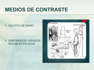
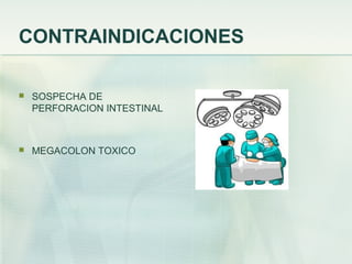
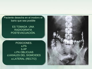
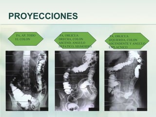
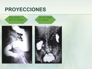
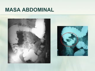
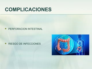
ENEMA BARITADOENEMA BARITADO
DR. ROBERTO CERONDR. ROBERTO CERON
INTERNISTAINTERNISTA
INTRODUCCION
 ENEMA BARITADO: ES LA EXPLORACION DEL COLON
MEDIANTE LA ADMINISTRACION DE UN MEDIO DE
CONTRASTE.
 LA EXPLORACION RADIOLOGICA DEL COLON CON MEDIO DE
CONTRASTE FUE DESCRITA POR PRIMERA VEZ EN 1904
 EN 1923 FISCHER DESCRIBIO POR PRIMERA VEZ EL USO DE
DOBLE CONTRASTE EN LA EVALUACION DEL COLON.
ANATOMIA
 EL COLON MIDE
APROXIMADAMENTE 1.5 M
DE LONGITUD.
 COMPRENDE: CIEGO,
APENDICE, COLON, RECTO,
SIGMOIDES Y CONDUCTO
ANAL
QUE VAMOS A OBSERVAR
MEDIOS DE CONTRASTE
 SULFATO DE BARIO
 PREPARADOS YODADOS
SOLUBLES EN AGUA
INDICACIONES DE ENEMA BARITADO
 TRASTORNO FUNCIONAL DEL COLON
 MALFORMACIONES ANORRECTALES
 ENFERMEDAD DE CROHN
 ENFERMEDAD DIVERTICULAR DEL COLON
 SOSPECHA DE POLIPOS
 COLITIS ULCEROSA
INDICACIONES DE ENEMA BARITADO
 DIARREA CRONICA
 SINDROME DE INTESTINO IRRITABLE
 PERDIDA DE PESO INEXPLICABLE
 CAMBIOS EN LOS HABITOS INTESTINALES
 SOSPECHA DE CANCER DE COLON
 COLITIS POR RADIACION
CONTRAINDICACIONES
 SOSPECHA DE
PERFORACION INTESTINAL
 MEGACOLON TOXICO
PREPARACION DEL PACIENTE
 DIETA DESDE 2 DIAS PREVIO AL PROCEDIMIENTO
 DIETA BLANDA 2 DIAS PREVIOS
 DIELTA DE LIQUIDOS 1 DIA PREVIO A ESTUDIO Y USO DE
LAXANTES
 UN ENEMA EVACUANTE 3 HORAS PREVIOS A
PROCEDIMIENTO, PARA LIMPIEZA DE AMPOLLA RECTAL
 8 HR DE AYUNO EL DIA DEL ESTUDIO
PROYECCIONES
 RX POSTEROANTERIOR O ANTEROPOSTERIOR
 PROYECCIONES OBLICUAS ANTERIOR DERECHA E
IZQUIERDA
 PROYECCIONES EN DECUBITO LATERAL DERECHO E
IZQUIERDO
 PROYECCION LATERAL DEL RECTO
INSERCION DE LA PUNTA DEL ENEMA
1. Colocar al paciente en posición de Sim’s.
2. Expone la región anal
3. Deja salir un poco de bario para eliminar
el aire, y se lubrica el tubo rectal con un
gel a base de agua.
4. Avisarle al paciente que respire profundo y se relaje.
5. Insertar el tubo rectal suavemente durante la espiración en el orificio anal.
Siguiendo el angulo de el canal anal, dirigiendolo anteriormente de 2.5 a
3.5 cm siguiendo la curvatura anal, No introducir mas de 10 cm.
6. No se debe forzar la entrada del tubo rectal.
7. Despues de que la punta del enema a sido insertada, sostenerla en su
posición para prevenir que se resbale.
ENEMA DE BARIO DE CONTRASTE
SIMPLE
Durante el estudio el radiólogo rotara en
diferentes posiciones al paciente para
examinar todas las porciones del colon,
por lo que el tubo del enema se retira
para que el paciente pueda maniobrar
mas facil.
Durante el estudio el radiólogo rotara en
diferentes posiciones al paciente para
examinar todas las porciones del colon,
por lo que el tubo del enema se retira
para que el paciente pueda maniobrar
mas facil.
Paciente desecha en el inodoro el
bario que sea posible
ES TOMADA UNA
RADIOGRAFIA
POSTEVACUACIÓN.
Paciente desecha en el inodoro el
bario que sea posible
ES TOMADA UNA
RADIOGRAFIA
POSTEVACUACIÓN.
POSICIONES.
a.PA
b.AP
c.PA OBLICUAS
d.AXIALES DEL SIGMOIDES
e.LATERAL (RECTO)
POSICIONES.
a.PA
b.AP
c.PA OBLICUAS
d.AXIALES DEL SIGMOIDES
e.LATERAL (RECTO)
ENEMA DE DOBLE CONTRASTE
APLICACIÓN DE BARIO Y AIREAPLICACIÓN DE BARIO Y AIRE
MÉTODO EN UN SOLO
TIEMPO
Colon limpio
Bario adecuado
Adecuado fluido del bario
MÉTODO EN 2 TIEMPOS
Llenar colon con bario
Aire u otros gases
MÉTODO EN UN SOLO
TIEMPO
Colon limpio
Bario adecuado
Adecuado fluido del bario
MÉTODO EN 2 TIEMPOS
Llenar colon con bario
Aire u otros gases
METODO EN UN TIEMPO
INTRODUCCION
DEL ENEMA
SE INSTALA
EL BARIO
EVACUACION
DEL BARIO
PROYECCIONES
PA, AP, TODO
EL COLON
PA, OBLICUA
DRECHA, COLON
ASCEND, ANGULO
HEPATICO, SIGMOIDES
PA, OBLICUA
IZQUIERDA, COLON
ASCENDENTE Y ANGULO
ESPLACNICO
PROYECCIONES
RX LATERAL:
RECTO Y SIGMOI
RX AP, TODO
EL COLON
PROYECCIONES
DECUBITO LATERAL DERECHO
LADO MEDIAL DEL COLON ASCENDENT
LADO LATERAL DEL COLON DESCEND
DECUBITO LATERAL IZQUIERDO
LADO LATERAL DEL COLON ASCENDENT
LADO MEDIAL DEL COLON ASCENDET
ENFERMEDAD DIVERTICULAR
MASA ABDOMINAL
COMPLICACIONES
 PERFORACION INTESTINAL
 RIESGO DE INFECCIONES

Presentacion de enema

  • 1.
    ENEMA BARITADOENEMA BARITADO DR.ROBERTO CERONDR. ROBERTO CERON INTERNISTAINTERNISTA
  • 2.
    INTRODUCCION  ENEMA BARITADO:ES LA EXPLORACION DEL COLON MEDIANTE LA ADMINISTRACION DE UN MEDIO DE CONTRASTE.  LA EXPLORACION RADIOLOGICA DEL COLON CON MEDIO DE CONTRASTE FUE DESCRITA POR PRIMERA VEZ EN 1904  EN 1923 FISCHER DESCRIBIO POR PRIMERA VEZ EL USO DE DOBLE CONTRASTE EN LA EVALUACION DEL COLON.
  • 3.
    ANATOMIA  EL COLONMIDE APROXIMADAMENTE 1.5 M DE LONGITUD.  COMPRENDE: CIEGO, APENDICE, COLON, RECTO, SIGMOIDES Y CONDUCTO ANAL
  • 4.
    QUE VAMOS AOBSERVAR
  • 5.
    MEDIOS DE CONTRASTE SULFATO DE BARIO  PREPARADOS YODADOS SOLUBLES EN AGUA
  • 6.
    INDICACIONES DE ENEMABARITADO  TRASTORNO FUNCIONAL DEL COLON  MALFORMACIONES ANORRECTALES  ENFERMEDAD DE CROHN  ENFERMEDAD DIVERTICULAR DEL COLON  SOSPECHA DE POLIPOS  COLITIS ULCEROSA
  • 7.
    INDICACIONES DE ENEMABARITADO  DIARREA CRONICA  SINDROME DE INTESTINO IRRITABLE  PERDIDA DE PESO INEXPLICABLE  CAMBIOS EN LOS HABITOS INTESTINALES  SOSPECHA DE CANCER DE COLON  COLITIS POR RADIACION
  • 8.
    CONTRAINDICACIONES  SOSPECHA DE PERFORACIONINTESTINAL  MEGACOLON TOXICO
  • 9.
    PREPARACION DEL PACIENTE DIETA DESDE 2 DIAS PREVIO AL PROCEDIMIENTO  DIETA BLANDA 2 DIAS PREVIOS  DIELTA DE LIQUIDOS 1 DIA PREVIO A ESTUDIO Y USO DE LAXANTES  UN ENEMA EVACUANTE 3 HORAS PREVIOS A PROCEDIMIENTO, PARA LIMPIEZA DE AMPOLLA RECTAL  8 HR DE AYUNO EL DIA DEL ESTUDIO
  • 10.
    PROYECCIONES  RX POSTEROANTERIORO ANTEROPOSTERIOR  PROYECCIONES OBLICUAS ANTERIOR DERECHA E IZQUIERDA  PROYECCIONES EN DECUBITO LATERAL DERECHO E IZQUIERDO  PROYECCION LATERAL DEL RECTO
  • 11.
    INSERCION DE LAPUNTA DEL ENEMA 1. Colocar al paciente en posición de Sim’s. 2. Expone la región anal 3. Deja salir un poco de bario para eliminar el aire, y se lubrica el tubo rectal con un gel a base de agua. 4. Avisarle al paciente que respire profundo y se relaje. 5. Insertar el tubo rectal suavemente durante la espiración en el orificio anal. Siguiendo el angulo de el canal anal, dirigiendolo anteriormente de 2.5 a 3.5 cm siguiendo la curvatura anal, No introducir mas de 10 cm. 6. No se debe forzar la entrada del tubo rectal. 7. Despues de que la punta del enema a sido insertada, sostenerla en su posición para prevenir que se resbale.
  • 12.
    ENEMA DE BARIODE CONTRASTE SIMPLE Durante el estudio el radiólogo rotara en diferentes posiciones al paciente para examinar todas las porciones del colon, por lo que el tubo del enema se retira para que el paciente pueda maniobrar mas facil. Durante el estudio el radiólogo rotara en diferentes posiciones al paciente para examinar todas las porciones del colon, por lo que el tubo del enema se retira para que el paciente pueda maniobrar mas facil.
  • 13.
    Paciente desecha enel inodoro el bario que sea posible ES TOMADA UNA RADIOGRAFIA POSTEVACUACIÓN. Paciente desecha en el inodoro el bario que sea posible ES TOMADA UNA RADIOGRAFIA POSTEVACUACIÓN. POSICIONES. a.PA b.AP c.PA OBLICUAS d.AXIALES DEL SIGMOIDES e.LATERAL (RECTO) POSICIONES. a.PA b.AP c.PA OBLICUAS d.AXIALES DEL SIGMOIDES e.LATERAL (RECTO)
  • 14.
    ENEMA DE DOBLECONTRASTE APLICACIÓN DE BARIO Y AIREAPLICACIÓN DE BARIO Y AIRE MÉTODO EN UN SOLO TIEMPO Colon limpio Bario adecuado Adecuado fluido del bario MÉTODO EN 2 TIEMPOS Llenar colon con bario Aire u otros gases MÉTODO EN UN SOLO TIEMPO Colon limpio Bario adecuado Adecuado fluido del bario MÉTODO EN 2 TIEMPOS Llenar colon con bario Aire u otros gases
  • 15.
    METODO EN UNTIEMPO INTRODUCCION DEL ENEMA SE INSTALA EL BARIO EVACUACION DEL BARIO
  • 16.
    PROYECCIONES PA, AP, TODO ELCOLON PA, OBLICUA DRECHA, COLON ASCEND, ANGULO HEPATICO, SIGMOIDES PA, OBLICUA IZQUIERDA, COLON ASCENDENTE Y ANGULO ESPLACNICO
  • 17.
    PROYECCIONES RX LATERAL: RECTO YSIGMOI RX AP, TODO EL COLON
  • 18.
    PROYECCIONES DECUBITO LATERAL DERECHO LADOMEDIAL DEL COLON ASCENDENT LADO LATERAL DEL COLON DESCEND DECUBITO LATERAL IZQUIERDO LADO LATERAL DEL COLON ASCENDENT LADO MEDIAL DEL COLON ASCENDET
  • 23.
  • 24.
  • 25.

Notas del editor

  • #12 Instruct the patient to tum onto the left side, lean forward about 35 to 40 de­ grees, and rest the flexed right knee on the table, above and in front of the slightly flexed left knee (Sims' posi­ tion). This position relaxes the abdomi­ nal muscle , which decreases i ntraab­ dominal pressure on the rectum and makes relaxation of the anal sphi ncter less difficult. Adjust the IV pole so that the enema contents are no higher than 24 inches (6 1 cm) above the level of the anus. • Adjust the overlapping back of the gown or other draping to expose the anal region only, but keep the patient otherwi e well covered. The anal ori­ fice is commonly partially obscured by distended hemorrhoids or a fringe of undistended hemorrhoids. Sometimes there is a contraction or other abnor­ mal ity of the orifice. It is therefore nec­ essary for the anus to be exposed and sufficiently well lighted for the orifice to be clearly visible so that the enema tip can be inserted without injury or discomfort. • Run a little of the barium mixture into a waste basin to free the tubing of air, and then lubricate the rectal tube well with a water-soluble lubricant. • Advise the patient to relax and take deep breaths so that no discomfort is felt when the tube is inserted. • Push the right buttock laterally to open the gluteal fold. • As the abdominal muscles and anal sphincter are relaxed during the expira­ tion phase of a deep breath, insert the rectal tube gently and slowly into the anal orifice. Following the angle of the anal canal, direct the tube anteriorly I to I Y; inches (2.5 to 3.8 cm). Then fol­ lowing the curve of the rectum, direct the tube slightly superiorly. • Insert the tube for a total distance of no more than 3Y; to 4 inches (8.9 to 10 cm). Insertion for a greater distance is not only unnecessary but may injure the rectum. • If the tube does not enter easily, ask the patient to assist if capable. • Never forcibly insert a rectal tube be­ cause the patient may have distended internal hemorrhoids or another condi­ tion that makes forced insertion of the tube dangerous. • After the enema tip is inserted, hold it in position to prevent it from slipping while the patient turns to the supine or prone position for fluoroscopy, according to the preference of the fluoroscopist. • Adjust the protective underpadding, and relieve any pressure on the tubing so that the enema mixture will flow freely.