Aparato digestivo
•Descargar como PPT, PDF•
1 recomendación•4,077 vistas

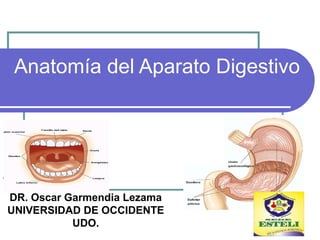
Descripción Anatómica del Aparato Digestivo. DOCENCIA A ESTUDIANTES DE UDO ESTELÍ. DR. OSCAR GARMENDIA UCATSE.

Recomendados
Más contenido relacionado
La actualidad más candente
La actualidad más candente (20)
Destacado
Destacado (17)
Similar a Aparato digestivo
Similar a Aparato digestivo (20)
Más de Oscar Garmendia Lezama
Más de Oscar Garmendia Lezama (14)
Último
Último (20)
Aparato digestivo
- 1. Anatomía del Aparato Digestivo DR. Oscar Garmendia Lezama UNIVERSIDAD DE OCCIDENTE UDO.
- 2. ÓRGANOS DEL SISTEMA DIGESTIVO. Se dividen en: 6. Intestino grueso o colon Ciego Apéndice Colon ascendente Colon transverso Colon sigmoides Recto Ano *Válvulas que regulan el paso del alimento A) TRACTO O TUBO DIGESTIVO 1.Boca 2.Faringe 3.Esófago Cardias* 1.Estómago Píloro* 1.Intestino delgado Duodeno Yeyuno Íleon Válvula íleocecal*
- 3. ÓRGANOS DEL SISTEMA DIGESTIVO. Se dividen en: B) ÓRGANOS ANEXOS O ACCESORIOS. Producen secreciones que se vacían al tubo digestivo 1.Glándulas salivales Parótidas (2) Submaxilares Sublinguales 1.Hígado (con la vesícula biliar) 2.Páncreas
- 5. Etapas del proceso digestivo Ingestión: Los alimentos son triturados por los dientes y mezclados con la saliva. Digestión: Las enzimas de los jugos descomponen los nutrientes en moléculas más sencillas. Absorción: Las moléculas sencillas atraviesan las paredes del tubo y son transportadas por la sangre. Asimilación: Las células utilizan los nutrientes para obtener energía o fabricar nuevas moléculas. Defecación: Las sustancias no digeridas o no absorbidas son eliminadas por el ano.
- 6. El aparato digestivo Tubo de 11 metros de largo, desde la boca hasta el ano. Cavidad bucal Esófago Estómago Intestino delgado Intestino Grueso Glándulas anexas Glándulas salivales Hígado Páncreas Glándulas gástricas Glándulas intestinales
- 7. Ingestión: Cavidad bucal Labios Lengua Dientes Glándulas salivares Istmo de las fauces Amígdalas
- 8. Lengua Órgano musculoso, muy móvil. Interviene en la masticación Interviene en la deglución Órgano del gusto
- 9. Dientes Estructura: Corona Raíz Cuello Tipos: Incisivos Caninos Premolares Molares
- 11. OmnívorosOmnívoros CarnívorosCarnívoros HerbívorosHerbívoros Dientes adaptados a diferentes dietas IncisivosIncisivos CaninosCaninos PremolaresPremolares MolaresMolares
- 13. Glándulas salivares Parótidas: Bajo la oreja. Vierten junto al segundo molar superior. Submaxilares: Bajo la base de la lengua. Sublinguales: Encima de las anteriores. Saliva: contiene amilasa (degrada almidón) y lipasa lingual (degrada grasas), agua, sales, lisozima (bactericida) y mucina (lubricante).
- 14. Faringe Tubo musculoso común a los aparatos digestivo y respiratorio. Comunica con: La boca a través del istmo de las fauces El esófago Las fosas nasales a través de las coanas La laringe a través de la glotis El oído medio a través de las trompas de Eustaquio.
- 15. Faringe
- 16. Ingestión: Esófago Tubo muscular de unos 30 cm que comunica la faringe con el estómago. Desciende por detrás de la tráquea y del corazón Atraviesa el diafragma por el hiato esofágico Tiene dos esfínteres, uno superior y otro inferior Termina en una válvula llamada cardias, que es a la vez la puerta de entrada al estómago
- 17. Esófago: Histología Capa mucosa: epitelio plano pluriestratificado no queratinizado. Capa submucosa: tejido conjuntivo Capa muscular: cálulas musculares lisas perimetrales y longitudinales, responsables de movimientos peristálticos Capa adventicia de tejido conjuntivo
- 18. Esófago: Ondas peristálticas Ondas de contracción de la musculatura lisa. Empujan el bolo hacia el estómago.
- 19. Digestión: El estómago saco muscular expandible (capacidad 1-1.5 litros) El cardias en la entrada y el píloro en la salida, regulan el paso del alimento El bolo alimenticio se transforma en una papilla llamada quimo El esfinter pilórico regula el vaciado gástrico
- 20. ANATOMIA. ESTOMAGO PRESENTA LAS SIGUIENTES PORCIONES CARDIAS FONDO CUERPO PILORO PRESENTA 2 CURVATURAS MAYOR Y MENOR.
- 22. ANATOMIA. LA GASTRICA DERECHA Y LA GASTRICA IZQUIERDA HACEN UN ARCO EN LA CURVATURA MENOR LA GASTRO EPIPLOICA Y LA GASTRICAS CORTAS HACEN UN ARCO EN LA CURVATURA MAYOR.
- 24. Intestino Delgado Ocurre la mayor parte de la digestión enzimática y casi toda la absorción. Es un tubo arrollado, de unos siete metros de longitud y de algo más de dos centímetros y medio de diámetro. El intestino delgado se subdivide en duodeno, yeyuno e íleon, que se continúa con el intestino grueso por medio de la válvula ileocecal.
- 26. ANATOMIA. EL COLEDOCO Y EL WIRSUNG DESEMBOCAN EN LA SEGUNDA PORCION DEL DUODENO EN LA CARUNCULA(PAPILA DUODENAL MAYOR) ESTA SITUADA MAS O MENOS 7 CM DEL PILORO Y SE CONOCE COMO LA DESEMBOCADURA DE LA AMPOLLA DE VATER.
- 28. Intestino delgado: digestión química La bilis y el jugo pancreático vierten en el duodeno a través de la ampolla de Vater, donde se mezclan con el quimo. Las glándulas intestinales segregan jugo intestinal
- 29. ANATOMIA. RIEGO SANGUINEO EL TRONCO CELIACO IRRIGA LA PRIMERA PORCION ATRAVEZ DE LA ARTERIA PILORICA(GASTRICA DERECHA)Y LA GASTRODUODENAL LA PORCION INFERIOR ES IRRIGADA POR LAS ARTERIAS PANCREATICOS DUODENALES INFERIORES RAMAS DE LA MESENTERICA SUPERIOR
- 32. ANATOMIA. YEYUNO E ILEON EL YEYUNO ES 2 QUINTAS PARTES Y EL ILEON ES 3 QUINTAS PARTES EL ILEON ES CON FRECUENCIA ESTA VACIO Y MAS VASCULARIZADO SUS PAREDES SON MAS GRUESAS Y SU MESENTERIO ES TRASLUCIDO POR LA AUSENCIA DE GRASA
- 34. ANATOMIA. REMANENTE ONFALOMESENTERICO DIVERTICULO DE MECKEL YEYUNO E ILEON ESTAN SUSPENDIDOS DE LA PARED ABDOMINAL CON EL MESENTERIO MESENTERIO EL BORDE UNIDO A LA PARED SE LLAMA RAIZ Y TIENE UNOS 15 CM DE LARGO Y SE DIRIGE HACIA ABAJO Y A LA DERECHA CONTIENE 2 LAMINAS PERITONEALES ENTRE ELLAS ESTAN LOS VASOS MESENTERICOS GANGLIOS NERVIOS Y UNA CANTIDAD VARIABLE DE GRASA
- 35. ANATOMIA. RIEGO SANGUINEO LA ARTERIA MESENTERICA SUPERIOR IRRIGA EL INTESTINO MEDIO OSEA EL INTESTINO DELGADO DESDE EL COLEDOCO HASTA LA CURVATURA COLICA IZQUIERDA LAS ARTERIAS YEYUNALES E ILEALES (DE 7 ) SE ORIGINAN EN EL LADO IZQUIERDO DE LA MESENTERICA SUPERIOR
- 37. Intestino grueso 1,5 m de longitud y 6,5 cm de diámetro En él se produce absorción de agua e iones inorgánicos, y formación y eliminación de heces fecales Contiene abundante flora bacteriana que fermenta residuos no digeridos, y sintetiza vitaminas K y B
- 38. Intestino grueso
- 39. ANATOMIA. INTESTINO GRUESO SE COMPONE DE CIEGO APENDICE COLON EL CUAL SE DIVIDE EN ASCENDENTE TRANSVERSO Y DESCENTE LUEGO SE CONTINUA CON EL SIGMOIDE RECTO Y CONDUCTO ANAL.
- 41. ANATOMIA. ESTRUCTURA EL INTESTINO GRUESO MENOS EL RECTO Y EL CONDUCTO ANAL SE CARACTERIZA POR TENER UNA MUCOSA CON CELULAS CALICIFORMES GLANDULAS Y CELULAS DE ABSORCION LOS APENDICES EPIPLOICOS SON PEQUEÑAS MASAS DE GRASA INCUIAS EN EL PERITONEO.
- 42. ANATOMIA. LA CAPA MUSCULAR EXTERNA ESTA ENGROSADA POR TRES BANDAS QUE SE LLAMAN LAS TENIAS DEL COLON.TIENEN APROXIMADAMENTE 1CM DE ANCHO ESTAN BIEN DELIMITADAS EN EL CIEGO Y EL COLON ASCENDENTE GRAN PARTE DEL INTESTINO GRUESO SE CARACTERIZA POR TENER PLIEGUES Y SACULACIONES LLAMADOS HAUSTRAS
- 44. ANATOMIA. CIEGO POR DEBAJO DEL ILEON ORIFICIO ILEOCOLICO (VALVULA) FORMA DE ESTRELLLA INTUSUSEPCION
- 46. ANATOMIA. APENDICE VERMIFORME 9-10CMTS ORGANO LINFOIDE POSICION IRRIGACION ILEOCECAL A MS APENDICITIS
Notas del editor
- (a) The varied, omnivorous diet of humans has fostered the evolution of an unspecialized dentition that includes flat incisors for biting, pointed canines for tearing, premolars for grinding, and molars for crushing and chewing. (b) Carnivores (such as the lion) have modest incisors but greatly enlarged canines for stabbing and tearing flesh. Carnivores also have a reduced set of cheek teeth (molars and premolars) that are specialized for shearing through tendon and bone. (c) Herbivores (such as the cow) have incisors that are specialized for snipping leaves, and their canines have been reduced in size and moved forward to help with that job. The cheek is filled with a full set of wide, flat premolars and molars that grind up the tough, cellulose-containing plant material.
- (a) The varied, omnivorous diet of humans has fostered the evolution of an unspecialized dentition that includes flat incisors for biting, pointed canines for tearing, premolars for grinding, and molars for crushing and chewing. (b) Carnivores (such as the lion) have modest incisors but greatly enlarged canines for stabbing and tearing flesh. Carnivores also have a reduced set of cheek teeth (molars and premolars) that are specialized for shearing through tendon and bone. (c) Herbivores (such as the cow) have incisors that are specialized for snipping leaves, and their canines have been reduced in size and moved forward to help with that job. The cheek is filled with a full set of wide, flat premolars and molars that grind up the tough, cellulose-containing plant material.