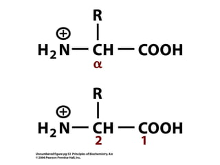
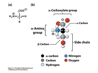
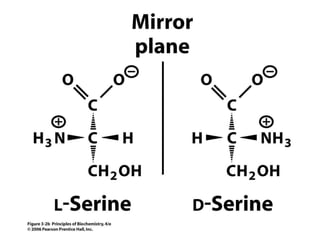
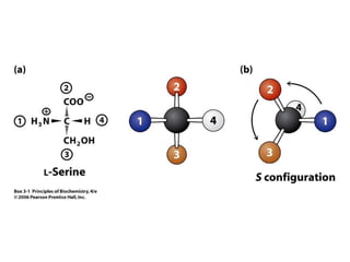
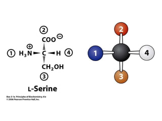
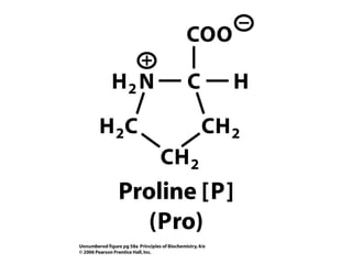
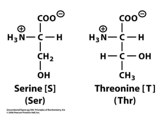
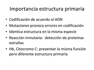
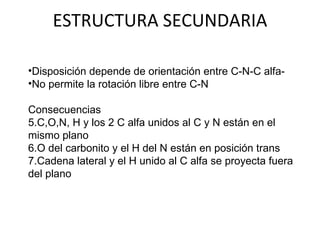
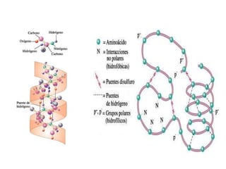
PROTEÍNAS Macromoléculas efectoras de la acción de los genes
Generalidades 50% en el peso de tejidos Esencial en procesos biológicos Eje: enzimas, hormonas, hemoglobina, anticuerpos, receptores, actina y miosina, colágeno Elementos: C, H, O, N, S N: 1 g por cada 6.25 g de N


NOMENCLATURA Resto de cada a.a : terminación en il Último residuo ( C terminal) :  nombre completo del aminoñacido Ej: Serina, ácido aspártico, tirosina, lisina, alanina y cisteína Nombre: seril-aspartil-tirosil-lisil-alanil-cisteína.
Péptidos de importancia biológica Glutatión: Tripéptido (ácido glutámico, cisteína y glicina) Participa en sistema enzimático de oxidación y reducción.  Previene daños oxidativos. Otros:  Hormonas o factores liberadores Encefalinas Antibióticos
PROTEÍNAS Propiedades ácido – base Carga eléctrica depende de ionización de grupos en cadenas laterales Residuo lisina, arginina o histidina: carácter básico
Residuos de aspartato y glutamato: carácter ácido Grupo fenolico (tirosina) y sulfihidrilo (cisteína): débilmente ácido Tirosina Cisteína Proteínas: Capacidad amortiguadora o buffer  por captar o liberar protones de acuerdo a concentración de H+
FORMAS MOLECULARES GLOBULARES Molécula se pliega sobre sí misma Forma ovoide Actividad funcional: enzimas, anticuerpos, hormonas, Hb FIBRILARES Se ordenan paralelamente Fibras o láminas Poco solubles o insolubles en gua Forman estructuras de sosten: tejido conjuntivo
ESTRUCTURA MOLECULAR Primaria:  estructuras lineales Secundaria:  Disposición espacial regular, repetitiva por enlace de Hidrógeno Terciaria:  Tridimensional Cuaternaria: 2 o más cadenas polipeptídicas en estructura espacial
ESTRUCTURA PRIMARIA Secuencia u Ordenamiento de aminoácidos De acuerdo a instrucciones en genes
Importancia estructura primaria Codificación de acuerdo al ADN Mutaciones provoca errores en codificación Identica estructura en la misma especie Reacción inmutaria:  detección de proteínas extrañas Hb, Citocromo C: presentan la misma función pero diferente estructura primaria
ESTRUCTURA SECUNDARIA Disposición depende de orientación entre C-N-C alfa- No permite la rotación libre entre C-N Consecuencias C,O,N, H y los 2 C alfa unidos al C y N están en el mismo plano O del carbonito y el H del N están en posición trans Cadena lateral y el H unido al C alfa se proyecta fuera del plano
ESTRUCTURA SECUNDARIA
 
Hélice alfa Enrollamiento sobre eje central Sentido de agujas de reloj Fuerza de unión: Puentes de Hidrógeno ( N y O) Disposición trans Presencia de ciertos aa impide formación de Helice alfa Prolina ( nucleo pirrolidina no puede rotar) Restos con carga ( arginina, lisina, ac. Glutámico) origina fuerzas electroestaticas
 
Lámina Beta Cada residuo de aa cubre un espacio mayor que en helice alfa 2 o más cadenas se aparean por puentes de hidróge3nos Forman plegamiento en zigzag
Disposición al azar Cadena no posee estructura regular Orientación termodinámicamente más favorable Moléculas con distintos tipos de estructuras secundarias Proteínas globular Segmentos con hélice alfa y plegamiento y al azar
ESTRUCTURA TERCIARIA Presente en estructuras globulares Segmentos dispuesto al azar Permite plegamientos para tomar formar esferoidal Estructura se mantiene debido a: Fuerzas electroestáticas Enlaces de hidrógeno Presencia de cadenas hidrofóbicas o hidrofílicas Puentes disulfuro
ESTRUCTURA CUATERNARIA Proteínas de tipo oligoméricas: formadas por más de una cadena polipéptidica Cada cadena es una subunidad Ej: insulina, Hb, enzima lactato deshidrogenasa Disposición espacial de estas subunidades Fuerzas responsables: las mismas que en terciaria
Función biológica en relación a secuencia y conformación Estructura primaria: determinante en formación de proteína Influye  en las fuerzas e interacciones  Cambios en secuencia provocan modificaciones conformacionales No todas las secuencias son viables Evolución ha ido escogiendo las secuencias adecuadas Elementos comunes en ciertas proteínas:  Dominio Dominios se relacionan con una función específica de la proteína Se relaciona con la existencia de un gen común.
HEMOGLOBINA Proteína conjugada de carácter básico Grupo prostético: hem o hemo Molécula tetramérica: 2 cadenas polipeptídicas de 141 aa y otras 2 de 146 aa 4 cadenas se ensamblan de forma esférica (estructura cuaternaria)


Hemoglobinas anormales Síntesis de cadenas controlada por genes diferent4es Alteraciones  en genes originan proteínas defectuosas  Provocan enfermedades hereditarias: Hemoglobinopatías Tipos de alteraciones sustitución de uno o dos aa por otro Falta de uno o más aa Presencia de cadena híbridas Presencia de cadenas más largas de lo normal Síntesis disminuida o nula de una cadena
Consecuencias Inestabilidad de la Hb:  Anemia falciforme acido glutámico esta sustituído por valina en cadena B Modificación de aa en el entorno Hemo oxidación Fe2+ en Fe 3+ :L Metahemoglobina Cambios en residuos aa en zona de contacto: relación con Oxígeno Sustitución de residuos en la unión de 2,3 bifosfosfoglicerato mayor afinidad con O2 con Hb
DESNATURALIZACIÓN DE PROTEÍNAS Provocada por calor radiaciones Congelamientos repetitivos grandes presiones agentes químicos Perdida de propiedades y funciones  Ruptura de fuerzas que mantienen las estructuras secundarias, terciarias y cuaternaria Proceso reversible e irreversible

Proteinas Efectoras De Genes

  • 1.
    PROTEÍNAS Macromoléculas efectorasde la acción de los genes
  • 2.
    Generalidades 50% enel peso de tejidos Esencial en procesos biológicos Eje: enzimas, hormonas, hemoglobina, anticuerpos, receptores, actina y miosina, colágeno Elementos: C, H, O, N, S N: 1 g por cada 6.25 g de N
  • 3.
  • 4.
  • 5.
  • 6.
  • 7.
  • 8.
  • 9.
  • 10.
  • 11.
  • 12.
  • 13.
  • 14.
  • 15.
  • 16.
  • 17.
  • 18.
  • 19.
  • 20.
  • 21.
  • 22.
  • 23.
  • 24.
  • 25.
  • 26.
  • 27.
  • 28.
  • 29.
  • 30.
  • 31.
  • 32.
  • 33.
  • 34.
  • 35.
  • 36.
  • 37.
    NOMENCLATURA Resto decada a.a : terminación en il Último residuo ( C terminal) : nombre completo del aminoñacido Ej: Serina, ácido aspártico, tirosina, lisina, alanina y cisteína Nombre: seril-aspartil-tirosil-lisil-alanil-cisteína.
  • 38.
    Péptidos de importanciabiológica Glutatión: Tripéptido (ácido glutámico, cisteína y glicina) Participa en sistema enzimático de oxidación y reducción. Previene daños oxidativos. Otros: Hormonas o factores liberadores Encefalinas Antibióticos
  • 39.
    PROTEÍNAS Propiedades ácido– base Carga eléctrica depende de ionización de grupos en cadenas laterales Residuo lisina, arginina o histidina: carácter básico
  • 40.
    Residuos de aspartatoy glutamato: carácter ácido Grupo fenolico (tirosina) y sulfihidrilo (cisteína): débilmente ácido Tirosina Cisteína Proteínas: Capacidad amortiguadora o buffer por captar o liberar protones de acuerdo a concentración de H+
  • 41.
    FORMAS MOLECULARES GLOBULARESMolécula se pliega sobre sí misma Forma ovoide Actividad funcional: enzimas, anticuerpos, hormonas, Hb FIBRILARES Se ordenan paralelamente Fibras o láminas Poco solubles o insolubles en gua Forman estructuras de sosten: tejido conjuntivo
  • 42.
    ESTRUCTURA MOLECULAR Primaria: estructuras lineales Secundaria: Disposición espacial regular, repetitiva por enlace de Hidrógeno Terciaria: Tridimensional Cuaternaria: 2 o más cadenas polipeptídicas en estructura espacial
  • 43.
    ESTRUCTURA PRIMARIA Secuenciau Ordenamiento de aminoácidos De acuerdo a instrucciones en genes
  • 44.
    Importancia estructura primariaCodificación de acuerdo al ADN Mutaciones provoca errores en codificación Identica estructura en la misma especie Reacción inmutaria: detección de proteínas extrañas Hb, Citocromo C: presentan la misma función pero diferente estructura primaria
  • 45.
    ESTRUCTURA SECUNDARIA Disposicióndepende de orientación entre C-N-C alfa- No permite la rotación libre entre C-N Consecuencias C,O,N, H y los 2 C alfa unidos al C y N están en el mismo plano O del carbonito y el H del N están en posición trans Cadena lateral y el H unido al C alfa se proyecta fuera del plano
  • 46.
  • 47.
  • 48.
    Hélice alfa Enrollamientosobre eje central Sentido de agujas de reloj Fuerza de unión: Puentes de Hidrógeno ( N y O) Disposición trans Presencia de ciertos aa impide formación de Helice alfa Prolina ( nucleo pirrolidina no puede rotar) Restos con carga ( arginina, lisina, ac. Glutámico) origina fuerzas electroestaticas
  • 49.
  • 50.
    Lámina Beta Cadaresiduo de aa cubre un espacio mayor que en helice alfa 2 o más cadenas se aparean por puentes de hidróge3nos Forman plegamiento en zigzag
  • 51.
    Disposición al azarCadena no posee estructura regular Orientación termodinámicamente más favorable Moléculas con distintos tipos de estructuras secundarias Proteínas globular Segmentos con hélice alfa y plegamiento y al azar
  • 52.
    ESTRUCTURA TERCIARIA Presenteen estructuras globulares Segmentos dispuesto al azar Permite plegamientos para tomar formar esferoidal Estructura se mantiene debido a: Fuerzas electroestáticas Enlaces de hidrógeno Presencia de cadenas hidrofóbicas o hidrofílicas Puentes disulfuro
  • 53.
    ESTRUCTURA CUATERNARIA Proteínasde tipo oligoméricas: formadas por más de una cadena polipéptidica Cada cadena es una subunidad Ej: insulina, Hb, enzima lactato deshidrogenasa Disposición espacial de estas subunidades Fuerzas responsables: las mismas que en terciaria
  • 54.
    Función biológica enrelación a secuencia y conformación Estructura primaria: determinante en formación de proteína Influye en las fuerzas e interacciones Cambios en secuencia provocan modificaciones conformacionales No todas las secuencias son viables Evolución ha ido escogiendo las secuencias adecuadas Elementos comunes en ciertas proteínas: Dominio Dominios se relacionan con una función específica de la proteína Se relaciona con la existencia de un gen común.
  • 55.
    HEMOGLOBINA Proteína conjugadade carácter básico Grupo prostético: hem o hemo Molécula tetramérica: 2 cadenas polipeptídicas de 141 aa y otras 2 de 146 aa 4 cadenas se ensamblan de forma esférica (estructura cuaternaria)
  • 56.
  • 57.
  • 58.
  • 59.
  • 60.
    Hemoglobinas anormales Síntesisde cadenas controlada por genes diferent4es Alteraciones en genes originan proteínas defectuosas Provocan enfermedades hereditarias: Hemoglobinopatías Tipos de alteraciones sustitución de uno o dos aa por otro Falta de uno o más aa Presencia de cadena híbridas Presencia de cadenas más largas de lo normal Síntesis disminuida o nula de una cadena
  • 61.
    Consecuencias Inestabilidad dela Hb: Anemia falciforme acido glutámico esta sustituído por valina en cadena B Modificación de aa en el entorno Hemo oxidación Fe2+ en Fe 3+ :L Metahemoglobina Cambios en residuos aa en zona de contacto: relación con Oxígeno Sustitución de residuos en la unión de 2,3 bifosfosfoglicerato mayor afinidad con O2 con Hb
  • 62.
    DESNATURALIZACIÓN DE PROTEÍNASProvocada por calor radiaciones Congelamientos repetitivos grandes presiones agentes químicos Perdida de propiedades y funciones Ruptura de fuerzas que mantienen las estructuras secundarias, terciarias y cuaternaria Proceso reversible e irreversible

Notas del editor

  • #4 Numbering conventions for amino acids. In traditional names, the carbon atoms adjacent to the carboxyl group are identified by the Greek letters  ,  ,  , etc. In the official IUPAC/IUBMB chemical names or systematic names, the carbon atom in the carboxyl group is number 1 and the adjacent carbons are numbered sequentially. Thus, the atom in traditional names is the carbon 2 atom in systematic names.
  • #5 Figure 3.1 Two representations of an L-amino acid at neutral pH. (a) General structure. An amino acid has a carboxylate group (whose carbon atom is designated C-1), an amino group, a hydrogen atom, and a side chain (or R group), all attached to C-2 (the  -carbon). Solid wedges indicate bonds above the plane of the paper; dashed wedges indicate bonds below the plane of the paper. The blunt ends of wedges are nearer the viewer than the pointed ends. (b) Ball-and-stick model of serine (whose R group is —CH 2 OH). Note the alternative numbering and lettering systems for the carbon atoms.
  • #6 Numbering conventions for amino acids. In traditional names, the carbon atoms adjacent to the carboxyl group are identified by the Greek letters  ,  ,  , etc. In the official IUPAC/IUBMB chemical names or systematic names, the carbon atom in the carboxyl group is number 1 and the adjacent carbons are numbered sequentially. Thus, the atom in traditional names is the carbon 2 atom in systematic names.
  • #7 Figure 3.1 Two representations of an L-amino acid at neutral pH. (a) General structure. An amino acid has a carboxylate group (whose carbon atom is designated C-1), an amino group, a hydrogen atom, and a side chain (or R group), all attached to C-2 (the  -carbon). Solid wedges indicate bonds above the plane of the paper; dashed wedges indicate bonds below the plane of the paper. The blunt ends of wedges are nearer the viewer than the pointed ends. (b) Ball-and-stick model of serine (whose R group is —CH 2 OH). Note the alternative numbering and lettering systems for the carbon atoms.
  • #8 Figure 3.1 Two representations of an L-amino acid at neutral pH. (a) General structure. An amino acid has a carboxylate group (whose carbon atom is designated C-1), an amino group, a hydrogen atom, and a side chain (or R group), all attached to C-2 (the  -carbon). Solid wedges indicate bonds above the plane of the paper; dashed wedges indicate bonds below the plane of the paper. The blunt ends of wedges are nearer the viewer than the pointed ends. (b) Ball-and-stick model of serine (whose R group is —CH 2 OH). Note the alternative numbering and lettering systems for the carbon atoms.
  • #9 Figure 3.2 Mirror-image pairs of amino acids. (a) Ball-and-stick models of L -serine and D -serine. Note that the two molecules are not identical; they cannot be superimposed. (b) L -Serine and D -serine.
  • #10 Figure 3.2 Mirror-image pairs of amino acids. (a) Ball-and-stick models of L -serine and D -serine. Note that the two molecules are not identical; they cannot be superimposed. (b) L -Serine and D -serine.
  • #11 Figure 3.2 Mirror-image pairs of amino acids. (a) Ball-and-stick models of L -serine and D -serine. Note that the two molecules are not identical; they cannot be superimposed. (b) L -Serine and D -serine.
  • #12 Assignment of configuration by the RS system. (a) Each group attached to a chiral carbon is assigned a priority based on atomic mass, 4 being the lowest priority. (b) By orienting the molecule with the priority 4 group pointing away (behind the chiral carbon) and tracing the path from the highest priority group to the lowest, the absolute configuration can be established. If the sequence 1, 2, 3 is clockwise, the configuration is R . If the sequence 1, 2, 3 is counterclockwise, the configuration is S . L -Serine has the S configuration.
  • #13 Assignment of configuration by the RS system. (a) Each group attached to a chiral carbon is assigned a priority based on atomic mass, 4 being the lowest priority. (b) By orienting the molecule with the priority 4 group pointing away (behind the chiral carbon) and tracing the path from the highest priority group to the lowest, the absolute configuration can be established. If the sequence 1, 2, 3 is clockwise, the configuration is R . If the sequence 1, 2, 3 is counterclockwise, the configuration is S . L-Serine has the S configuration.
  • #14 Assignment of configuration by the RS system. (a) Each group attached to a chiral carbon is assigned a priority based on atomic mass, 4 being the lowest priority. (b) By orienting the molecule with the priority 4 group pointing away (behind the chiral carbon) and tracing the path from the highest priority group to the lowest, the absolute configuration can be established. If the sequence 1, 2, 3 is clockwise, the configuration is R . If the sequence 1, 2, 3 is counterclockwise, the configuration is S . L-Serine has the S configuration.
  • #15 Aliphatic R Groups
  • #16 Figure 3.3 Stereoisomers of isoleucine.
  • #19 Figure 3.4 Formation of cystine. When oxidation links the sulfhydryl groups of two cysteine molecules, the resulting compound is a disulfide called cystine.
  • #24 Figure 3.5 Compounds derived from common amino acids. (a)  -Aminobutyrate a derivative of glutamate. (b) Histamine, a derivative of histidine. (c) Epinephrine, a derivative of tyrosine. (d) Thyroxine and triiodothyronine, derivatives of tyrosine. Thyroxine contains one more atom of iodine (in parentheses) than does triiodothyronine.
  • #25 Figure 3.5 Compounds derived from common amino acids. (a)  -Aminobutyrate a derivative of glutamate. (b) Histamine, a derivative of histidine. (c) Epinephrine, a derivative of tyrosine. (d) Thyroxine and triiodothyronine, derivatives of tyrosine. Thyroxine contains one more atom of iodine (in parentheses) than does triiodothyronine.
  • #26 Figure 3.5 Compounds derived from common amino acids. (a)  -Aminobutyrate a derivative of glutamate. (b) Histamine, a derivative of histidine. (c) Epinephrine, a derivative of tyrosine. (d) Thyroxine and triiodothyronine, derivatives of tyrosine. Thyroxine contains one more atom of iodine (in parentheses) than does triiodothyronine.
  • #27 Figure 3.5 Compounds derived from common amino acids. (a)  -Aminobutyrate a derivative of glutamate. (b) Histamine, a derivative of histidine. (c) Epinephrine, a derivative of tyrosine. (d) Thyroxine and triiodothyronine, derivatives of tyrosine. Thyroxine contains one more atom of iodine (in parentheses) than does triiodothyronine.
  • #28 Figure 3.5 Compounds derived from common amino acids. (a)  -Aminobutyrate a derivative of glutamate. (b) Histamine, a derivative of histidine. (c) Epinephrine, a derivative of tyrosine. (d) Thyroxine and triiodothyronine, derivatives of tyrosine. Thyroxine contains one more atom of iodine (in parentheses) than does triiodothyronine.
  • #30 Figure 3.6 Titration curve for alanine. The first p K a value is 2.4; the second is 9.9. pI Ala represents the isoelectric point of alanine.
  • #31 Figure 3.7 Ionization of histidine. (a) Titration curve for histidine. The three p K a values are 1.8, 6.0, and 9.3. pI His represents the isoelectric point of histidine. (b) Deprotonation of the imidazolium ring of the side chain of histidine.
  • #32 Figure 3.7 Ionization of histidine. (a) Titration curve for histidine. The three p K a values are 1.8, 6.0, and 9.3. pI His represents the isoelectric point of histidine. (b) Deprotonation of the imidazolium ring of the side chain of histidine.
  • #34 Figure 3.8 Ionization of amino acid side chains. (a) Ionization of the protonated  -carboxyl group of glutamate. The negative charge of the carboxylate anion is delocalized. (b) Deprotonation of the guanidinium group of the side chain of arginine. The positive charge is delocalized.
  • #35 Figure 3.8 Ionization of amino acid side chains. (a) Ionization of the protonated  -carboxyl group of glutamate. The negative charge of the carboxylate anion is delocalized. (b) Deprotonation of the guanidinium group of the side chain of arginine. The positive charge is delocalized.
  • #37 Figure 3.9 Peptide bond between two amino acids. The structure of the peptide linkage can be viewed as the product of a condensation reaction in which the  -carboxyl group of one amino acid condenses with the  -amino group of another amino acid. The result is a dipeptide in which the amino acids are linked by a peptide bond. Here, alanine is condensed with serine to form alanylserine.
  • #57 Figure 4.41 Scanning electron micrograph of mammalian erythrocytes. Each cell contains approximately 300 million hemoglobin molecules.
  • #58 Figure 4.42 Human (Homo sapiens) oxyhemoglobin. (a) Structure of human oxyhemoglobin showing two  and two  subunits. Heme groups are shown as stick models. [PDB 1 HND]. (b) Schematic diagram of the hemoglobin tetramer. The heme groups are red.
  • #59 Figure 4.42 Human (Homo sapiens) oxyhemoglobin. (a) Structure of human oxyhemoglobin showing two  and two  subunits. Heme groups are shown as stick models. [PDB 1 HND]. (b) Schematic diagram of the hemoglobin tetramer. The heme groups are red.
  • #60 Figure 4.42 Human (Homo sapiens) oxyhemoglobin. (a) Structure of human oxyhemoglobin showing two  and two  subunits. Heme groups are shown as stick models. [PDB 1 HND]. (b) Schematic diagram of the hemoglobin tetramer. The heme groups are red.