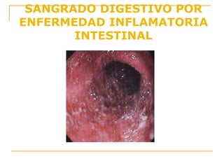
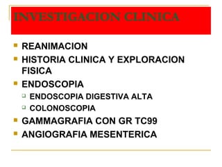
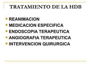
SlideShare una empresa de Scribd logo
1 de 42
Encuentra las videosclases en: http://www.youtube.com/channel/UCgZGxUTlxGuZV3MYDcLWEBg?feature=watch
HEMORRAGIA DIGESTIVA
BAJA (HDB)
HECTOR PAUCAR SOTOMAYOR
GASTROENTEROLOGIA
FMH - UNSAAC
HEMORRAGIA DIGESTIVA BAJAHEMORRAGIA DIGESTIVA BAJA
HDBHDB
SANGRADO POR DEBAJO DEL
ANGULO DE TREITZ
EPIDEMIOLOGIA:
 Incidencia: 20,5-27 x 100,000 Hab.
 Incrementa con la edad
 10 - 24 % de todos los episodios
de sangrado
CASO CLINICO
 Mujer de 76 años que acude por presentar
deposiciones sanguinolentas abundantes (> 1
lt) hace 2 horas, sus familiares la encontraron
desmayada y la trajeron inmediatamente al
hospital
 No tiene antecedentes patologicos de
importancia
 PA: 50/0 FC: 120 x min
 Palidez importante de piel y mucosas.
COLONOSCOPIA
HEMORRAGIA DIGESTIVA BAJA
FORMAS DE PRESENTACION CLINICAFORMAS DE PRESENTACION CLINICA
 HEMATOQUEZIA
 SIGNOS DE DESCOMPENSACION
HEMODINAMICA
CAUSAS DE HDB
 Diverticulosis
 Angiodisplasia
 Hemorroides
 Fisuras anales
 Neoplasias
 Enfermedad intestinal inflamatoria
 Colitis Isquemica
 Colitis Infecciosa
 Colitis inducida por radiacion
 Diverticulo de Meckel
 Intusucepcion intestinal
 Fistula aortointestinal
 Ulceras rectales solitarias
 Ulceras cecales inducidas por AINEs
CAUSAS DE HDB EN EL PERU
DIVERTICULOS DE COLON 50,4%
FIEBRE TIFOIDEA 8,8%
HEMORROIDES INTERNAS 5,9%
ANGIODISPLASIA DEL COLON 3,7%
CANCER DE COLON 2,9%
OTROS 32,0%
HNERM LIMA
HEMORRAGIA
DIVERTICULAR
Afecta mas a poblacion geriatrica
 Complicacion (3%) de los pacientes que
sufren diverticulosis
 Preponderante en colon sigmoides
 Hematoquezia masiva
 No hay dolor abdominal
SANGRADO DIVERTICULAR
COLONOSCOPIA
SANGRADO DIVERTICULAR
ARTERIOGRAFIA MESENTERICA
SCAN CON TECNECIO 99m
HEMORRAGIA POR
CANCER DE COLON
HEMORRAGIA POR
ANGIODISPLASIA DEL
COLON
LOCALIZACION ANATOMICA DE LA
ANGIODISPLASIA COLONICA
ANGIODISPLASIA O ECTASIA
VASCULAR
Mayor frecuencia en poblacion geriatrica
Las lesiones suelen ser multiples y pequeñas (< 5 mm) y se
ubican preferentemente en hemicolon derecho y ciego
Se relacionan con enfermedad aortica (?)
FISIOPATOLOGIA
COLONOSCOPIA
COLONOSCOPIA
COLONOSCOPIA
COLONOSCOPIA
ANGIOGRAFIA
ANGIOGRAFIA
ANGIOGRAFIA
HEMORRAGIA POR
COLITIS ISQUEMICA
SANGRADO DIGESTIVO POR
COLITIS ISQUEMICA
SANGRADO DIGESTIVO POR
COLITIS ISQUEMICA
HEMORRAGIA POR
COLITIS ACTINICA
SANGRADO DIGESTIVO POR
ENFERMEDAD INFLAMATORIA
INTESTINAL
CAUSAS DIVERSAS DE HDB
COLITIS
INFECCIOSA
HEMORROIDES
INTERNAS
INVESTIGACION CLINICA
 REANIMACION
 HISTORIA CLINICA Y EXPLORACION
FISICA
 ENDOSCOPIA
 ENDOSCOPIA DIGESTIVA ALTA
 COLONOSCOPIA
 GAMMAGRAFIA CON GR TC99
 ANGIOGRAFIA MESENTERICA
HDB – TRATAMIENTO
CONSIDERACIONES GENERALES:
Las mismas que en HDA
CONSIDERACIONES ESPECIFICAS:
60% dejan de sangrar en las 48 hs, 20% cesa la hemorragia pero
se reanuda a las 48-72 hs, 20% continuan sangrando
1. Si continua sangrando y necesita mas de 2 unidades de sangre
c/8 hs por 24 hs debe considerarse cirugia de emergencia con
o sin diagnostico etiologico
2. Si vuelve a sangrar luego de 48-72 hs de haber cesado,
compensar hemodinamicamente y luego pasar a cirugia.
3. Si ceso el sangrado, pero no tenemos diagnostico etiologico,
re-evaluar con estudios diagnosticos adecuados o repetirlos.
TRATAMIENTO DE LA HDB
 REANIMACION
 MEDICACION ESPECIFICA
 ENDOSCOPIA TERAPEUTICA
 ANGIOGRAFIA TERAPEUTICA
 INTERVENCION QUIRURGICA
HEMORRAGIA DIGESTIVA BAJA
Estimar volumen de la perdida sanguinea
Hb,Hto, Hem, Pruebas de coagulacion, GS y rH
Via central, PVC,
Sonda Foley (volumen urinario)
PROCTOSIGMOIDOSCOPIA
NEGATIVA
TRATAMIENTO
POSITIVA
SNG – ENDOSCOPIA
DIGESTIVA ALTA
NEGATIVA
CESO LA
HEMORRAGIA
DIGESTIVA
CONTINUA LA
HEMORRAGIA
DIGESTIVA
HEMORRAGIA DIGESTIVA BAJA
MASIVO
COLONOSCOPIA
Rx DE COLON
ARTERIOGRAFIA
SCAN CON TC99
LENTO
CESO LA
HEMORRAGIA
DIGESTIVA
CONTINUA LA
HEMORRAGIA
DIGESTIVA
POSITIVA NEGATIVA
TRATAMIENTO
ARTERIOGRAFIA
POSITIVA NEGATIVA
RE-EVALUACION
TRATAMIENTO
QUIRURGICO
POSITIVO NEGATIVO
TRATAMIENTO CIRUGIA
COLONOSCOPIA
INTRAOPERATO
RIA

Más contenido relacionado

La actualidad más candente

Clasificación de Christmann
Clasificación de ChristmannClasificación de Christmann
Clasificación de ChristmannEveGuevara3
 
Fisiopatologia Dolor Abdominal
Fisiopatologia Dolor AbdominalFisiopatologia Dolor Abdominal
Fisiopatologia Dolor Abdominalfisipato13
 
Signos clínicos en apendicitis aguda
Signos clínicos en  apendicitis agudaSignos clínicos en  apendicitis aguda
Signos clínicos en apendicitis agudaJaime dehais
 
Enfermedad diverticular del colon
Enfermedad diverticular del colon Enfermedad diverticular del colon
Enfermedad diverticular del colon carlos west
 
Enfermedad Diverticular: Diverticulosis, Diverticulitis, Hemorragia diverticular
Enfermedad Diverticular: Diverticulosis, Diverticulitis, Hemorragia diverticularEnfermedad Diverticular: Diverticulosis, Diverticulitis, Hemorragia diverticular
Enfermedad Diverticular: Diverticulosis, Diverticulitis, Hemorragia diverticularBryan Priego
 
Isquemia mesenterica diagnostico y tratamiento
Isquemia mesenterica  diagnostico y tratamientoIsquemia mesenterica  diagnostico y tratamiento
Isquemia mesenterica diagnostico y tratamientoIvan Vojvodic Hernández
 
Apendicitis aguda
Apendicitis agudaApendicitis aguda
Apendicitis agudaariverarodr
 
Coledocolitiasis y colangitis aguda
Coledocolitiasis y colangitis agudaColedocolitiasis y colangitis aguda
Coledocolitiasis y colangitis agudaVaneska Suarez
 
Presentación de caso clínico final
Presentación de caso clínico finalPresentación de caso clínico final
Presentación de caso clínico finalRigoberto Lozano
 
Enfermedad diverticular y complicaciones
Enfermedad diverticular y complicacionesEnfermedad diverticular y complicaciones
Enfermedad diverticular y complicacionesCirugias
 

La actualidad más candente (20)

Vasculitis 2019 - Sesión Académica del CRAIC
Vasculitis 2019 - Sesión Académica del CRAICVasculitis 2019 - Sesión Académica del CRAIC
Vasculitis 2019 - Sesión Académica del CRAIC
 
Quiste hidatidico
Quiste hidatidicoQuiste hidatidico
Quiste hidatidico
 
Clasificación de Christmann
Clasificación de ChristmannClasificación de Christmann
Clasificación de Christmann
 
Fisiopatologia Dolor Abdominal
Fisiopatologia Dolor AbdominalFisiopatologia Dolor Abdominal
Fisiopatologia Dolor Abdominal
 
Vólvulo de colon
Vólvulo de colonVólvulo de colon
Vólvulo de colon
 
Signos clínicos en apendicitis aguda
Signos clínicos en  apendicitis agudaSignos clínicos en  apendicitis aguda
Signos clínicos en apendicitis aguda
 
ENFERMEDAD DE CROHN
ENFERMEDAD DE CROHNENFERMEDAD DE CROHN
ENFERMEDAD DE CROHN
 
Hernia
HerniaHernia
Hernia
 
Apendicitis - cirugía
Apendicitis - cirugía Apendicitis - cirugía
Apendicitis - cirugía
 
Enfermedad diverticular del colon
Enfermedad diverticular del colon Enfermedad diverticular del colon
Enfermedad diverticular del colon
 
Apendicitis aguda final
Apendicitis aguda finalApendicitis aguda final
Apendicitis aguda final
 
Enfermedad Diverticular: Diverticulosis, Diverticulitis, Hemorragia diverticular
Enfermedad Diverticular: Diverticulosis, Diverticulitis, Hemorragia diverticularEnfermedad Diverticular: Diverticulosis, Diverticulitis, Hemorragia diverticular
Enfermedad Diverticular: Diverticulosis, Diverticulitis, Hemorragia diverticular
 
Enfermedad diverticular
Enfermedad diverticularEnfermedad diverticular
Enfermedad diverticular
 
Isquemia mesenterica diagnostico y tratamiento
Isquemia mesenterica  diagnostico y tratamientoIsquemia mesenterica  diagnostico y tratamiento
Isquemia mesenterica diagnostico y tratamiento
 
Apendicitis aguda
Apendicitis agudaApendicitis aguda
Apendicitis aguda
 
Coledocolitiasis y colangitis aguda
Coledocolitiasis y colangitis agudaColedocolitiasis y colangitis aguda
Coledocolitiasis y colangitis aguda
 
Neoplasias colon
Neoplasias colonNeoplasias colon
Neoplasias colon
 
Presentación de caso clínico final
Presentación de caso clínico finalPresentación de caso clínico final
Presentación de caso clínico final
 
Apendice normal
Apendice normalApendice normal
Apendice normal
 
Enfermedad diverticular y complicaciones
Enfermedad diverticular y complicacionesEnfermedad diverticular y complicaciones
Enfermedad diverticular y complicaciones
 

Destacado

Aprendizaje auotonomo
Aprendizaje auotonomoAprendizaje auotonomo
Aprendizaje auotonomoMario Lopez
 
Damo Consulting INFORMS 2015 cloud based analytics for healthcare
Damo Consulting INFORMS 2015 cloud based analytics for healthcare Damo Consulting INFORMS 2015 cloud based analytics for healthcare
Damo Consulting INFORMS 2015 cloud based analytics for healthcare Damo Consulting Inc.
 
Introduction KJS-JKT 2016 - Vessels
Introduction KJS-JKT 2016 - VesselsIntroduction KJS-JKT 2016 - Vessels
Introduction KJS-JKT 2016 - VesselsYunand R. Ridwan
 
Tips for successful interview
Tips for successful  interviewTips for successful  interview
Tips for successful interviewAJAL A J
 
Mario Benedetti-Vida y obra
Mario Benedetti-Vida y obra Mario Benedetti-Vida y obra
Mario Benedetti-Vida y obra Michael Chac
 
Maestria activ. para trabajar el tema la familia
Maestria activ. para trabajar el tema la familiaMaestria activ. para trabajar el tema la familia
Maestria activ. para trabajar el tema la familiaAguilas Powell
 

Destacado (7)

Library spaces
Library spacesLibrary spaces
Library spaces
 
Aprendizaje auotonomo
Aprendizaje auotonomoAprendizaje auotonomo
Aprendizaje auotonomo
 
Damo Consulting INFORMS 2015 cloud based analytics for healthcare
Damo Consulting INFORMS 2015 cloud based analytics for healthcare Damo Consulting INFORMS 2015 cloud based analytics for healthcare
Damo Consulting INFORMS 2015 cloud based analytics for healthcare
 
Introduction KJS-JKT 2016 - Vessels
Introduction KJS-JKT 2016 - VesselsIntroduction KJS-JKT 2016 - Vessels
Introduction KJS-JKT 2016 - Vessels
 
Tips for successful interview
Tips for successful  interviewTips for successful  interview
Tips for successful interview
 
Mario Benedetti-Vida y obra
Mario Benedetti-Vida y obra Mario Benedetti-Vida y obra
Mario Benedetti-Vida y obra
 
Maestria activ. para trabajar el tema la familia
Maestria activ. para trabajar el tema la familiaMaestria activ. para trabajar el tema la familia
Maestria activ. para trabajar el tema la familia
 

Similar a HDB

Hemorragia digestiva f ebrero 2010 corregido
Hemorragia digestiva f ebrero 2010 corregidoHemorragia digestiva f ebrero 2010 corregido
Hemorragia digestiva f ebrero 2010 corregidoHNAL
 
Hemorragia Gastrointestinal.pptx
Hemorragia Gastrointestinal.pptxHemorragia Gastrointestinal.pptx
Hemorragia Gastrointestinal.pptxPaolaLizeth7
 
Seminario hemorragia digestiva baja
Seminario hemorragia digestiva bajaSeminario hemorragia digestiva baja
Seminario hemorragia digestiva bajaGuillermo Gonzales
 
hemorragia digestiva alta
hemorragia digestiva altahemorragia digestiva alta
hemorragia digestiva altajunior alcalde
 
sangrado de tubo digestivo alto y bajo
sangrado de tubo digestivo alto y bajosangrado de tubo digestivo alto y bajo
sangrado de tubo digestivo alto y bajoKateMagaa1
 
H.d.a.curs oddddd
H.d.a.curs odddddH.d.a.curs oddddd
H.d.a.curs odddddGuido A MP
 
Hemorragia digestiva alta no variceal
Hemorragia digestiva alta no varicealHemorragia digestiva alta no variceal
Hemorragia digestiva alta no varicealJorgeYahir1
 
Complicaciones de la Úlcera Péptica
Complicaciones de la Úlcera PépticaComplicaciones de la Úlcera Péptica
Complicaciones de la Úlcera PépticaRodrigo Díaz
 
SHOCK HIPOVOLEMICO EN MEDICINA INTERNA Dr.Luis Troncoso Castro
SHOCK HIPOVOLEMICO  EN  MEDICINA  INTERNA Dr.Luis Troncoso  CastroSHOCK HIPOVOLEMICO  EN  MEDICINA  INTERNA Dr.Luis Troncoso  Castro
SHOCK HIPOVOLEMICO EN MEDICINA INTERNA Dr.Luis Troncoso CastroLuis Troncosocc
 
Hemorragia digestiva alta no varicial
Hemorragia digestiva alta no varicialHemorragia digestiva alta no varicial
Hemorragia digestiva alta no varicialFundacinCiencias
 

Similar a HDB (20)

Hemorragia digestiva f ebrero 2010 corregido
Hemorragia digestiva f ebrero 2010 corregidoHemorragia digestiva f ebrero 2010 corregido
Hemorragia digestiva f ebrero 2010 corregido
 
Hemorragia Gastrointestinal.pptx
Hemorragia Gastrointestinal.pptxHemorragia Gastrointestinal.pptx
Hemorragia Gastrointestinal.pptx
 
Seminario hemorragia digestiva baja
Seminario hemorragia digestiva bajaSeminario hemorragia digestiva baja
Seminario hemorragia digestiva baja
 
01 hemorragia digestiva
01 hemorragia digestiva01 hemorragia digestiva
01 hemorragia digestiva
 
hemorragia digestiva alta
hemorragia digestiva altahemorragia digestiva alta
hemorragia digestiva alta
 
Hemorragia digestiva
Hemorragia digestivaHemorragia digestiva
Hemorragia digestiva
 
sangrado de tubo digestivo alto y bajo
sangrado de tubo digestivo alto y bajosangrado de tubo digestivo alto y bajo
sangrado de tubo digestivo alto y bajo
 
H.d.a.curs oddddd
H.d.a.curs odddddH.d.a.curs oddddd
H.d.a.curs oddddd
 
Hemorragia digestiva alta no variceal
Hemorragia digestiva alta no varicealHemorragia digestiva alta no variceal
Hemorragia digestiva alta no variceal
 
Sangrado de tubo digestivo bajo
Sangrado de tubo digestivo bajoSangrado de tubo digestivo bajo
Sangrado de tubo digestivo bajo
 
Sangrado de tubo digestivo bajo
Sangrado de tubo digestivo bajoSangrado de tubo digestivo bajo
Sangrado de tubo digestivo bajo
 
Hemorragias digestivas
Hemorragias digestivasHemorragias digestivas
Hemorragias digestivas
 
Htda ok
Htda okHtda ok
Htda ok
 
Hemorragia Digestiva Alta
Hemorragia Digestiva AltaHemorragia Digestiva Alta
Hemorragia Digestiva Alta
 
HEMORRAGIA DIGESTIVA ALTA
HEMORRAGIA DIGESTIVA ALTAHEMORRAGIA DIGESTIVA ALTA
HEMORRAGIA DIGESTIVA ALTA
 
Complicaciones de la Úlcera Péptica
Complicaciones de la Úlcera PépticaComplicaciones de la Úlcera Péptica
Complicaciones de la Úlcera Péptica
 
SHOCK HIPOVOLEMICO EN MEDICINA INTERNA Dr.Luis Troncoso Castro
SHOCK HIPOVOLEMICO  EN  MEDICINA  INTERNA Dr.Luis Troncoso  CastroSHOCK HIPOVOLEMICO  EN  MEDICINA  INTERNA Dr.Luis Troncoso  Castro
SHOCK HIPOVOLEMICO EN MEDICINA INTERNA Dr.Luis Troncoso Castro
 
Hemorragia digestiva
Hemorragia digestivaHemorragia digestiva
Hemorragia digestiva
 
Hemorr
HemorrHemorr
Hemorr
 
Hemorragia digestiva alta no varicial
Hemorragia digestiva alta no varicialHemorragia digestiva alta no varicial
Hemorragia digestiva alta no varicial
 

Más de Genry German Aguilar Tacusi

1. fisiologia de la gestacion-binestar fetal-perfilbiofisico ecografico
1.  fisiologia de la gestacion-binestar fetal-perfilbiofisico ecografico1.  fisiologia de la gestacion-binestar fetal-perfilbiofisico ecografico
1. fisiologia de la gestacion-binestar fetal-perfilbiofisico ecograficoGenry German Aguilar Tacusi
 
18. cardiopatias-heg-higado graso- farmacos en el embarazo
18.  cardiopatias-heg-higado graso- farmacos en el embarazo18.  cardiopatias-heg-higado graso- farmacos en el embarazo
18. cardiopatias-heg-higado graso- farmacos en el embarazoGenry German Aguilar Tacusi
 
16. torch-vih-antiretrovirales-lactancia-cesarea
16.  torch-vih-antiretrovirales-lactancia-cesarea16.  torch-vih-antiretrovirales-lactancia-cesarea
16. torch-vih-antiretrovirales-lactancia-cesareaGenry German Aguilar Tacusi
 
14. rpm-infe intrauterina corioamnionitis-inf puerperal - isoinmunizacion
14.  rpm-infe intrauterina corioamnionitis-inf puerperal - isoinmunizacion14.  rpm-infe intrauterina corioamnionitis-inf puerperal - isoinmunizacion
14. rpm-infe intrauterina corioamnionitis-inf puerperal - isoinmunizacionGenry German Aguilar Tacusi
 
10. acretismo placentario-placenta previa-liquido anniotico
10.  acretismo placentario-placenta previa-liquido anniotico10.  acretismo placentario-placenta previa-liquido anniotico
10. acretismo placentario-placenta previa-liquido annioticoGenry German Aguilar Tacusi
 

Más de Genry German Aguilar Tacusi (20)

the best for you
the best for youthe best for you
the best for you
 
8. patologia colo rectal
8.  patologia colo rectal8.  patologia colo rectal
8. patologia colo rectal
 
7. apendicitis aguda-obstruccion intestinal
7.  apendicitis aguda-obstruccion intestinal7.  apendicitis aguda-obstruccion intestinal
7. apendicitis aguda-obstruccion intestinal
 
5. patologia digestiva-pat intestinal
5.  patologia digestiva-pat intestinal5.  patologia digestiva-pat intestinal
5. patologia digestiva-pat intestinal
 
4. hdb
4.  hdb4.  hdb
4. hdb
 
1. trastornos motores de esogago - acalasia
1.  trastornos  motores de esogago - acalasia1.  trastornos  motores de esogago - acalasia
1. trastornos motores de esogago - acalasia
 
1. fisiologia de la gestacion-binestar fetal-perfilbiofisico ecografico
1.  fisiologia de la gestacion-binestar fetal-perfilbiofisico ecografico1.  fisiologia de la gestacion-binestar fetal-perfilbiofisico ecografico
1. fisiologia de la gestacion-binestar fetal-perfilbiofisico ecografico
 
18. cardiopatias-heg-higado graso- farmacos en el embarazo
18.  cardiopatias-heg-higado graso- farmacos en el embarazo18.  cardiopatias-heg-higado graso- farmacos en el embarazo
18. cardiopatias-heg-higado graso- farmacos en el embarazo
 
17. diabetes gestacional-enf cardiovasculares
17.  diabetes gestacional-enf cardiovasculares17.  diabetes gestacional-enf cardiovasculares
17. diabetes gestacional-enf cardiovasculares
 
16. torch-vih-antiretrovirales-lactancia-cesarea
16.  torch-vih-antiretrovirales-lactancia-cesarea16.  torch-vih-antiretrovirales-lactancia-cesarea
16. torch-vih-antiretrovirales-lactancia-cesarea
 
15. anemia en el embarazo y torch
15.  anemia en el embarazo y torch15.  anemia en el embarazo y torch
15. anemia en el embarazo y torch
 
14. rpm-infe intrauterina corioamnionitis-inf puerperal - isoinmunizacion
14.  rpm-infe intrauterina corioamnionitis-inf puerperal - isoinmunizacion14.  rpm-infe intrauterina corioamnionitis-inf puerperal - isoinmunizacion
14. rpm-infe intrauterina corioamnionitis-inf puerperal - isoinmunizacion
 
13. embarazo prolongado-hemorragia post parto
13.  embarazo prolongado-hemorragia post parto13.  embarazo prolongado-hemorragia post parto
13. embarazo prolongado-hemorragia post parto
 
12. parto pretermino - tocolisis
12.  parto pretermino - tocolisis12.  parto pretermino - tocolisis
12. parto pretermino - tocolisis
 
11. embarazo multiple
11.  embarazo multiple11.  embarazo multiple
11. embarazo multiple
 
10. acretismo placentario-placenta previa-liquido anniotico
10.  acretismo placentario-placenta previa-liquido anniotico10.  acretismo placentario-placenta previa-liquido anniotico
10. acretismo placentario-placenta previa-liquido anniotico
 
9. presentacion podalica y transversa
9.  presentacion podalica y transversa9.  presentacion podalica y transversa
9. presentacion podalica y transversa
 
8. distosias - desproporcion feto - macrosomia
8.  distosias - desproporcion feto - macrosomia8.  distosias - desproporcion feto - macrosomia
8. distosias - desproporcion feto - macrosomia
 
7. puereprio normal
7.  puereprio normal7.  puereprio normal
7. puereprio normal
 
6. factores del parto-periodos del parto
6.  factores del parto-periodos del parto6.  factores del parto-periodos del parto
6. factores del parto-periodos del parto
 

HDB

Notas del editor

  1. Figure 7-20. Most bleeding from colorectal polyps and cancer is occult or mild and intermittent. Rarely polyps and cancer may cause significant lower gastrointestinal hemorrhage. Left-sided and rectal neoplasms are more likely to cause gross bleeding than right-sided lesions. Diagnosis is made by endoscopy or barium enema. Treatment for bleeding polyps or cancer is usually by colonoscopic removal or surgery. This figure depicts a pedunculated sigmoid polyp (A). A descending colon adenocarcinoma (B) and an annular rectal adenocarcinoma (C) are also shown. (A, Courtesy of J. Lappas, Indianapolis, IN)
  2. Figure 7-20. Most bleeding from colorectal polyps and cancer is occult or mild and intermittent. Rarely polyps and cancer may cause significant lower gastrointestinal hemorrhage. Left-sided and rectal neoplasms are more likely to cause gross bleeding than right-sided lesions. Diagnosis is made by endoscopy or barium enema. Treatment for bleeding polyps or cancer is usually by colonoscopic removal or surgery. This figure depicts a pedunculated sigmoid polyp (A). A descending colon adenocarcinoma (B) and an annular rectal adenocarcinoma (C) are also shown. (A, Courtesy of J. Lappas, Indianapolis, IN)
  3. Figure 7-14. Angiodysplasia are the most frequently encountered vascular abnormalities of the gastrointestinal tract and are a common etiology of lower gastrointestinal bleeding [14]. In patients with lower gastrointestinal bleeding, the prevalence of angiodysplasia ranges from 6% to 30%. Colonic angiodysplasia are more commonly found in the cecum and ascending colon. Pictured is the distribution of colonic angiodysplasia in one series of 59 patients. Multiple lesions are seen in 40% to 75% of patients. Bleeding is usually subacute and recurrent, but 15% of patients present with massive hemorrhage. In 90% of cases, bleeding stops spontaneously. With the advent of colonoscopy, angiodysplasia are often found in persons without evidence of bleeding. Of 964 asymptomatic patients undergoing colonoscopy for colorectal cancer screening, Foutch [19] found the prevalence of angiodysplasia to be 0.93%. (From Hochter et al. [20]; with permission.) References: [14]. Reinus JF, Brandt LJ, Vascular ectasias and diverticulosis. Common causes of lower intestinal bleeding. Gastroenterol Clin North Am 1994 23 1-20 [19]. Foutch PG, Angiodysplasia of the gastrointestinal tract. Am J Gastroenterol 1993 88 807-818 [20]. Hochter W, Weingart J, Kuhner W, et al. Angiodysplasia in the colon and rectum. Endoscopic morphology, localization, and frequency. Endoscopy 1985 17 182-185
  4. Figure 7-15. Colonic angiodysplasia are thought to be acquired and associated with aging. Boley et al. [21] proposed that repeated intermittent low-grade obstruction of submucosal veins at the point where they crossed colonic muscle layers led to the development of angiodysplasia. A normal vein perforating the muscular layer is shown (A). The vein is partially obstructed because of muscular contraction or increased intraluminal pressure (B). Over a period of years, the submucosal vein becomes dilated and tortuous (C), the veins and venules draining into the submucosal vein become dilated (D), and the capillary ring becomes dilated, the precapillary sphincter becomes incompetent, and a small arteriovenous communication arises (E). Boley et al. [21] proposed that angiodysplasia were more prevalent in the right colon because of the increased diameter and wall tension. The factors responsible for the initiation of bleeding are unknown. (From Boley et al. [21]; with permission.) References: [21]. Boley SJ, Sammartano R, Adams A, On the nature and etiology of vascular ectasias of the colon. Degenerative lesions of aging. Gastroenterology 1977 72 650-660
  5. Figure 7-16. Colonoscopy detects approximately 80% of colonic angiodysplasia. Lesions appear flat or slightly raised, red, and 2 to 10 mm in diameter. They may be round, stellate, or fernlike [19]. There may be a prominent feeding vessel or a pale mucosal halo (A-D). Many lesions may mimic angiodysplasia, including lesions of hereditary hemorrhagic telangiectasia, ischemia, radiation colitis, and suction artifacts. Lesions may be missed if the patient is anemic or volume depleted. The effect of narcotics on the endoscopic appearance of angiodysplasia is controversial. Narcotics may lead to vasoconstriction and decreased mucosal blood flow, thus obscuring angiodysplasia, whereas naloxone hydrochloride may act to reverse these vasoconstrictive effects [22]. References: [19]. Foutch PG, Angiodysplasia of the gastrointestinal tract. Am J Gastroenterol 1993 88 807-818 [22]. Deal SE, Zfass AM, Duckworth PF, et al. Arteriovenous malformation (AVMs). Are they concealed by meperidine [Abstract]? Am J Gastroenterol 1991 86 1351-1351
  6. Figure 7-16. Colonoscopy detects approximately 80% of colonic angiodysplasia. Lesions appear flat or slightly raised, red, and 2 to 10 mm in diameter. They may be round, stellate, or fernlike [19]. There may be a prominent feeding vessel or a pale mucosal halo (A-D). Many lesions may mimic angiodysplasia, including lesions of hereditary hemorrhagic telangiectasia, ischemia, radiation colitis, and suction artifacts. Lesions may be missed if the patient is anemic or volume depleted. The effect of narcotics on the endoscopic appearance of angiodysplasia is controversial. Narcotics may lead to vasoconstriction and decreased mucosal blood flow, thus obscuring angiodysplasia, whereas naloxone hydrochloride may act to reverse these vasoconstrictive effects [22]. References: [19]. Foutch PG, Angiodysplasia of the gastrointestinal tract. Am J Gastroenterol 1993 88 807-818 [22]. Deal SE, Zfass AM, Duckworth PF, et al. Arteriovenous malformation (AVMs). Are they concealed by meperidine [Abstract]? Am J Gastroenterol 1991 86 1351-1351
  7. Figure 7-16. Colonoscopy detects approximately 80% of colonic angiodysplasia. Lesions appear flat or slightly raised, red, and 2 to 10 mm in diameter. They may be round, stellate, or fernlike [19]. There may be a prominent feeding vessel or a pale mucosal halo (A-D). Many lesions may mimic angiodysplasia, including lesions of hereditary hemorrhagic telangiectasia, ischemia, radiation colitis, and suction artifacts. Lesions may be missed if the patient is anemic or volume depleted. The effect of narcotics on the endoscopic appearance of angiodysplasia is controversial. Narcotics may lead to vasoconstriction and decreased mucosal blood flow, thus obscuring angiodysplasia, whereas naloxone hydrochloride may act to reverse these vasoconstrictive effects [22]. References: [19]. Foutch PG, Angiodysplasia of the gastrointestinal tract. Am J Gastroenterol 1993 88 807-818 [22]. Deal SE, Zfass AM, Duckworth PF, et al. Arteriovenous malformation (AVMs). Are they concealed by meperidine [Abstract]? Am J Gastroenterol 1991 86 1351-1351
  8. Figure 7-16. Colonoscopy detects approximately 80% of colonic angiodysplasia. Lesions appear flat or slightly raised, red, and 2 to 10 mm in diameter. They may be round, stellate, or fernlike [19]. There may be a prominent feeding vessel or a pale mucosal halo (A-D). Many lesions may mimic angiodysplasia, including lesions of hereditary hemorrhagic telangiectasia, ischemia, radiation colitis, and suction artifacts. Lesions may be missed if the patient is anemic or volume depleted. The effect of narcotics on the endoscopic appearance of angiodysplasia is controversial. Narcotics may lead to vasoconstriction and decreased mucosal blood flow, thus obscuring angiodysplasia, whereas naloxone hydrochloride may act to reverse these vasoconstrictive effects [22]. References: [19]. Foutch PG, Angiodysplasia of the gastrointestinal tract. Am J Gastroenterol 1993 88 807-818 [22]. Deal SE, Zfass AM, Duckworth PF, et al. Arteriovenous malformation (AVMs). Are they concealed by meperidine [Abstract]? Am J Gastroenterol 1991 86 1351-1351
  9. Figure 7-17. The diagnostic yield of angiography for angiodysplasia is 17% to 20%. Classic findings include late draining veins (85% to 90%) representing obstructed submucosal veins (A), vascular tufts (70% to 75%) (B), early filling veins (60% to 80%) indicative of arteriovenous communication (C), and extravasation of contrast (6% to 20%) (not pictured) [19]. (Courtesey of N. Harris, Indianapolis, IN) References: [19]. Foutch PG, Angiodysplasia of the gastrointestinal tract. Am J Gastroenterol 1993 88 807-818
  10. Figure 7-17. The diagnostic yield of angiography for angiodysplasia is 17% to 20%. Classic findings include late draining veins (85% to 90%) representing obstructed submucosal veins (A), vascular tufts (70% to 75%) (B), early filling veins (60% to 80%) indicative of arteriovenous communication (C), and extravasation of contrast (6% to 20%) (not pictured) [19]. (Courtesey of N. Harris, Indianapolis, IN) References: [19]. Foutch PG, Angiodysplasia of the gastrointestinal tract. Am J Gastroenterol 1993 88 807-818
  11. Figure 7-17. The diagnostic yield of angiography for angiodysplasia is 17% to 20%. Classic findings include late draining veins (85% to 90%) representing obstructed submucosal veins (A), vascular tufts (70% to 75%) (B), early filling veins (60% to 80%) indicative of arteriovenous communication (C), and extravasation of contrast (6% to 20%) (not pictured) [19]. (Courtesey of N. Harris, Indianapolis, IN) References: [19]. Foutch PG, Angiodysplasia of the gastrointestinal tract. Am J Gastroenterol 1993 88 807-818
  12. Figure 7-23. Most patients with ischemic colitis present with abdominal pain, however, 15% to 25% of patients may present with overt rectal bleeding. Complete absence of bleeding, including occult bleeding, is uncommon. Bleeding is occasionally massive. Endoscopy is the diagnostic procedure of choice. Endoscopically, rectal sparing is typical because of the middle rectal artery collateral circulation. Ischemic proctitis has primarily been reported in patients with extensive pelvic surgery and presumed disruption of rectal blood flow. A, The mucosa may be pale, edematous, and friable in early disease. B and C, The mucosa may also show a blue-black discoloration signifying mucosal necrosis and submucosal hemorrhage if the disease is extensive. The areas most commonly affected are the watershed areas of the splenic flexure and sigmoid colon, which are at the periphery of the inferior mesenteric artery circulation. Most patients with ischemic colitis have a self-limited course with spontaneous recovery. However, transmural necrosis and peritonitis may develop, and this possibility necessitates careful clinical observation. In some cases, healing may occur with subsequent stricture formation. (A, Courtesy of D. Johnson, Norfolk, VA) (B and C, Courtesy of R. Goulet, Indianapolis, IN)
  13. Figure 7-23. Most patients with ischemic colitis present with abdominal pain, however, 15% to 25% of patients may present with overt rectal bleeding. Complete absence of bleeding, including occult bleeding, is uncommon. Bleeding is occasionally massive. Endoscopy is the diagnostic procedure of choice. Endoscopically, rectal sparing is typical because of the middle rectal artery collateral circulation. Ischemic proctitis has primarily been reported in patients with extensive pelvic surgery and presumed disruption of rectal blood flow. A, The mucosa may be pale, edematous, and friable in early disease. B and C, The mucosa may also show a blue-black discoloration signifying mucosal necrosis and submucosal hemorrhage if the disease is extensive. The areas most commonly affected are the watershed areas of the splenic flexure and sigmoid colon, which are at the periphery of the inferior mesenteric artery circulation. Most patients with ischemic colitis have a self-limited course with spontaneous recovery. However, transmural necrosis and peritonitis may develop, and this possibility necessitates careful clinical observation. In some cases, healing may occur with subsequent stricture formation. (A, Courtesy of D. Johnson, Norfolk, VA) (B and C, Courtesy of R. Goulet, Indianapolis, IN)
  14. Figure 7-27. Approximately 2% to 5% of patients receiving radiation therapy for gynecologic, prostate, bladder, or rectal cancer will develop chronic radiation injury to the rectum and sigmoid colon. The extent and severity of the injury depends on the total dose of radiation and the volume of the bowel exposed. The pathogenesis of the injury is believed to involve radiation-induced vascular changes of the bowel wall with resultant tissue ischemia. Because of its fixed position in the pelvis, the rectum is damaged in 70% to 90% of all patients with intestinal radiation injuries. Patients usually present with intermittent rectal bleeding, tenesmus, and altered bowel habits. The bleeding may be slow, resulting in iron deficiency anemia, or occasionally, massive. A and B, Endoscopically there is mucosal erythema, edema, friability, and diffuse telangiectasias. An effective therapy for hemorrhage from radiation proctitis is endoscopic laser ablation or electrocautery of telangiectasias. Decreased transfusion requirements can be achieved using the Nd:YAG (neodymium:yttrium-aluminum-garnet) laser or argon laser. Medical therapy of radiation proctitis has met with limited success. Oral or rectal steroids and sulfasalazine, sucralfate enemas, intrarectal formalin, and hyperbaric oxygen have all been used, but no controlled trials of these modalities have been performed. Surgery is seldom used for radiation injury because of a substantial risk of complications.
  15. Figure 7-28. Inflammatory bowel disease is a common cause of lower gastrointestinal bleeding in the younger patient. Rectal bleeding is a common manifestation of ulcerative colitis. Patients with Crohn&apos;s disease usually present with abdominal pain and diarrhea but may have bleeding. Bleeding in inflammatory bowel disease is usually recurrent and minor. Profuse bleeding develops in up to 6% of patients with ulcerative colitis or Crohn&apos;s disease. Robert et al. [41] evaluated 21 patients with Crohn&apos;s disease and severe gastrointestinal hemorrhage. They found that 66% of the patients had ileocolitis, 19% had entries alone, and 14% had colitis alone. The bleeding stopped spontaneously in 50% of the patients, but 33% had rebleeding. The authors advocated early surgical intervention in patients who have a high rebleeding rate. A, An endoscopic view of severe Crohn&apos;s colitis. B, Enteroclysis revealed terminal ileum involvement of Crohn&apos;s disease. C, A positive bleeding scan in a patient with Crohn&apos;s disease. D, Surgical resection specimen from a patient with Crohn&apos;s disease and severe bleeding. (B and C, Courtesy of J. Lappas, Indianapolis, IN) (D, Courtesy of R. Goulet, Indianapolis, IN) References: [41]. Robert JR, Sachar DB, Greenstein AJ, et al. Severe gastrointestinal hemorrhage in Crohn&apos;s disease. Ann Surg 1991 213 207-211