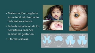
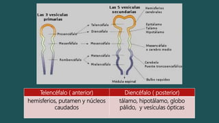
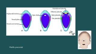
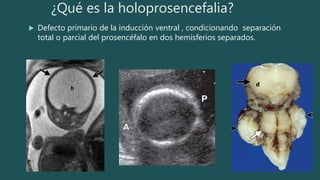
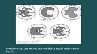
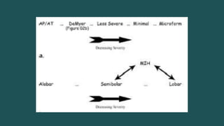
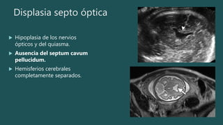
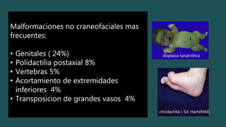
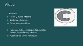
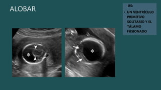
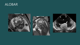
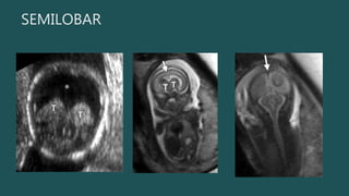
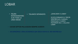
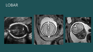
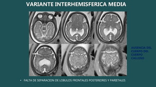
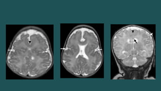
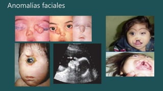
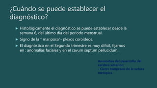
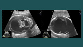
HOLOPROSENCEFALIA
DRA. ANA PAULA CASTILLO GONZÁLEZ
R2 IMAGENOLOGÍA DIAGNÓSTICA Y TERAPÉUTICA
HRAEPY
MIÉRCOLES 29 DE MARZO DEL 2017
• Malformación congénita
estructural más frecuente
del cerebro anterior.
• Falta de separación de los
hemisferios en la 5ta
semana de gestación.
• 3 formas clínicas.
Embriología
 Prosencéfalo: proceso de
inducción ventral.
1. Formación ( aprosencefalia y
atelecenfalia).
2. Formación de la hendidura
(HPE).
3. Desarrollo línea media (agenesia
del cuerpo calloso, displasia septo
óptica, ausencia aislada de septum
pellucidum).
Telencéfalo ( anterior) Diencéfalo ( posterior)
hemisferios, putamen y núcleos
caudados
tálamo, hipotálamo, globo
pálido, y vesículas ópticas
Platillo precordal
¿Qué es la holoprosencefalia?
 Defecto primario de la inducción ventral , condicionando separación
total o parcial del prosencéfalo en dos hemisferios separados.
h
d
Syntelencefalia: una variante interhemisférica medial, recientemente
descrita.
46-50%
18 % 10%
Displasia septo óptica
 Hipoplasia de los nervios
ópticos y del quiasma.
 Ausencia del septum cavum
pellucidum.
 Hemisferios cerebrales
completamente separados.
Epidemiologia
 Malformación de cara y cerebro mas común en humanos.
 < 1 en 10,000 nacidos vivos y nacidos muertos.
 50 en 10, 000 embriones abortados.
 Se postula que una preponderancia mayor en mujeres nacidas
vivas sea secundario a una mayor letalidad en los hombres.
Malformaciones no craneofaciales mas
frecuentes:
• Genitales ( 24%)
• Polidactilia postaxial 8%
• Vertebras 5%
• Acortamiento de extremidades
inferiores 4%
• Transposicion de grandes vasos 4%
displasia tanatrófica
ctrodactilia ( Sd. Hartsfield)
Etiología
 Anomalías cromosómicas ( trisomía 13 24-45%)
 Genéticas (13 genes , no existe una relación genotipo fenotípica
estricta),
 Mutación 20%, una de las fuentes mas importantes de señales de
ventralización durante el desarrollo del cerebro anterior es el gen :
proteína Sónic Hedgehhog.
 Teratógenos
 Diabetes materna, alcohol, tabaco, y ácido retinoico
Alobar
Ausentes:
 Tracto y bulbo olfatorio
 Septum pellucidum
 Fisura interhemisférica
 Fusión en la línea media de los ganglios
basales, hipotálamo y tálamos
 Ausencia del tercer ventrículo.
ALOBAR
US:
• UN VENTRÍCULO
PRIMITIVO
SOLITARIO Y EL
TÁLAMO
FUSIONADO
ALOBAR
 SIGNO “ SERPIENTE DEBAJO DEL
CRÁNEO”.
Semilobar y Lobar
 El único hallazgo
obvio por US a las
18-20 sdg sería la
incapacidad de
detectar el cavum
septum pellucidum.
• La malformación es
semilobar si los
lóbulos frontales
están fusionados
> 50%.
• Hoz y fisura interhemisférica
posterior y parte posterior del cuerpo calloso
Hipotálamo y tálamo
siempre fusionados
SEMILOBAR
SEMILOBAR
SEMILOBAR
LOBAR
• FISURA
INTERHEMISFERIC
A EN TODA LA
LÍNEA MEDIA
• TÁLAMOS SEPARADOS
• CAVUM SEPTUM PELLUCIDUM SIEMPRE AUSENTE
¿SEMILOBAR O LOBAR?
SI ESTÁ FORMADO EL TERCER
VENTRÍCULO, CUERNOS
FRONTALES Y CUERPO
POSTERIOR DEL C. CALLOSO:
LOBAR
• DX ESPECÍFICO: LÍNEA ECOGÉNICA QUE SIGUE HASTA EL 3ER VENTRÍCULO
LOBAR
• FALTA DE SEPARACION DE LOBULOS FRONTALES POSTERIORES Y PARIETALES
AUSENCIA DEL
CUERPO DEL
CUERPO
CALLOSO
VARIANTE INTERHEMISFERICA MEDIA
Anomalías faciales
¿Cuándo se puede establecer el
diagnóstico?
 Histológicamente el diagnóstico se puede establecer desde la
semana 6, del último día del periodo menstrual.
 Signo de la “ mariposa”- plexos coroideos.
 El diagnóstico en el Segundo trimestre es muy difícil, fijarnos
en : anomalías faciales y en el cavum septum pellucidum.
Anomalías del desarrollo del
cerebro anterior:
- Cierre temprano de la sutura
metópica
PRONÓSTICO
 Severidad de malformaciones faciales y del cerebro
 Anomalías cromosómicas
 Involucro de órganos
 ALOBAR: mueren antes del año de edad, 50% antes de los 5 meses.
 LOBAR: 50% pueden deambular, leve función de manos, decir algunas
palabras
 Disfunción hipotalámica: alteraciones en la homeostasis, regulación de la
temperatura, sed y apetito.
MANEJO PRENATAL
 BUSCAR MAS ANOMALÍAS
 CARIOTIPO
 TERMINAR EL EMBARAZO O TIEMPO Y FORMA DE NACIMIENTO PARA EVITAR LA
MORBILIDAD MATERNA.
 FIN

Holoprosencefalia

  • 1.
    HOLOPROSENCEFALIA DRA. ANA PAULACASTILLO GONZÁLEZ R2 IMAGENOLOGÍA DIAGNÓSTICA Y TERAPÉUTICA HRAEPY MIÉRCOLES 29 DE MARZO DEL 2017
  • 2.
    • Malformación congénita estructuralmás frecuente del cerebro anterior. • Falta de separación de los hemisferios en la 5ta semana de gestación. • 3 formas clínicas.
  • 4.
    Embriología  Prosencéfalo: procesode inducción ventral. 1. Formación ( aprosencefalia y atelecenfalia). 2. Formación de la hendidura (HPE). 3. Desarrollo línea media (agenesia del cuerpo calloso, displasia septo óptica, ausencia aislada de septum pellucidum).
  • 5.
    Telencéfalo ( anterior)Diencéfalo ( posterior) hemisferios, putamen y núcleos caudados tálamo, hipotálamo, globo pálido, y vesículas ópticas
  • 6.
  • 7.
    ¿Qué es laholoprosencefalia?  Defecto primario de la inducción ventral , condicionando separación total o parcial del prosencéfalo en dos hemisferios separados. h d
  • 8.
    Syntelencefalia: una varianteinterhemisférica medial, recientemente descrita. 46-50% 18 % 10%
  • 10.
    Displasia septo óptica Hipoplasia de los nervios ópticos y del quiasma.  Ausencia del septum cavum pellucidum.  Hemisferios cerebrales completamente separados.
  • 11.
    Epidemiologia  Malformación decara y cerebro mas común en humanos.  < 1 en 10,000 nacidos vivos y nacidos muertos.  50 en 10, 000 embriones abortados.  Se postula que una preponderancia mayor en mujeres nacidas vivas sea secundario a una mayor letalidad en los hombres.
  • 12.
    Malformaciones no craneofacialesmas frecuentes: • Genitales ( 24%) • Polidactilia postaxial 8% • Vertebras 5% • Acortamiento de extremidades inferiores 4% • Transposicion de grandes vasos 4% displasia tanatrófica ctrodactilia ( Sd. Hartsfield)
  • 13.
    Etiología  Anomalías cromosómicas( trisomía 13 24-45%)  Genéticas (13 genes , no existe una relación genotipo fenotípica estricta),  Mutación 20%, una de las fuentes mas importantes de señales de ventralización durante el desarrollo del cerebro anterior es el gen : proteína Sónic Hedgehhog.  Teratógenos  Diabetes materna, alcohol, tabaco, y ácido retinoico
  • 14.
    Alobar Ausentes:  Tracto ybulbo olfatorio  Septum pellucidum  Fisura interhemisférica  Fusión en la línea media de los ganglios basales, hipotálamo y tálamos  Ausencia del tercer ventrículo.
  • 15.
  • 16.
  • 17.
     SIGNO “SERPIENTE DEBAJO DEL CRÁNEO”.
  • 18.
    Semilobar y Lobar El único hallazgo obvio por US a las 18-20 sdg sería la incapacidad de detectar el cavum septum pellucidum. • La malformación es semilobar si los lóbulos frontales están fusionados > 50%. • Hoz y fisura interhemisférica posterior y parte posterior del cuerpo calloso Hipotálamo y tálamo siempre fusionados SEMILOBAR
  • 19.
  • 20.
  • 21.
    LOBAR • FISURA INTERHEMISFERIC A ENTODA LA LÍNEA MEDIA • TÁLAMOS SEPARADOS • CAVUM SEPTUM PELLUCIDUM SIEMPRE AUSENTE ¿SEMILOBAR O LOBAR? SI ESTÁ FORMADO EL TERCER VENTRÍCULO, CUERNOS FRONTALES Y CUERPO POSTERIOR DEL C. CALLOSO: LOBAR • DX ESPECÍFICO: LÍNEA ECOGÉNICA QUE SIGUE HASTA EL 3ER VENTRÍCULO
  • 22.
  • 23.
    • FALTA DESEPARACION DE LOBULOS FRONTALES POSTERIORES Y PARIETALES AUSENCIA DEL CUERPO DEL CUERPO CALLOSO VARIANTE INTERHEMISFERICA MEDIA
  • 25.
  • 26.
    ¿Cuándo se puedeestablecer el diagnóstico?  Histológicamente el diagnóstico se puede establecer desde la semana 6, del último día del periodo menstrual.  Signo de la “ mariposa”- plexos coroideos.  El diagnóstico en el Segundo trimestre es muy difícil, fijarnos en : anomalías faciales y en el cavum septum pellucidum. Anomalías del desarrollo del cerebro anterior: - Cierre temprano de la sutura metópica
  • 27.
    PRONÓSTICO  Severidad demalformaciones faciales y del cerebro  Anomalías cromosómicas  Involucro de órganos  ALOBAR: mueren antes del año de edad, 50% antes de los 5 meses.  LOBAR: 50% pueden deambular, leve función de manos, decir algunas palabras  Disfunción hipotalámica: alteraciones en la homeostasis, regulación de la temperatura, sed y apetito.
  • 28.
    MANEJO PRENATAL  BUSCARMAS ANOMALÍAS  CARIOTIPO  TERMINAR EL EMBARAZO O TIEMPO Y FORMA DE NACIMIENTO PARA EVITAR LA MORBILIDAD MATERNA.
  • 30.

Notas del editor

  • #4 Cyclopic pig, an engraving by N. F. Regnault, in Morceau de la Sarthe, 1808. Arrow = proboscis. (Reprinted, with permission, from reference 2.)
  • #7 La línea media se forma desde una estructura en la línea media bajo la infuencia del paltillo precodal , el campo visual debe separarse en derecho e izquerdi.
  • #8 Aprosencephaly. Coronal in utero magnetic resonance (MR) image shows failure of normal division of the prosencephalon (forebrain) into telencephalon (cerebrum) and diencephalon (thalamus, hypothalamus). Note the intact calvaria and scalp (arrows) with no supratentorial brain. The key observation is that the falx is absent (* = expected locationn of normal falx). Figure 4. Atelencephaly. (a) Transverse ultrasonographic (US) image shows a featureless appearance with no identifiable brain architecture at 21 weeks gestational age. A = anterior, P = posterior. (b) Autopsy specimen confirms the diagnosis of atelencephaly, with formation of the diencephalon (black arrow) only and absence of telencephalic structures. Normal cerebella (arrowheads) and brainstem structures (white arrow) are present. (Fig 4b reprinted, with permission, from Amirsys [Salt Lake City, Utah].)
  • #10 Minimal y microforma: These cannot be diagnosed in the fetus and will not be further discussed other than to note that identification of these forms in first-degree relatives (including parents) supports an underlying genetic component with potential for higher recurrence risk than in isolated sporadic cases.
  • #11 Coronal endovaginal US image shows SOD in a fetus. Presence of the corpus callosum (arrowhead) with absence of the cavum helped identify SOD, with postnatal confirmation of the diagnosis. Axial T2-weighted half-Fourier rapid acquisition with relaxation enhancement (RARE) fetal MR image shows SOD. Communication between the frontal horns with an absent cavum septum pellucidum (arrow) is seen. No other abnormality was seen.
  • #13 Saber el riesgo de recurrencua de HPE es imposible debido a su etilogia multifactorial-
  • #15 The brain in alobar HPE can be described as ball-, cup-, or pancake-like in shape on the basis of the appearance on sagittal views. In the ball-like form, which is the most common, a relatively generous volume of cerebral cortex completely encircles the monoventricle. In the intermediate stage, the monoventricle is not completely encircled and the remaining cortex has a “cup” configuration. In the “pancake” forremaining cortex is flattened at the base of the skull. m, the small volume of
  • #16 Alobar HPE. (a) Axial oblique US image obtained at 18 weeks shows fused thalami (arrowheads) and a monoventricle (*). (b) Axial oblique US scan obtained at a higher level shows the cuplike morphology of the noncleaved hemispheres (arrows) “cupping” the monoventricle (*). middle and anterior cerebral arteries may be absent, replaced by a network of vessels arising from the internal carotid and basilar vessels
  • #17 Figure 12. Alobar HPE. (a) Coronal fetal MR image shows a monoventricle (*), fused thalami (arrows), and minimal overlying brain tissue (arrowheads). (b) Sagittal T2- weighted half-Fourier RARE MR image shows a flattened midface (arrow) and the “ball” configuration of the remaining cortex (arrowheads) surrounding the monoventricle (*). (c) Coronal US image in the neonate confirms the single monoventricle (*), fused thalami (arrows), and minimal overlying brain tissue (arrowheads).
  • #18 Figure 13. Alobar HPE. Sagittal color Doppler US image of a fetal head shows the “snake under the skull” sign: a single anterior cerebral artery (arrow), also known as an azygous anterior cerebral artery, displaced anteriorly to run under the frontal bones, instead of between the frontal lobes as in a normally formed brain.
  • #19 El diagnóstico prenatal de HPE alobar es bastante sencillo Semilobar y lobar pueden ser un desafío. In semilobar HPE, the interhemispheric fissure and falx cerebri may be present posteriorly and part of the corpus callosum may be seen in the region of the posteriorly sepa rated hemispheres , with the anterior extent corresponding to that of the interhemispheric fissure. somewhat arbitrary criterion used in postnatal imaging is that the malformation is semilobar if the frontal lobes are more than 50% fused
  • #20 Semilobar HPE. (a) Axial oblique US image shows a continuous mantle of brain anteriorly (arrowheads) but posterior division into hemispheres (arrows). (b) More inferior axial US image in the same patient shows normal posterior fossa structures (arrow) below the tentorium but fused anterior supratentorial brain (arrowheads).
  • #21 Semilobar HPE. (a) Coronal US image shows a monoventricle (*) and partial fusion of the thalami (T). (b) Coronal T2-weighted half-Fourier RARE MR image shows the continuous anterior brain parenchyma (arrow), monoventricle (*), and partially fused thalami (T). (c) Coronal T2-weighted half-Fourier RARE MR image more posteriorly shows two occipital lobes separated by the falx (arrow).
  • #23 Lobar HPE. (a) Axial US image shows a continuous midline echo anteriorly (arrow). We were not able to demonstrate the cavum septum pellucidum in any plane. (b) Axial fetal MR image shows apparent complete division of the cerebral hemispheres (arrowheads). (c) Coronal fetal MR image shows anterior inferior gyral continuity between the frontal lobes (arrow). This was not demonstrable at US; the index finding was inability to demonstrate a normal cavum septum pellucidum.
  • #24 MIH. Axial T2-weighted half-Fourier RARE MR images obtained at 33 weeks estimated gestational age. Note the lack of midline separation in the posterior frontal and parietal areas (*), while rostral and posterior regions of the cerebrum are cleaved normally, and the orbits are spaced normally. Also, note the vertically oriented sylvian fissures (arrows), which abnormally connect across the vertex.
  • #25 MIH. Axial (a, b) and coronal (c) T2-weighted MR images in a neonate diagnosed prenatally with MIH show a lack of separation in the posterior frontal lobes (black arrowhead). Also note the abnormal appearance of the vertically oriented sylvian fissures (arrows in a and b), which extend more toward the midline than expected, and the cortical dysplasia (white arrowhead in c) and gray matter heterotopia (arrow in c).
  • #27 The butterfly sign may be reliably seen in a cross section of the normal first-trimester brain. The choroid plexus is narrow in the middle but thicker at both ends, and when seen side by side the choroids create a butterfly appearance.