Descargar como PPS, PPTX


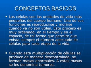
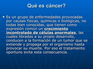


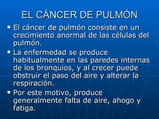

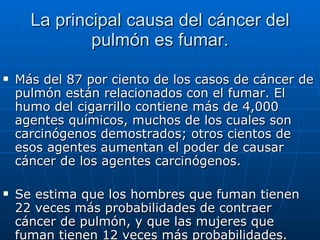

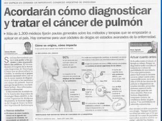


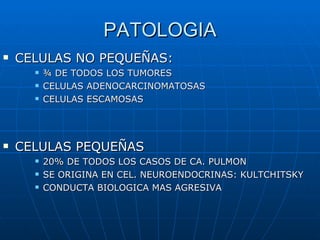


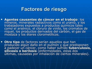
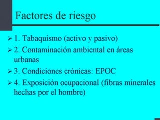


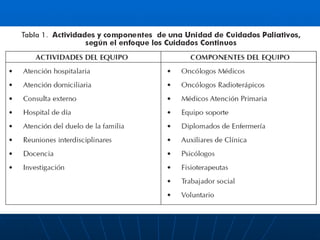


Este documento trata sobre conceptos básicos del cáncer de pulmón. Explica que el cáncer de pulmón se produce por un crecimiento anormal e incontrolado de las células pulmonares, y que el principal factor de riesgo es fumar tabaco. Describe los dos principales tipos de cáncer de pulmón, de células pequeñas y de células no pequeñas, y sus características. Finalmente, analiza las causas y factores de riesgo asociados al desarrollo de cáncer de pulmón.


