

Recomendados
Recomendados
Más contenido relacionado
La actualidad más candente
La actualidad más candente (20)
Destacado
Destacado (20)
Similar a Sangre 2
Similar a Sangre 2 (20)
Más de Michelle Ceseña
Más de Michelle Ceseña (20)
Sangre 2
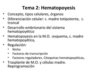
- 1. Tema 2: Hematopoyesis • Concepto, tipos celulares, órganos • Diferenciación celular: c. madre totipotente, c. troncal • Desarrollo embrionario del sistema hematopoyético • Hematopoyesis en la M.O. :esquema, c. madre hematopoyética, • Regulación: • Nicho • Factores de transcripción • Factores reguladores. Citoquinas hematopoyéticas, • Trasplante de M.O. y células madre. Reprogramación
- 2. Concepto • Formación de las células sanguíneas: – Eritropoyesis – Trombopoyesis – Leucopoyesis – Linfopoyesis • Periodos – Desarrollo embrionario – Posembrionario (hematopoyesis sensu estricto)
- 3. Tipos celulares básicos dependiendo de origen • Mieloide: –Eritrocitos (hematíes) –Leucocitos (excepto linfocitos) –Plaquetas. • Linfoide –Linfocitos T y B. –NK
- 4. ÓRGANOS HEMATOPOYÉTICOS primarios • Saco vitelino (bolsa amniótica): 0-3 meses feto: hematíes nucleados • AGM (Mesonefros aórtico-gonadal) • Hígado: 1 mes vida fetal - nacimiento • Bazo : 2,5 meses vida fetal - nacimiento • Huesos largos: 3,5 meses vida fetal- 25 años • Huesos esponjosos: 3,5 meses vida fetal - resto de la vida • Timo (LT)
- 5. ESQUEMA BÁSICO DIFERENCIACION CELULAR Célula madre (troncal) (pluripotente) ↓ Células progenitoras ↓ Células diferenciadas ↓ Muerte (programada o por envejecimiento) en días o meses Lodish y cols.: ”Molecular Cell Biology”.5º Ed. W.H. Freeman 2004
- 6. Célula madre totipotente Es la que tiene capacidad de originar mesodermo, endodermo y ectodermo: sólo pueden obtenerse de embriones (blastocisto) y de las gonadas de fetos – Célula madre embrionaria – Célula germinal fetal Lodish y cols.: ”Molecular Cell Biology”.5º Ed. W.H. Freeman 2004 Masa celular interna
- 7. Células troncales en el desarrollo Martinez-Agosto et al. Genes Dev. 2007 21: 3044-3060
- 8. Célula troncal (stem cell) • Es una célula del embrión o del adulto que tiene la capacidad, en circunstancias determinadas, de dar lugar a células iguales a ella, o a células especializadas que originan los distintos tejidos y órganos. Lodish y cols.: ”Molecular Cell Biology”.5º Ed. W.H. Freeman 2004
- 9. • Célula troncal célula diferenciada presente en un tejido diferenciado, que se renueva y que puede dar lugar a células especializadas: Médula ósea –Sangre –Córnea –Cerebro –Músculo –Pulpa dental –Hígado –Piel –Tubo digestivo –Páncreas • Célula progenitora o precursora: da lugar a células especializadas
- 10. Stuart H. Orkin, and Leonard I. Zon: Cell 132, 631–644, February 22, 2008 • La colonización de células madre (troncales) se produce en olas • La producción celular embrionaria está dirigida sobre todo a la formación de eritrocitos. Desarrollo embrionario
- 11. Médula ósea • Roja • Amarilla (grasa) • Es uno de los órganos más voluminosos del cuerpo humano. • Estroma y células sanguíneas – 75% leucocitos – 25% eritrocitos – 1 célula madre/104 células
- 12. • Células sanguíneas: 2x10¹¹ / día • Epitelio intestinal : 1011 / día • Otras células con alto recambio: epidermis, espermatozoides Producción celular
- 13. Tipos morfológicos de las células generadas: • nucleadas : leucocitos • no nucleadas : eritrocitos • partículas celulares: plaquetas.
- 14. CD34+, CD38- , c-kit+ (CD117) CD34+, CD38- c-kit+ CD38+ , CD3, CD4, CD8, ……. Lodish y cols.: ”Molecular Cell Biology”.5º Ed. W.H. Freeman 2004
- 15. Célula madre hematopoyética • CD34+ , CD38-, c-kit+ • Alta capacidad regenerativa • Criopreservable • Regenera (trasplante): eritrocitos, plaquetas, leucocitos, macrófagos tisulares, osteoclastos, células de Langerhans de la piel
- 16. Hematopoyesis: REGULACIÓN ERITROCITOS LEUCOCITOS F. ESTIMULADORES Citoquinas hematopoyéticas F. APOPTÓTICOS ENVEJECIMIENTO
- 17. Hematopoyesis: regulación • Localización célula madre ML: Factores locales “nicho adecuado”. Factores derivados del estroma, factores derivados de los osteoblastos • Estimulación proliferación y supervivencia : citoquinas – Clásicas (IL1, IL3; IL6) – Específicas: Factores estimuladores de colonias (CSF) – Hormonas-citoquinas: EPO, trombopoyetina
- 18. C.Troncal Hematop. duradera CTH de vida corta Precusror mieloide común Precursor linfoide común Prec. Megacariocitos/eritro, Prec. Granulocitos/macrófagos
- 19. Factores Reguladores De La Hematopoyesis • Factores Estimuladores – SF (Steel factor, CD117)-cKit • Factores estimuladores de colonias (CSF) – CSF-GM (granulocitos-macrófagos): CSF 2(sargramostim) – CSF-G (granulocitos): CSF 3 (lenograstim) – CSF-M (monocitos, macrofagos): CSF1(lanimostim) – Eritropoyetina (EPO) – Trombopoyetina • Citoquinas – Interleuquinas – Quimioquinas • Hormonas: tiroideas, insulina..
- 20. c-kit / SF
- 21. Importancia Citoquinas Hematopoyéticas Donald Metcalf :BLOOD, 15 JANUARY 2008 VOLUME 111, NUMBER 2
- 22. Lodish y cols.: ”Molecular Cell Biology”.5º Ed. W.H. Freeman 2004 Importancia Citoquinas Hematopoyéticas
- 23. Origen de las citoquinas hematopoyéticas Donald Metcalf :BLOOD, 15 JANUARY 2008 VOLUME 111, NUMBER 2
- 24. Tipos de regulación por citoquinas hematopoyéticas Restringida a estirpe celular Actividad plural Actividad secuencial Acción de las citoquinas hematopoyéticas Donald Metcalf :BLOOD, 15 JANUARY 2008 VOLUME 111, NUMBER 2
- 25. Funciones múltiples de las citoquinas hematopoyéticas Donald Metcalf :BLOOD, 15 JANUARY 2008 VOLUME 111, NUMBER 2
- 26. Mecanismo de acción citoquinas Lodish y cols.: ”Molecular Cell Biology”.5º Ed. W.H. Freeman 2004
- 27. Trasplante M.O. TIPOS • Auto: no rechazo – Peligro: trasplante de células tumorales. • Alo (misma especie) : rechazo ↑ FUENTE: M.O. Sangre circulante: escasas células madre. Aumentan con la administración de factores hematopoyéticos (G-CSF, GM-CSF). El Alo es más peligroso por la administración conjunta de LT Cordón umbilical: muchas células. Bancos.
- 28. Reprogramación células diferenciadas: celulas pluripotentes inducidas Induction of Pluripotent Stem Cells from Adult Human Fibroblasts by Defined Factors. Kazutoshi Takahashi,Koji Tanabe, Mari Ohnuki, Megumi Narita, Tomoko Ichisaka, Kiichiro Tomoda, and Shinya Yamanaka. Cell 2007), doi:10.1016/j .cell.2007.11.019 SUMMARY Successful reprogramming of differentiated human somatic cells into a pluripotent state would allow creation of patient- and disease-specific stem cells. We previously reported generation of induced pluripotent stem (iPS) cells, capable of germline transmission, from mouse somatic cells by transduction of four defined transcription factors. Here, we demonstrate the generation of iPS cells from adult human dermal fibroblasts with the same four factors: Oct3/4, Sox2, Klf4, and c-Myc. Human iPS cells were similar to human embryonic stem (ES) cells in morphology, proliferation, surface antigens, gene expression, epigenetic status of pluripotent cell-specific genes, and telomerase activity. Furthermore, these cells could differentiate into cell types of the three germ layers in vitro and in teratomas. These findings demonstrate that iPS cells can be generated from adult human fibroblasts.
Notas del editor
- Evidence of a Pluripotent Human Embryonic Stem Cell Line Derived from a Cloned Blastocyst [Reports: Developmental Biology] Hwang, Woo Suk1,2*; Ryu, Young June1; Park, Jong Hyuk3; Park, Eul Soon1; Lee, Eu Gene1; Koo, Ja Min4; Jeon, Hyun Yong1; Lee, Byeong Chun1; Kang, Sung Keun1; Kim, Sun Jong3; Ahn, Curie5; Hwang, Jung Hye6; Park, Ky Young7; Cibelli, Jose B.8; Moon, Shin Yong5* Somatic cell nuclear transfer (SCNT) technology has recently been used to generate animals with a common genetic composition. In this study, we report the derivation of a pluripotent embryonic stem (ES) cell line (SCNT-hES-1) from a cloned human blastocyst. The SCNT-hES-1 cells displayed typical ES cell morphology and cell surface markers and were capable of differentiating into embryoid bodies in vitro and of forming teratomas in vivo containing cell derivatives from all three embryonic germ layers in severe combined immunodeficient mice. After continuous proliferation for more than 70 passages, SCNT-hES-1 cells maintained normal karyotypes and were genetically identical to the somatic nuclear donor cells. Although we cannot completely exclude the possibility that the cells had a parthenogenetic origin, imprinting analyses support a SCNT origin of the derived human ES cells.
- Figure 1. Stem cells in the context of development. ( A–C ) Embryos consist of mitotically dividing cells called progenitors. Progenitors can be pluripotent (e.g., blastomeres in mammalian embryos) or multipotent (e.g., ectoderm or mesoderm). ( D ) At later developmental stages, cells exit the mitotic cycle. Generally called precursors, these cells can still be multipotent (e.g.,cells of imaginal discs in Drosophila ). At some point precursors become committed to a particular fate and differentiate. ( E ) Stem cells (e.g., HSCs) develop from embryonic progenitors that are prevented from exiting the mitotic cycle by specific microenvironments, called niches. ( F ) In the adult organism, stem cells undergo asymmetric cell divisions and produce mitotically active daughter cells also called progenitors (“transient amplifying cells”).
- Figure 1. Developmental Regulation of Hematopoiesis in the Mouse (A) Hematopoiesis occurs first in the yolk sac (YS) blood islands and later at the aorta-gonad mesonephros (AGM) region, placenta, and fetal liver (FL). YS blood islands are visualized by LacZ staining of transgenic embryo expression GATA-1- driven LacZ. AGM and FL are stained by LacZ in Runx1-LacZ knockin mice. (Photos courtesy of Y. Fujiwara and T. North.) (B) Hematopoiesis in each location favors the production of specific blood lineages. Abbreviations:ECs, endothelial cells; RBCs, red blood cells; LTHSC, long-term hematopoietic stem cell; ST-HSC, short-termhematopoietic stemcell;CMP,common myeloid progenitor; CLP, common lymphoid progenitor; MEP,megakaryocyte/erythroid progenitor; GMP, granulocyte/macrophage progenitor. (C) Developmental timewindows for shifting sites of hematopoiesis
- Figure 3. Stem Cell Niche in the Adult Bone Marrow HSCs are found adjacent to osteoblasts that are under the regulation of bone morphogenetic protein (BMP) (the osteobast niche). HSCs are also found adjacent to blood vessels (the vascular niche). The chemokine CXCL12 regulates the migration of HSCs from the circulation to the bone marrow. The osteoblast vascular niches in vivo lie in close proximity or may be interdigitated. The marrow space also contains stromal cells that support hematopoiesis including the production of cytokines, such as c-Kit ligand, that stimulate stem cells and progenitors. Cytokines, including interleukins, thrombopoietin (Tpo), and erythropoietin (Epo), also influence progenitor function and survival Stuart H. Orkin, and Leonard I. Zon:Cell 132, 631–644, February 22, 2008
- Figure 4. Requirements of Transcription Factors in Hematopoiesis The stages at which hematopoietic development is blocked in the absence of a given transcription factor, as determined through conventional gene knockouts, are indicated by red bars. The factors depicted in black have been associated with oncogenesis. Those factors in light font have not yet been found translocated or mutated in human/mouse hematologic malignancies. Abbreviations: LT-HSC, long-term hematopoietic stem cell; ST-HSC, short-term hematopoietic stem cell; CMP, common myeloid progenitor; CLP, common lymphoid progenitor; MEP, megakaryocyte/erythroid progenitor; GMP, granulocyte/macrophage progenitor; RBCs, red blood cells. Stuart H. Orkin, and Leonard I. Zon: Cell 132, 631–644, February 22, 2008
- The EMBO Journal Vol. 19, pp. 1312-1326, 2000 Point mutation in Kit receptor tyrosine kinase reveals essential roles for Kit signaling in spermatogenesis and oogenesis without affecting other Kit responses Holger Kissel , Inna Timokhina 1, Matthew P. Hardy , Gerson Rothschild 1, Youichi Tajima 1, Vera Soares 1, Michael Angeles , Scott R. Whitlow , Katia Manova 1 and Peter Besmer 1,2 1 Molecular Biology Program, Memorial Sloan-Kettering Cancer Center, 6 Molecular Cytology Core Facility, Memorial Sloan-Kettering Cancer Center, 1275 York Avenue, 2 Cornell University Graduate School of Medical Sciences and 3 Population Council, Center for Biomedical Research, New York, NY 10021, USA 4 Present address: Department of Biochemical Cell Research, Tokyo Metropolitan Institute of Medical Sciences, Tokyo 113, Japan 5 Present address: University of Iowa, 440 Eckstein Medical Research Building, Iowa City, IA 52242, USA Abstract The Kit receptor tyrosine kinase functions in hemato- poiesis, melanogenesis and gametogenesis. Kit receptor-mediated cellular responses include proliferation, survival, adhesion, secretion and differentiation. In mast cells, Kit-mediated recruitment and activation of phosphatidylinositol 3'-kinase (PI 3-kinase) produces phosphatidylinositol 3'-phosphates, plays a critical role in mediating cell adhesion and secretion and has contributory roles in mediating cell survival and proliferation. To investigate the consequences in vivo of blocking Kit-mediated PI 3-kinase activation we have mutated the binding site for the p85 subunit of PI 3-kinase in the Kit gene, using a knock-in strategy. Mutant mice have no pigment deficiency or impairment of steady-state hematopoiesis. However, gametogenesis is affected in several ways and tissue mast cell numbers are affected differentially. While primordial germ cells during embryonic development are not affected, KitY719F/KitY719F males are sterile due to a block at the premeiotic stages in spermatogenesis. Furthermore, adult males develop Leydig cell hyperplasia. The Leydig cell hyperplasia implies a role for Kit in Leydig cell differentiation and/or steroido- genesis. In mutant females follicle development is impaired at the cuboidal stages resulting in reduced fertility. Also, adult mutant females develop ovarian cysts and ovarian tubular hyperplasia. Therefore, a block in Kit receptor-mediated PI 3-kinase signaling may be compensated for in hematopoiesis, melano- genesis and primordial germ cell development, but is critical in spermatogenesis and oogenesis.
- Figure 4. The importance of a hematopoietic cytokine such as G-CSF can be validated in several ways. (A) By injecting G-CSF to elevate neutrophil levels and (B) by deleting the gene, a procedure resulting in low neutrophil levels and poor neutrophil responses to challenge infections.
- Figure 2. Varying tissue origin of hematopoietic cytokines. (A) EPO is mainly a product of kidney tissue. (B) GM-CSF is a product of multiple tissues and cell types. (C) M-CSF, CSF-1 can be a humoral factor and the product of many tissues or a membrane-displayed factor on local stromal cells.
- Figure 1. Three types of action of hematopoietic cytokines. (A) Lineage restricted. (B) Action on multiple lineages; broken line shows actions only at high concentrations. (C) Sequential actions; SCF acts on stem and early erythroid progenitors, while EPO acts on more mature precursors. The notion of sequential actions was later found to be incorrect.
- Figure 3. Hematopoietic cytokines are polyfunctional. Hematopoietic cytokines such as G-CSF are not simply mandatory proliferative stimuli but also act on cell survival, differentiation commitment, maturation induction, and the functional stimulation of mature cells.