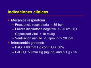
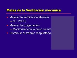
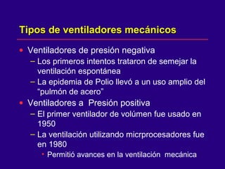
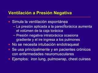
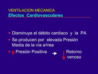
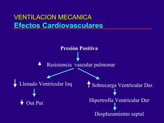
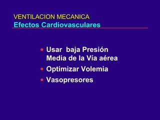
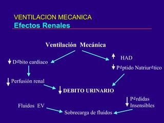
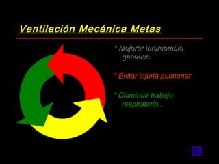
SlideShare una empresa de Scribd logo
VENTILACION MECANICAVENTILACION MECANICA
Indicaciones de Ventilación mecánicaIndicaciones de Ventilación mecánica
• Primarias
– Ventilación espontánea inadecuada
• disminución del pH
– Hipoxemia refractaria a alto flujo de oxígeno
• PaO2 < 60 mm Hg con FiO2 > 50%
• Los gases arteriales indican la insuficiencia
respiratoria
Indicaciones de Ventilación mecánicaIndicaciones de Ventilación mecánica
• Expansión pulmonar inadecuada
• Fatiga de los músculos respiratorios
• Trabajo respiratorio excesivo (WOB)
• Protección en el post operatorio
• TEC grave
– Disminuir el PaCO2
• Tórax inestable
Ventilación MecánicaVentilación Mecánica
INDICACIONESINDICACIONES
• Falla de la Ventilación Alveolar
• Hipertensión endocraneana
• Hipoxemia severa
• Profilxis frente a inestabilidad
hemodinámica
• Aumento del trabajo respiratorio
• Tórax inestable
• FR > 30 a 35
Indicaciones clínicasIndicaciones clínicas
• Mecánica respiratoria
– Frecuencia respiratoria > 35 bpm
– Fuerza inspiratoria negativa < -25 cm H2O
– Capacidad vital < 10 ml/kg
– Ventilación minuto < 3 lpm or > 20 lpm
• Intercambio gaseoso
– PaO2 < 60 mm Hg con FiO2 > 50%
– PaCO2 > 50 mm Hg (agudo) and pH < 7.25
Metas de la Ventilación mecánicaMetas de la Ventilación mecánica
• Mejorar la ventilación alveolar
– pH, PaCO2
• Mejorar la oxigenación
– Monitorizar con la pulso oximetria
• Disminuir el trabajo respiratorio
Tipos de ventiladores mecánicosTipos de ventiladores mecánicos
• Ventiladores de presión negativa
– Los primeros intentos trataron de semejar la
ventilación espontánea
– La epidemia de Polio llevó a un uso amplio del
“pulmón de acero”
• Ventiladores a Presión positiva
– El primer ventilador de volúmen fue usado en
1950
– La ventilación utilizando micrprocesadores fue
en 1980
• Permitió avances en la ventilación mecánica
Ventilación a Presión NegativaVentilación a Presión Negativa
• Simula la ventilación espontánea
– La presión aplicada a la pared torácica aumenta
el volúmen de la caja torácica
– Presión negativa intratorácica ocasiona
gradiente y el ire ingresa a los pulmones
• No se necesita intubación endotraqueal
• Se usa principalmente y en pacientes crónicos
con enfermedades neuromusculares
• Ejemplos: iron lung, pulmowrap, chest cuirass
Ventilación a presión PositivaVentilación a presión Positiva
• El aire es aplicado a presión positiva y se
produce el flujo de gas a los pulmones
• Los cambios de la presión intratorácica son
opuestos a la respiración espontánea
• Impide el retorno venoso
• El paciente puede necesitar aporte de volúmen
intravenoso
Ventilación a Presión PositivaVentilación a Presión Positiva
• La presión intratorácica
permanece positiva durante el
ciclo respiratorio
• El flujo de gas se dirige a las
zonas de menor resistencia
• El gas se distribuye a zonas
menor perfundidas
• Disbalance
Ventilación/Perfusión
Respiración espontánea vs PresiónRespiración espontánea vs Presión
positivapositiva
I E I E
Presión
Volúmen
Espontáneo Presión Positiva
I E I E
VENTILACION MECANICAVENTILACION MECANICA
Efectos CardiovascularesEfectos Cardiovasculares
• Disminuye el débito cardiaco y la PA
• Se producen por elevada Presión
Media de la vía a₫rea
• Presión Positiva Retorno
venoso
VENTILACION MECANICAVENTILACION MECANICA
Efectos CardiovascularesEfectos Cardiovasculares
Presión Positiva
Resistencia vascular pulmonar
Llenado Ventricular Izq
Out Put
Sobrecarga Ventricular Der.
Hipertrofia Ventricular Der
Desplazamiento septal
VENTILACION MECANICAVENTILACION MECANICA
Efectos CardiovascularesEfectos Cardiovasculares
• Usar baja Presión
Media de la Vía aérea
• Optimizar Volemia
• Vasopresores
VENTILACION MECANICAVENTILACION MECANICA
Efectos RenalesEfectos Renales
Ventilación Mecánica
D₫bito cardiaco
Perfusión renal
DEBITO URINARIO
HAD
P₫ptido Natriur₫tico
Sobrecarga de fluidos
Fluidos EV
P₫rdidas
Insensibles
VENTILACION MECANICAVENTILACION MECANICA
Efectos GastrointestinalesEfectos Gastrointestinales
• Distensión ( meteorismo )
• Ulceras de estres
• HDA
VENTILACION MECANICAVENTILACION MECANICA
Efectos NeurológicosEfectos Neurológicos
Ventilación Mecánica
Retorno Venoso
Presión Intracerebral
Presión de Perfusión Cerebral
Debito
PA
PPC = PAM - PIC
VENTILACION MECANICAVENTILACION MECANICA
Efectos NeurológicosEfectos Neurológicos
• Usar baja Presión Media
de la Vía aérea
• Usar Bajo PEEP
VENTILACION MECANICAVENTILACION MECANICA
Efectos NutricionalesEfectos Nutricionales
Subalimentados
• Catabolismo muscular
• Neumonía
• Edema pulmonar
VENTILACION MECANICAVENTILACION MECANICA
Efectos NutricionalesEfectos Nutricionales
Sobrealimentados
• Mayor producción de C02
* Calorimetría Indirecta
Programación Básica del VentiladorProgramación Básica del Ventilador
• FiO2
– Graduar con
pulsooximetría
– Meta: < 50%
• Frecuencia
– 10 to 20 bpm
• Volúmen Tidal (VT)
– 7 to 12 cc/kg
• Sensibilidad
– Disparo por presión o
flujo
• Flujo pico, Tiempo
inspiratorio o Relación
I/E
– Controla cuan rápido el
volúmen tidal es
entregado , o cuanto
tiempo la presión
iinspiratoria programada
es aplicada
• Patrón de flujo
– Cuadrado Vs
desacelerada
• Mode of ventilation
– A/C, SIMV, Espontánea
– Volume o Presión
CMV
CMV
IPPVIPPV
SIMV
SIMV
MMV
MMV
BIPAPBIPAP
CPAP
CPAP
SPONT
SPONT
PCVPCV
VCV
VCV
APRV
APRV
PLV
PLV
PSPS
ASBASB
ILV
PRVCPRVC
VAPS
VAPS
PAVPAV
Que estratégia debería utilizar?Que estratégia debería utilizar?
Auto ModeAuto Mode
AutoFlowAutoFlow
PPSPPS
VSVS
Ventilación Mecánica MetasVentilación Mecánica Metas
* Mejorar intercambio* Mejorar intercambio
gaseoso.gaseoso.
* Evitar injuria pulmonar* Evitar injuria pulmonar
* Disminuir trabajo* Disminuir trabajo
respiratorio .respiratorio .
Ventilación MecánicaVentilación Mecánica
• Injuria Bioquímica:
• Mediadores InflamatoriosMediadores Inflamatorios
• MacrófagosMacrófagos
• NeutrófilosNeutrófilos
• Translocación Bacteriana.Translocación Bacteriana.
•⇓⇓
• Falla MultisistémicaFalla Multisistémica
• Injuria BiofísicaInjuria Biofísica
• Sobredistensión AlveolarSobredistensión Alveolar
• Apertura y Colapso CíclicoApertura y Colapso Cíclico
• ↑↑ Presión IntratorácicaPresión Intratorácica
• ↑↑ Permeabilidad alveolo-Permeabilidad alveolo-
capilarcapilar
• ↓↓Gasto CardiacoGasto Cardiaco
• ↓↓Perfusión sistémicaPerfusión sistémica
•⇓⇓
• Falla MultisistémicaFalla Multisistémica
Metas en SDRAMetas en SDRA
• Controlar y minimizar el daño pulmonarControlar y minimizar el daño pulmonar
• Literatura sugiereLiteratura sugiere
– Presiones alveolares bajaasPresiones alveolares bajaas
– Mas preocupación por el daño pulmonarMas preocupación por el daño pulmonar
causado por sobredistensióncausado por sobredistensión
– Prevenir colapso y reapertura alveolarPrevenir colapso y reapertura alveolar
repetidarepetida
Recomendaciones para VentilaciónRecomendaciones para Ventilación
MecánicaMecánica
• ACCP ConcensusACCP Concensus
Conference. Chest 1993.Conference. Chest 1993.
• Modo: El mas familiar.
• Oxigenación: Sp02 ⇒ 90%
• Plateau: 〈 35 mmHg (↓ VT)
• Hipercapnia Permisiva
• PEEP : Siempre. (Menor posible)
• FiO2: Menor Posible (Sp02)
• Hipoxemia;
Sedación/Paralisis/Prono
• Protocolo del Dr. M. Amato
• ↓ Vt
• ↓ Frecuencia Respiratoria
• Limitación de Presión Pico
• PEEP: (⇒ Pflex) 2cm H2O
• Maniobra de Reclutamiento 40 / 40.
Upper And Lower Inflection PointsUpper And Lower Inflection Points
0 20 40 602040-60
0.2
LITERS
0.4
0.6
Paw
cmH2O
VT
Upper AndUpper And Lower Inflection PointsLower Inflection Points
0 20 40 602040-60
0.2
LITERS
0.4
0.6
Paw
cmH2O
VT
Alveolar collapse
P
T
Lower inflection points are thought to be a point of critical opening pressure
SensibilidadSensibilidad
• Su programación establece la variable de
disparo
• El trigger determina cuando el ventilador
reconocerá el esfuerzo inspiratorio del
paciente
• Cuando el esfuerzo del paciente es
reconocido el ventilador entregará una
respíración.
• El trigger puede ser un cambio en presión o
flujo
Sensibilidad por PresiónSensibilidad por Presión
• El esfuerzo inspiratorio del paciente se inicia
con la contracción del diafragma
• Este esfuerzo disminuye la presión en el
circuito del ventilador (sistema cerrado)
X X
Sensibilidad por PresiónSensibilidad por Presión
• Cuando la presión disminuye y alcanza la
sensibilidad programada, el ventilador
dispara una respiración .
• Hay un pequeño retardo de tiempo desde el
inicio del esfuerzo del paciente hasta que el
ventilador reconoce y entrega una
respiración.
Baseline
Trigger
Patient effort
Pressure
Sensibilidad por PresiónSensibilidad por Presión
• Sensibilidad por Presión programada a -2 cm
H2O
• Los primeros 2 esfuerzos del paciente
alcanzan la sensibilidad por presión y el
ventilador dispara la respiración programada.
• El tercer esfuerzo del paciente no alcanza la
sensibilidad, el ventilador no reconoce el
esfuerzo
-2 cm H2O
Disparo por FlujoDisparo por Flujo
• El ventilador entrega un flujo constante en el
circuito del paciente (sistema abierto)
Delivered flowReturned flow
No patient effort
Disparo por FlujoDisparo por Flujo
• El esfuerzo inspiratorio del paciente se inicia
con la contracción del diafragma
• Al iniciar la inspiración , algo de este flujo
constante es desviado al paciente
Delivered flowLess flow returned
Disparo por FlujoDisparo por Flujo
• El bajo nivel de flujo necesario satisface el
esfuerzo inspiratorio inicial del paciente
• Hay un retardo mínimo entre el esfuerzo del
paciente y la respiración entregada
• Mejor tiempo de respuesta del ventilador
cuando se compara con disparo por presión
All inspiratory efforts recognized
Tim
e
Pressure
Modos VentilatoriosModos Ventilatorios
• Ventilación Asistida/Controlada (A/C)
• Ventilación Mandatoria Intermitente
Sincronizada (SIMV)
• Ventilación controlada por presión (PCV)
• Espontánea
– Ventilación con Soporte de Presión (PSV)
– Presión Positiva continua en vías aéreas /
Presión Positiva al final de la espiración
(CPAP/PEEP)
Asistida / ControladaAsistida / Controlada
• Las respiraciones se entregan según lo
programado :
– Volúmen tidal
– Flujo pico y forma de la onda
– Frecuencia respiratoria base
• Las respiraciones iniciadas por la máquina o
el paciente se entregan con estos parámetros
Time
Pressure
Ventilación AsistidaVentilación Asistida
• PC
• PS
• BiPAP/BiLevel
• APRV
• Volume Assist/Control
• Volume SIMV
• PRVC/AutoFlow
• VS
• VAPS/ Pres Aug
Presión constante Volumen Constante
PAVPAV
Sincronía Paciente/VentiladorSincronía Paciente/Ventilador
• Alcanzar las demandas de flujo del paciente y mejorar
la sincronía paciente / ventilador
– Demandas de flujo variable / tiempos inspiratorios
variables
• PCV permite al paciente tener el flujo que necesita
pero controlamos el Tiempo inspiratorio
• PS permite al paciente tener el flujo que quieran y el
tiempo inspiratorio que deseen
Asistida / ControladaAsistida / Controlada
• Ventajas
– Proporciona soporte ventilatorio completo
– El paciente controla la frecuencia respiratoria
• Desventajas
– La programación puede no estar sincronizada con
las demandas ventilatorias del paciente
– Al aumentar la frecuencia respiratoria , aumenta la
ventilación minuto proporcionalmente
• Causa hiperventilación
VCV: ONDAS DE FLUJO INSPIRATORIO
AFECTAN ONDAS DE PRESION
VCV
A: C normal
B: C alta
C: C baja
SIMVSIMV
• Combinación de respiración de la máquina y
espontánea
• La respiración mandatoria se entrega cuando
se sensa el esfuerzo del paciente
(sincronizada)
• El paciente determina el volúmen tidal y la
frecuencia de la respiración espontánea
Time
Pressure
Synchronized machine breath
Patient effort
SIMVSIMV
• Ventajas
– Las respiraciones sincronizadas mejoran el
comfort del paciente
– Se reduce la competencia entre el paciente y
el ventilador
– Ocasiona menos hiperventilación, comparado
con A/C
SIMVSIMV
• Desventajas
– Puede ocasionar soporte insuficiente si la
frecuencia o el Vt programado es muy bajo
– Puede aumentar WOB
• Hay espacio de tiempo entre el esfuerzo del
paciente y el flujo entregado
• Resistencia del TET y el circuito
SIMV
Ventilación Control Volumen Vs. PresiónVentilación Control Volumen Vs. Presión
Ventilación Volúmen
• El Volúmen entregado es
consrtante
• La presión inspiratoria
varía
• Flujo inspiratorio
constante
• Tiempo inspiratorio
determinado por el flujo y
el volúmen programado
Ventilación Presión
• Varía el volúmen entregado
• Presión inspiratoria
constante
• Varía el flujo inspiratorio
• El tiempo inspiratorio es
programado por el médico
Ventilación Control de PresiónVentilación Control de Presión
• Definición
– Es la aplicación de una presión inspiratoria y
un tiempo inspiratorio programado por el
médico. El flujo entregado varía de acuerdo a
la demanda del paciente .
• El médico programa la presión inspiratoria,
tiempo inspiratorio o relación I:E y FR
• El volúmen tidal varía con cambios en la
compliance y la resistencia
• El flujo entregado es desacelerante
Ventilación Control de PresiónVentilación Control de Presión
• Puede ser usado en modos A/C y SIMV
• En A/C - todas las respiraciones (iniciadas
por la máquina o el paciente ) son cicladas
por tiempo y limitadas por presión
• En SIMV - solo las respiraciones iniciadas por
la máquina son cicladas por tiempo y
limitadas por presión
– Las respiraciones espontáneas pueden ser
soportadas por presión
Pressure Control VentilationPressure Control Ventilation
P
R
E
S
S
U
R
E
I-time
F
L
O
W
Pressure constant
Ventilación control PresiónVentilación control Presión
• Ventajas
– Limita el riesgo de barotrauma
– Puede reclutar alveolos colapsados y
congestivos
– Mejora la distribución de gases
• Desventajas
– Los volúmenes tidales varían cuando cambia
la compliance (e.j. SDRA, , edema pulmonar )
– Con aumentos en el tiempo inspiratorio, el
paciente puede requerir sedación o parálisis
Indicaciones de PCVIndicaciones de PCV
• Mejorar sincronía paciente / ventilador
– El paciente determina el flujo
• Estrategia de protección pulmonar
– Presiones inspiratorias bajas con flujo
desacelerante pueden mejorar relación V/Q
– Ajustando el tiempo inspiratorio aumenta la
presión media de las vías aéreas y puede
mejorar la oxigenación
• En las enfermedades alveolares que producen
tiempos constantes variables
– Se pueden reclutar alveolos al aumentar el
tiempo inspiratorio
VCP
A: C normal
B: C alta
C: C baja
VCP
VCP-IMV
PC-SIMV
VENTILACION I:E INVERSAVENTILACION I:E INVERSA
• Normal I : E = 1 : 3 o 1 : 2
• El incremento del tiempo I y el acortamiento del
tiempo E aumenta la presión media de la vía
aérea y mejora la oxigenación
• Requiere sedación y analgesia
• Debe hacerse monitoreo hemodinámico y
determinación de auto PEEP
PCV
RELACION I:E
INVERSA
VCV
Relación I:E inversa
•Flujo inspiratorio bajo
•Pausa inspiratoria
Ventilación Presión SoporteVentilación Presión Soporte
• Definición
– Es la aplicación de una presión positiva
programada a un esfuerzo inspiratorio
espontáneo. El flujo entregado es
desacelerante, lo cual es inherente a la
ventilación por presión.
• Se requiere estímulo respiratorio intacto
• El esfuerzo inspiratorio espontáneo es
asistido a un nivel de presión programado.
• El paciente determina la frecuencia resp., el
tiempo inspiratorio, flujo pico y volúmen tidal
Ventilación Presión soporteVentilación Presión soporte
• Metas
– Superar el trabajo de respirar al mover el flujo
inspiratorio a través de una vía aérea artificial
y el circuito respiratorio.
– Meorar sincronía paciente / ventilador
– Aumentar el volúmen tidal espontáneo
10cm
Time
Pressure
Ventilación Presión soporteVentilación Presión soporte
• PSV de bajo nivel
– 5 to 10 cm H2O PSV aplicado a la respiración
espontánea durante otros modos ventilatorios
(SIMV, PCV)
– Disminuye el trabajo requerido para mover el aire a
través del TET y circuito del ventilador
– Puede ser el nivel final de soporte antes de la
extubación
• PSV máxima
– La PS se incrementa a un nivel que aumente el
esfuerzo inspiratorio espontáneo a un Vt de 10 ml/Kg
– Se alcanzan las necesidades ventilatorias totales del
paciente.
Ventilación Presión SoporteVentilación Presión Soporte
• Ventajas
– El paciente controla la frecuencia, volúmen y
duración de la respiración.
– Da comfort al paciente
– Puede superar WOB
• Desventajas
– Puede no ser soporte ventilatorio suficiente si
cambian las condiciones del paciente
• Fatiga o cambios en compliance/resistencia
– El nivel de soporte permanece constante sin
importar el esfuerzo del paciente
Ventilación Presión SoporteVentilación Presión Soporte
• Evaluación del paciente
– Monitorizar el Vt exhalado
– Mantener sistema libre de fugas de aire
• El criterio de término del flujo varía entre los
ventiladores
– Monitorizar un aumento de la FR con
disminucíón del Vt
• Candidatos para PSV
– Pacientes con respiración espontánea y
centro respiratorio intacto.
VPS
VENTILACION
PRESION
SOPORTE
VPS
A: Sensibilidad
B: Rise time
C: Límite de presión
D: Ciclo
overshoot Tiempo inspiratorio
prolongado
VPS
CICLO POR
PRESION, NO
CRITERIO DE
FLUJO
VPS
Efectos al cambiar
Rise time y
Disminuir criterio de ciclo
PEEPPEEP
• Definición
– Aplicación de una presión positiva constante,
al final de la exhalación, la presión no retorna
a la atmosférica Se utiliza con otro modos
ventilatorios tales como A/C, SIMV or PCV
• Cuando se aplica a las respiraciones
espontáneas se denomina como CPAP
PEEPPEEP
• Aumenta la Capacidad residual funcional
(FRC) y mejora la oxigenación
– Recluta alveolos colapsados
– Estabiliza y distiende alveolos
– Redestribuye el agua pulmonar del alveolo al
espacio perivascular
5 cm H2O
PEEP
CPAPCPAP
• Definición
– Es la aplicación de una presión positiva
constante en un ciclo respiratorio espontáneo
– Presión positiva continua de las vías aéreas
• No se proporciona asistencia inspiratoria
– Se necesita de un estímulo respiratorio
espontáneo activo
• Los mismos efectos fisiológicos que el PEEP
CPAPCPAP
• Puede disminuir WOB
• El volúmen tidal y la frecuencia son
determinados por el paciente
• Con frecuencia modo final de ventilación
antes de extubación
10 cm
H2O
PEEP
Time
CPAP
PEEP / CPAPPEEP / CPAP
• Indicaciones
– Prevenir y/o revertir atelectasisas
– Mejorar la oxigenación
• Efectos adversos potenciales
– Disminuye el gasto cardiaco debido a un
aumento en presión positiva intratorácica
– Barotrauma
– Aumento de la Presión intracraneal
VENTILACION MANDATORIA M INUTOVENTILACION MANDATORIA M INUTO
MMVMMV
• Permite al paciente respiraciones espontáneas
• Asegura un nivel mínimo de ventilación
predeterminada
• Automáticamente ofrece el soporte necesario
para cumplir con la ventilación programada al
minuto
APRV
VENTILACION
CON
LIBERACION
DE PRESION
EN LAS
VIAS AEREAS
BiLevelBiLevel
• APRV es similar pero utiliza un Tiempo
espiratorio muy corto
– Este corto tiempo a bajas presiones
permite la ventilación
• Bilevel combina los atributos del BiPAP
(Biphasic) con APRV.
BiLevelBiLevel
• BiLevel combina las capacidades de APRV y BiPAP
• Se pueden programar 2 niveles de presión
• Es posible la respiración espontánea en cualquiera de
esos niveles .
• La Presión soporte está disponible en ambos niveles
de presión
BiLevel PerformanceBiLevel Performance
• Programar directamente Palta, Pbaja o la
relación Pa / Pb
• El tiempo de transición de un nivel de PEEP a
otro será sincronizado con la respiración del
paciente
BiLevelBiLevel
Synchronized Transitions
Spontaneous Breaths
P
T
Pressure Support
PL
PH
BiLevel con Presión SoporteBiLevel con Presión Soporte
PEEPHigh Pressure Support
P
T
PEEPL
PEEPH
Pressure Support
BiLevel / APRVBiLevel / APRV
Synchronized Transition
Spontaneous Breath
P
T
VAPS : PRESION SOPORTE VOLUMEN ASEGURADO
PS
Vt prog =
Vt calculado
Volumen control
Vtc < Vtp
Tiempo insp. largo
Compl baja
Resist alta
Ti hasta 3 seg.
Esfuerzo paciente
Permite Vt mayores
VENTILACION CICLADO POR FLUJO LIMITADO POR PRESION = VS
VENTILACION CON PRESION SOPORTE QUE UTILIZA
EL VOLUMEN TIDAL COMO CONTROL DE RETROALIMENTACION
PARA REGULAR EN FORMA CONTINUA EL NIVEL DE PRESION
DE SOPORTE
MODOS VENTILATORIOS CONTROL DUAL
ESTOS MODOS VENTILATORIOS CON CONTROL DUAL
(PRESION – VOLUMEN) EN CADA CICLO RESPIRATORIO
MANTIENEN LA MENOR PRESION PICO QUE CONSIGA UN
VOLUMEN TIDAL PROGRAMADO, CONDICIONANDO UNA
DISMINUCION AUTOMATICA DE LA PRESION CUANDO LA
CONDICION DEL PACIENTE MEJORE.
VENTILACION CICLADO POR TIEMPO- LIMITADO POR PRESION
(PRVC)
VOLUMEN
PROGRAMADO
AUTOMODO (Siemens 300A)
COMBINA SOPORTE DE VOLUMEN (VS) CON PRVC
EN UN MODO UNICO, UTILIZANDO UN ALGORITMO.
SI EL PACIENTE ESTA PARALIZADO SE UTILIZA PRVC
DONDE LAS RESPIRACIONES SON MANDATORIAS , CICLADAS
POR TIEMPO Y LIMITADAS POR PRESION. MANTENIENDO UN
VOLUMEN TIDAL PROGRAMADO.
SI EL PACIENTE RESPIRA ESPONTANEAMENTE LA VENTILACION
CAMBIA A SOPORTE DE VOLUMEN (VS)
VENTILACION DE SOPORTE ADPATATIVO (ASV)
(Hamilton Galileo)
COMBINA EL CONTROL DUAL DE CICLADO POR TIEMPO Y EL
CICLADO POR FLUJO, SE PERMITE AL VENTILADOR ESCOGER
LA PROGRAMACION INICIAL, BASADO EN EN EL PESO IDEAL
Y UN PORCENTAJE DEL VOLUMEN MINUTO.
ES EL PROGRMA MAS SOFISTICADO DE CONTROL EN ASA CERRADA.
EL VENTILADOR PROGRAMA LA FR, Vt, LIMITE DE PRESION DE LAS
RESPIRACIONES MANDATORIAS Y ESPONTANEAS, Ti DE LAS RESP.
MANDATORIAS Y CUANDO ESTA EN CONTROLADA PROGRMA LA
RELACION I:E.
VENTILACION DE SOPORTE ADPATATIVO (ASV)
(Hamilton Galileo)
ASV ESTA BASADO EN EL CONCEPTO DEL MINIMO TRABAJO RESPIRATORIO
(Otis 1950). EL PACIENTE RESPIRA CON UN VOLUMEN TIDAL Y UNA FRECUENCIA
RESPIRATORIA QUE MINIMIZA LAS FUERZAS ELASTICAS Y DE RESISTENCIA,
MANTENIENDO LA OXIGENACION Y EL EQUILIBRIO ACIDO BASE.
RR =
1 – 4 2 RC (VA/VD) - 1
2 RC
2
EL MEDICO INGRESA EL PESO IDEAL, PROGRAMA LA ALARMA DE ALTA
PRESION, PEEP, FiO2, RISE TIME Y LA VARIABLE DE CICLADO POR FLUJO
ENTRE 10 Y 40% DEL FLUJO PICO INICIAL.
EL VENTILADOR ADMINISTRA UN VOLUMEN MINUTO DE 100 ml/Kg O UN %
(20 A 200%)
VENTILACION ASISTIDA PROPORCIONAL
(PAV)
PAV PERMITE AL VENTILADOR CAMBIAR LA PRESION ADMINISTRADA
PARA SIEMPRE REALIZAR UNTRABAJO PROPORCIONAL AL ESFUERZO
DEL PACIENTE, MEDIANTE LA MEDICION EN CADA CICLO RESPIRATORIO
DE LA ELASTANCIA Y LA RESISTENCIA.
SE REQUIERE PROGRAMAR PEEP Y FiO2 Y EL % DE ASISTENCIA DE VOLUMEN
ASI COMO EL % ASISTENCIA DE FLUJO (80% TRABAJO RESPIRATORIO)
PAV ES UNA VENTILACION INICIADA POR EL PACIENTE, CONTROLADA POR
PRESION Y CICLADA POR FLUJO.
PAV
VENTILACION
ASISTIDA
PROPORCIONAL

Más contenido relacionado

La actualidad más candente

Curvas y bucles en la ventilación mecánica
Curvas y bucles en la ventilación mecánicaCurvas y bucles en la ventilación mecánica
Curvas y bucles en la ventilación mecánicaGuillermo Beltrán Ríos
 
Monitoreo en Ventilación Mecánica
Monitoreo en Ventilación MecánicaMonitoreo en Ventilación Mecánica
Monitoreo en Ventilación MecánicaJack Moreno
 
MODOS VENTILATORIOS NO CONVENCIONALES
MODOS VENTILATORIOS NO CONVENCIONALESMODOS VENTILATORIOS NO CONVENCIONALES
MODOS VENTILATORIOS NO CONVENCIONALESAnaMiryamPrezZavala
 
Ventilación mecánica no invasiva CPAP - CICAT-SALUD
Ventilación mecánica no invasiva  CPAP - CICAT-SALUDVentilación mecánica no invasiva  CPAP - CICAT-SALUD
Ventilación mecánica no invasiva CPAP - CICAT-SALUDCICAT SALUD
 
Asincronías.pptx
Asincronías.pptxAsincronías.pptx
Asincronías.pptxJesus Garcia
 
Fisiología Aplicada a Ventilación Mecánica
Fisiología Aplicada a Ventilación Mecánica Fisiología Aplicada a Ventilación Mecánica
Fisiología Aplicada a Ventilación Mecánica drmelgar
 
Arm 2 variables
Arm 2 variablesArm 2 variables
Arm 2 variablesmemmerich
 
Modos ventilatorios convencionales
Modos ventilatorios convencionalesModos ventilatorios convencionales
Modos ventilatorios convencionalesRaul Porras
 
Interacciones corazón-pulmón en el paciente sometido a ventilación mecánica
Interacciones corazón-pulmón en el paciente sometido a ventilación mecánicaInteracciones corazón-pulmón en el paciente sometido a ventilación mecánica
Interacciones corazón-pulmón en el paciente sometido a ventilación mecánicaItzetl Rios Perez
 
Asincronias segunda expo
Asincronias segunda expoAsincronias segunda expo
Asincronias segunda expodenis vasquez
 
Ventilación mecánica residencia
Ventilación mecánica   residenciaVentilación mecánica   residencia
Ventilación mecánica residenciaFabián Ahumada
 
Fisiología pulmonar aplicada a la ventilación mecánica
Fisiología pulmonar aplicada a la ventilación mecánicaFisiología pulmonar aplicada a la ventilación mecánica
Fisiología pulmonar aplicada a la ventilación mecánicaRafael Eduardo Herrera Elizalde
 
Interacciones cardiopulmonares de la ventilación mecánica
Interacciones cardiopulmonares de la ventilación mecánicaInteracciones cardiopulmonares de la ventilación mecánica
Interacciones cardiopulmonares de la ventilación mecánicaedgardo_md_neo
 
VM en UCI de obesos
VM en UCI de obesosVM en UCI de obesos
VM en UCI de obesosUci Grau
 

La actualidad más candente (20)

Curvas y bucles en la ventilación mecánica
Curvas y bucles en la ventilación mecánicaCurvas y bucles en la ventilación mecánica
Curvas y bucles en la ventilación mecánica
 
Monitoreo en Ventilación Mecánica
Monitoreo en Ventilación MecánicaMonitoreo en Ventilación Mecánica
Monitoreo en Ventilación Mecánica
 
MODOS VENTILATORIOS NO CONVENCIONALES
MODOS VENTILATORIOS NO CONVENCIONALESMODOS VENTILATORIOS NO CONVENCIONALES
MODOS VENTILATORIOS NO CONVENCIONALES
 
Ventilación mecánica
Ventilación mecánicaVentilación mecánica
Ventilación mecánica
 
Ventilación mecánica no invasiva CPAP - CICAT-SALUD
Ventilación mecánica no invasiva  CPAP - CICAT-SALUDVentilación mecánica no invasiva  CPAP - CICAT-SALUD
Ventilación mecánica no invasiva CPAP - CICAT-SALUD
 
Ventilacion mecanica en anestesiologia
Ventilacion mecanica en anestesiologiaVentilacion mecanica en anestesiologia
Ventilacion mecanica en anestesiologia
 
Asincronías.pptx
Asincronías.pptxAsincronías.pptx
Asincronías.pptx
 
Fisiología Aplicada a Ventilación Mecánica
Fisiología Aplicada a Ventilación Mecánica Fisiología Aplicada a Ventilación Mecánica
Fisiología Aplicada a Ventilación Mecánica
 
Arm 2 variables
Arm 2 variablesArm 2 variables
Arm 2 variables
 
Ventilacion mecanica invasiva
Ventilacion mecanica invasiva   Ventilacion mecanica invasiva
Ventilacion mecanica invasiva
 
Interacciones cardiopulmonares
Interacciones cardiopulmonaresInteracciones cardiopulmonares
Interacciones cardiopulmonares
 
Modos ventilatorios convencionales
Modos ventilatorios convencionalesModos ventilatorios convencionales
Modos ventilatorios convencionales
 
Interacciones corazón-pulmón en el paciente sometido a ventilación mecánica
Interacciones corazón-pulmón en el paciente sometido a ventilación mecánicaInteracciones corazón-pulmón en el paciente sometido a ventilación mecánica
Interacciones corazón-pulmón en el paciente sometido a ventilación mecánica
 
VENTILACIÓN PROTECTIVA.pptx
VENTILACIÓN PROTECTIVA.pptxVENTILACIÓN PROTECTIVA.pptx
VENTILACIÓN PROTECTIVA.pptx
 
Asincronias segunda expo
Asincronias segunda expoAsincronias segunda expo
Asincronias segunda expo
 
VENTILACION MECANICA
VENTILACION MECANICAVENTILACION MECANICA
VENTILACION MECANICA
 
Ventilación mecánica residencia
Ventilación mecánica   residenciaVentilación mecánica   residencia
Ventilación mecánica residencia
 
Fisiología pulmonar aplicada a la ventilación mecánica
Fisiología pulmonar aplicada a la ventilación mecánicaFisiología pulmonar aplicada a la ventilación mecánica
Fisiología pulmonar aplicada a la ventilación mecánica
 
Interacciones cardiopulmonares de la ventilación mecánica
Interacciones cardiopulmonares de la ventilación mecánicaInteracciones cardiopulmonares de la ventilación mecánica
Interacciones cardiopulmonares de la ventilación mecánica
 
VM en UCI de obesos
VM en UCI de obesosVM en UCI de obesos
VM en UCI de obesos
 

Similar a Ventilaciomecanica

Ventilación mecánica dr jose portugal . lobitoferoz13
Ventilación mecánica dr jose portugal . lobitoferoz13Ventilación mecánica dr jose portugal . lobitoferoz13
Ventilación mecánica dr jose portugal . lobitoferoz13unlobitoferoz
 
Ventilación mecánica dr jose portugal . lobitoferoz13
Ventilación mecánica dr jose portugal . lobitoferoz13Ventilación mecánica dr jose portugal . lobitoferoz13
Ventilación mecánica dr jose portugal . lobitoferoz13unlobitoferoz
 
Vm modos y curvas sabogal
Vm  modos y curvas sabogalVm  modos y curvas sabogal
Vm modos y curvas sabogalClaudio Mercado
 
Ventilación mecánica modos y curvas
Ventilación mecánica modos y curvasVentilación mecánica modos y curvas
Ventilación mecánica modos y curvasClaudio Mercado
 
Copia-de-Ventilacion_mecanica-UAM-Abbott.ppt
Copia-de-Ventilacion_mecanica-UAM-Abbott.pptCopia-de-Ventilacion_mecanica-UAM-Abbott.ppt
Copia-de-Ventilacion_mecanica-UAM-Abbott.pptEstebanAndradeJavis
 
Falla respiratoria aguda 2014
Falla respiratoria aguda 2014Falla respiratoria aguda 2014
Falla respiratoria aguda 2014fredywatts
 
VENTILACION MECANICA EN NEONATOLOGIA
VENTILACION MECANICA EN NEONATOLOGIAVENTILACION MECANICA EN NEONATOLOGIA
VENTILACION MECANICA EN NEONATOLOGIAMonica Solis
 
mecanica ventilatoria bioetica uds......
mecanica ventilatoria bioetica uds......mecanica ventilatoria bioetica uds......
mecanica ventilatoria bioetica uds......Pablovaz9
 
Copia-de-Ventilacion_mecanica-UAM-Abbott.pptx
Copia-de-Ventilacion_mecanica-UAM-Abbott.pptxCopia-de-Ventilacion_mecanica-UAM-Abbott.pptx
Copia-de-Ventilacion_mecanica-UAM-Abbott.pptxLauRamrezOlvera
 
Destete de la Ventilación Mecánica
Destete de la Ventilación MecánicaDestete de la Ventilación Mecánica
Destete de la Ventilación Mecánicakeran2503
 
APOYO VENTILATORIO MECÁNICO.pptx
APOYO VENTILATORIO MECÁNICO.pptxAPOYO VENTILATORIO MECÁNICO.pptx
APOYO VENTILATORIO MECÁNICO.pptxLuisAngelOchoa3
 
Ventilación Mecánica en el Paciente Crítico
Ventilación Mecánica en el Paciente CríticoVentilación Mecánica en el Paciente Crítico
Ventilación Mecánica en el Paciente CríticoPedro Barajas Campa
 
Ventilacion Mecanica 2009
Ventilacion Mecanica 2009Ventilacion Mecanica 2009
Ventilacion Mecanica 2009guest942d1b
 
Ventilacion mechanical
Ventilacion mechanicalVentilacion mechanical
Ventilacion mechanicalhospital
 
modosventilatoriosconvencionales en ventilación
modosventilatoriosconvencionales en ventilaciónmodosventilatoriosconvencionales en ventilación
modosventilatoriosconvencionales en ventilaciónKatherineQuirogaQuis
 
modosventilatoriosconvencionales en ventilacion mecanica
modosventilatoriosconvencionales  en ventilacion mecanicamodosventilatoriosconvencionales  en ventilacion mecanica
modosventilatoriosconvencionales en ventilacion mecanicaKatherineQuirogaQuis
 
Ventilación mecanica
Ventilación mecanicaVentilación mecanica
Ventilación mecanicaSergio Butman
 

Similar a Ventilaciomecanica (20)

Ventilación mecánica dr jose portugal . lobitoferoz13
Ventilación mecánica dr jose portugal . lobitoferoz13Ventilación mecánica dr jose portugal . lobitoferoz13
Ventilación mecánica dr jose portugal . lobitoferoz13
 
Ventilación mecánica dr jose portugal . lobitoferoz13
Ventilación mecánica dr jose portugal . lobitoferoz13Ventilación mecánica dr jose portugal . lobitoferoz13
Ventilación mecánica dr jose portugal . lobitoferoz13
 
Ventilación mecánica
Ventilación mecánicaVentilación mecánica
Ventilación mecánica
 
Vm modos y curvas sabogal
Vm  modos y curvas sabogalVm  modos y curvas sabogal
Vm modos y curvas sabogal
 
Ventilación mecánica modos y curvas
Ventilación mecánica modos y curvasVentilación mecánica modos y curvas
Ventilación mecánica modos y curvas
 
Copia-de-Ventilacion_mecanica-UAM-Abbott.ppt
Copia-de-Ventilacion_mecanica-UAM-Abbott.pptCopia-de-Ventilacion_mecanica-UAM-Abbott.ppt
Copia-de-Ventilacion_mecanica-UAM-Abbott.ppt
 
Falla respiratoria aguda 2014
Falla respiratoria aguda 2014Falla respiratoria aguda 2014
Falla respiratoria aguda 2014
 
VENTILACION MECANICA EN NEONATOLOGIA
VENTILACION MECANICA EN NEONATOLOGIAVENTILACION MECANICA EN NEONATOLOGIA
VENTILACION MECANICA EN NEONATOLOGIA
 
Ventilación mecánica invasiva
Ventilación mecánica invasivaVentilación mecánica invasiva
Ventilación mecánica invasiva
 
mecanica ventilatoria bioetica uds......
mecanica ventilatoria bioetica uds......mecanica ventilatoria bioetica uds......
mecanica ventilatoria bioetica uds......
 
Copia-de-Ventilacion_mecanica-UAM-Abbott.pptx
Copia-de-Ventilacion_mecanica-UAM-Abbott.pptxCopia-de-Ventilacion_mecanica-UAM-Abbott.pptx
Copia-de-Ventilacion_mecanica-UAM-Abbott.pptx
 
Destete de la Ventilación Mecánica
Destete de la Ventilación MecánicaDestete de la Ventilación Mecánica
Destete de la Ventilación Mecánica
 
APOYO VENTILATORIO MECÁNICO.pptx
APOYO VENTILATORIO MECÁNICO.pptxAPOYO VENTILATORIO MECÁNICO.pptx
APOYO VENTILATORIO MECÁNICO.pptx
 
Ventilación Mecánica en el Paciente Crítico
Ventilación Mecánica en el Paciente CríticoVentilación Mecánica en el Paciente Crítico
Ventilación Mecánica en el Paciente Crítico
 
Ventilacion Mecanica 2009
Ventilacion Mecanica 2009Ventilacion Mecanica 2009
Ventilacion Mecanica 2009
 
Ventilacion mechanical
Ventilacion mechanicalVentilacion mechanical
Ventilacion mechanical
 
Ventilacion mecanica en el servicio de urgencias
Ventilacion mecanica en el servicio de urgenciasVentilacion mecanica en el servicio de urgencias
Ventilacion mecanica en el servicio de urgencias
 
modosventilatoriosconvencionales en ventilación
modosventilatoriosconvencionales en ventilaciónmodosventilatoriosconvencionales en ventilación
modosventilatoriosconvencionales en ventilación
 
modosventilatoriosconvencionales en ventilacion mecanica
modosventilatoriosconvencionales  en ventilacion mecanicamodosventilatoriosconvencionales  en ventilacion mecanica
modosventilatoriosconvencionales en ventilacion mecanica
 
Ventilación mecanica
Ventilación mecanicaVentilación mecanica
Ventilación mecanica
 

Ventilaciomecanica

  • 2. Indicaciones de Ventilación mecánicaIndicaciones de Ventilación mecánica • Primarias – Ventilación espontánea inadecuada • disminución del pH – Hipoxemia refractaria a alto flujo de oxígeno • PaO2 < 60 mm Hg con FiO2 > 50% • Los gases arteriales indican la insuficiencia respiratoria
  • 3. Indicaciones de Ventilación mecánicaIndicaciones de Ventilación mecánica • Expansión pulmonar inadecuada • Fatiga de los músculos respiratorios • Trabajo respiratorio excesivo (WOB) • Protección en el post operatorio • TEC grave – Disminuir el PaCO2 • Tórax inestable
  • 4. Ventilación MecánicaVentilación Mecánica INDICACIONESINDICACIONES • Falla de la Ventilación Alveolar • Hipertensión endocraneana • Hipoxemia severa • Profilxis frente a inestabilidad hemodinámica • Aumento del trabajo respiratorio • Tórax inestable • FR > 30 a 35
  • 5. Indicaciones clínicasIndicaciones clínicas • Mecánica respiratoria – Frecuencia respiratoria > 35 bpm – Fuerza inspiratoria negativa < -25 cm H2O – Capacidad vital < 10 ml/kg – Ventilación minuto < 3 lpm or > 20 lpm • Intercambio gaseoso – PaO2 < 60 mm Hg con FiO2 > 50% – PaCO2 > 50 mm Hg (agudo) and pH < 7.25
  • 6. Metas de la Ventilación mecánicaMetas de la Ventilación mecánica • Mejorar la ventilación alveolar – pH, PaCO2 • Mejorar la oxigenación – Monitorizar con la pulso oximetria • Disminuir el trabajo respiratorio
  • 7. Tipos de ventiladores mecánicosTipos de ventiladores mecánicos • Ventiladores de presión negativa – Los primeros intentos trataron de semejar la ventilación espontánea – La epidemia de Polio llevó a un uso amplio del “pulmón de acero” • Ventiladores a Presión positiva – El primer ventilador de volúmen fue usado en 1950 – La ventilación utilizando micrprocesadores fue en 1980 • Permitió avances en la ventilación mecánica
  • 8. Ventilación a Presión NegativaVentilación a Presión Negativa • Simula la ventilación espontánea – La presión aplicada a la pared torácica aumenta el volúmen de la caja torácica – Presión negativa intratorácica ocasiona gradiente y el ire ingresa a los pulmones • No se necesita intubación endotraqueal • Se usa principalmente y en pacientes crónicos con enfermedades neuromusculares • Ejemplos: iron lung, pulmowrap, chest cuirass
  • 9. Ventilación a presión PositivaVentilación a presión Positiva • El aire es aplicado a presión positiva y se produce el flujo de gas a los pulmones • Los cambios de la presión intratorácica son opuestos a la respiración espontánea • Impide el retorno venoso • El paciente puede necesitar aporte de volúmen intravenoso
  • 10.
  • 11. Ventilación a Presión PositivaVentilación a Presión Positiva • La presión intratorácica permanece positiva durante el ciclo respiratorio • El flujo de gas se dirige a las zonas de menor resistencia • El gas se distribuye a zonas menor perfundidas • Disbalance Ventilación/Perfusión
  • 12. Respiración espontánea vs PresiónRespiración espontánea vs Presión positivapositiva I E I E Presión Volúmen Espontáneo Presión Positiva I E I E
  • 13. VENTILACION MECANICAVENTILACION MECANICA Efectos CardiovascularesEfectos Cardiovasculares • Disminuye el débito cardiaco y la PA • Se producen por elevada Presión Media de la vía a₫rea • Presión Positiva Retorno venoso
  • 14. VENTILACION MECANICAVENTILACION MECANICA Efectos CardiovascularesEfectos Cardiovasculares Presión Positiva Resistencia vascular pulmonar Llenado Ventricular Izq Out Put Sobrecarga Ventricular Der. Hipertrofia Ventricular Der Desplazamiento septal
  • 15. VENTILACION MECANICAVENTILACION MECANICA Efectos CardiovascularesEfectos Cardiovasculares • Usar baja Presión Media de la Vía aérea • Optimizar Volemia • Vasopresores
  • 16. VENTILACION MECANICAVENTILACION MECANICA Efectos RenalesEfectos Renales Ventilación Mecánica D₫bito cardiaco Perfusión renal DEBITO URINARIO HAD P₫ptido Natriur₫tico Sobrecarga de fluidos Fluidos EV P₫rdidas Insensibles
  • 17. VENTILACION MECANICAVENTILACION MECANICA Efectos GastrointestinalesEfectos Gastrointestinales • Distensión ( meteorismo ) • Ulceras de estres • HDA
  • 18. VENTILACION MECANICAVENTILACION MECANICA Efectos NeurológicosEfectos Neurológicos Ventilación Mecánica Retorno Venoso Presión Intracerebral Presión de Perfusión Cerebral Debito PA PPC = PAM - PIC
  • 19. VENTILACION MECANICAVENTILACION MECANICA Efectos NeurológicosEfectos Neurológicos • Usar baja Presión Media de la Vía aérea • Usar Bajo PEEP
  • 20. VENTILACION MECANICAVENTILACION MECANICA Efectos NutricionalesEfectos Nutricionales Subalimentados • Catabolismo muscular • Neumonía • Edema pulmonar
  • 21. VENTILACION MECANICAVENTILACION MECANICA Efectos NutricionalesEfectos Nutricionales Sobrealimentados • Mayor producción de C02 * Calorimetría Indirecta
  • 22. Programación Básica del VentiladorProgramación Básica del Ventilador • FiO2 – Graduar con pulsooximetría – Meta: < 50% • Frecuencia – 10 to 20 bpm • Volúmen Tidal (VT) – 7 to 12 cc/kg • Sensibilidad – Disparo por presión o flujo • Flujo pico, Tiempo inspiratorio o Relación I/E – Controla cuan rápido el volúmen tidal es entregado , o cuanto tiempo la presión iinspiratoria programada es aplicada • Patrón de flujo – Cuadrado Vs desacelerada • Mode of ventilation – A/C, SIMV, Espontánea – Volume o Presión
  • 24. Ventilación Mecánica MetasVentilación Mecánica Metas * Mejorar intercambio* Mejorar intercambio gaseoso.gaseoso. * Evitar injuria pulmonar* Evitar injuria pulmonar * Disminuir trabajo* Disminuir trabajo respiratorio .respiratorio .
  • 25. Ventilación MecánicaVentilación Mecánica • Injuria Bioquímica: • Mediadores InflamatoriosMediadores Inflamatorios • MacrófagosMacrófagos • NeutrófilosNeutrófilos • Translocación Bacteriana.Translocación Bacteriana. •⇓⇓ • Falla MultisistémicaFalla Multisistémica • Injuria BiofísicaInjuria Biofísica • Sobredistensión AlveolarSobredistensión Alveolar • Apertura y Colapso CíclicoApertura y Colapso Cíclico • ↑↑ Presión IntratorácicaPresión Intratorácica • ↑↑ Permeabilidad alveolo-Permeabilidad alveolo- capilarcapilar • ↓↓Gasto CardiacoGasto Cardiaco • ↓↓Perfusión sistémicaPerfusión sistémica •⇓⇓ • Falla MultisistémicaFalla Multisistémica
  • 26. Metas en SDRAMetas en SDRA • Controlar y minimizar el daño pulmonarControlar y minimizar el daño pulmonar • Literatura sugiereLiteratura sugiere – Presiones alveolares bajaasPresiones alveolares bajaas – Mas preocupación por el daño pulmonarMas preocupación por el daño pulmonar causado por sobredistensióncausado por sobredistensión – Prevenir colapso y reapertura alveolarPrevenir colapso y reapertura alveolar repetidarepetida
  • 27. Recomendaciones para VentilaciónRecomendaciones para Ventilación MecánicaMecánica • ACCP ConcensusACCP Concensus Conference. Chest 1993.Conference. Chest 1993. • Modo: El mas familiar. • Oxigenación: Sp02 ⇒ 90% • Plateau: 〈 35 mmHg (↓ VT) • Hipercapnia Permisiva • PEEP : Siempre. (Menor posible) • FiO2: Menor Posible (Sp02) • Hipoxemia; Sedación/Paralisis/Prono • Protocolo del Dr. M. Amato • ↓ Vt • ↓ Frecuencia Respiratoria • Limitación de Presión Pico • PEEP: (⇒ Pflex) 2cm H2O • Maniobra de Reclutamiento 40 / 40.
  • 28. Upper And Lower Inflection PointsUpper And Lower Inflection Points 0 20 40 602040-60 0.2 LITERS 0.4 0.6 Paw cmH2O VT
  • 29. Upper AndUpper And Lower Inflection PointsLower Inflection Points 0 20 40 602040-60 0.2 LITERS 0.4 0.6 Paw cmH2O VT Alveolar collapse P T Lower inflection points are thought to be a point of critical opening pressure
  • 30. SensibilidadSensibilidad • Su programación establece la variable de disparo • El trigger determina cuando el ventilador reconocerá el esfuerzo inspiratorio del paciente • Cuando el esfuerzo del paciente es reconocido el ventilador entregará una respíración. • El trigger puede ser un cambio en presión o flujo
  • 31. Sensibilidad por PresiónSensibilidad por Presión • El esfuerzo inspiratorio del paciente se inicia con la contracción del diafragma • Este esfuerzo disminuye la presión en el circuito del ventilador (sistema cerrado) X X
  • 32. Sensibilidad por PresiónSensibilidad por Presión • Cuando la presión disminuye y alcanza la sensibilidad programada, el ventilador dispara una respiración . • Hay un pequeño retardo de tiempo desde el inicio del esfuerzo del paciente hasta que el ventilador reconoce y entrega una respiración. Baseline Trigger Patient effort Pressure
  • 33. Sensibilidad por PresiónSensibilidad por Presión • Sensibilidad por Presión programada a -2 cm H2O • Los primeros 2 esfuerzos del paciente alcanzan la sensibilidad por presión y el ventilador dispara la respiración programada. • El tercer esfuerzo del paciente no alcanza la sensibilidad, el ventilador no reconoce el esfuerzo -2 cm H2O
  • 34. Disparo por FlujoDisparo por Flujo • El ventilador entrega un flujo constante en el circuito del paciente (sistema abierto) Delivered flowReturned flow No patient effort
  • 35. Disparo por FlujoDisparo por Flujo • El esfuerzo inspiratorio del paciente se inicia con la contracción del diafragma • Al iniciar la inspiración , algo de este flujo constante es desviado al paciente Delivered flowLess flow returned
  • 36. Disparo por FlujoDisparo por Flujo • El bajo nivel de flujo necesario satisface el esfuerzo inspiratorio inicial del paciente • Hay un retardo mínimo entre el esfuerzo del paciente y la respiración entregada • Mejor tiempo de respuesta del ventilador cuando se compara con disparo por presión All inspiratory efforts recognized Tim e Pressure
  • 37. Modos VentilatoriosModos Ventilatorios • Ventilación Asistida/Controlada (A/C) • Ventilación Mandatoria Intermitente Sincronizada (SIMV) • Ventilación controlada por presión (PCV) • Espontánea – Ventilación con Soporte de Presión (PSV) – Presión Positiva continua en vías aéreas / Presión Positiva al final de la espiración (CPAP/PEEP)
  • 38. Asistida / ControladaAsistida / Controlada • Las respiraciones se entregan según lo programado : – Volúmen tidal – Flujo pico y forma de la onda – Frecuencia respiratoria base • Las respiraciones iniciadas por la máquina o el paciente se entregan con estos parámetros Time Pressure
  • 39. Ventilación AsistidaVentilación Asistida • PC • PS • BiPAP/BiLevel • APRV • Volume Assist/Control • Volume SIMV • PRVC/AutoFlow • VS • VAPS/ Pres Aug Presión constante Volumen Constante PAVPAV
  • 40. Sincronía Paciente/VentiladorSincronía Paciente/Ventilador • Alcanzar las demandas de flujo del paciente y mejorar la sincronía paciente / ventilador – Demandas de flujo variable / tiempos inspiratorios variables • PCV permite al paciente tener el flujo que necesita pero controlamos el Tiempo inspiratorio • PS permite al paciente tener el flujo que quieran y el tiempo inspiratorio que deseen
  • 41. Asistida / ControladaAsistida / Controlada • Ventajas – Proporciona soporte ventilatorio completo – El paciente controla la frecuencia respiratoria • Desventajas – La programación puede no estar sincronizada con las demandas ventilatorias del paciente – Al aumentar la frecuencia respiratoria , aumenta la ventilación minuto proporcionalmente • Causa hiperventilación
  • 42. VCV: ONDAS DE FLUJO INSPIRATORIO AFECTAN ONDAS DE PRESION
  • 43. VCV A: C normal B: C alta C: C baja
  • 44. SIMVSIMV • Combinación de respiración de la máquina y espontánea • La respiración mandatoria se entrega cuando se sensa el esfuerzo del paciente (sincronizada) • El paciente determina el volúmen tidal y la frecuencia de la respiración espontánea Time Pressure Synchronized machine breath Patient effort
  • 45. SIMVSIMV • Ventajas – Las respiraciones sincronizadas mejoran el comfort del paciente – Se reduce la competencia entre el paciente y el ventilador – Ocasiona menos hiperventilación, comparado con A/C
  • 46. SIMVSIMV • Desventajas – Puede ocasionar soporte insuficiente si la frecuencia o el Vt programado es muy bajo – Puede aumentar WOB • Hay espacio de tiempo entre el esfuerzo del paciente y el flujo entregado • Resistencia del TET y el circuito
  • 47. SIMV
  • 48. Ventilación Control Volumen Vs. PresiónVentilación Control Volumen Vs. Presión Ventilación Volúmen • El Volúmen entregado es consrtante • La presión inspiratoria varía • Flujo inspiratorio constante • Tiempo inspiratorio determinado por el flujo y el volúmen programado Ventilación Presión • Varía el volúmen entregado • Presión inspiratoria constante • Varía el flujo inspiratorio • El tiempo inspiratorio es programado por el médico
  • 49. Ventilación Control de PresiónVentilación Control de Presión • Definición – Es la aplicación de una presión inspiratoria y un tiempo inspiratorio programado por el médico. El flujo entregado varía de acuerdo a la demanda del paciente . • El médico programa la presión inspiratoria, tiempo inspiratorio o relación I:E y FR • El volúmen tidal varía con cambios en la compliance y la resistencia • El flujo entregado es desacelerante
  • 50. Ventilación Control de PresiónVentilación Control de Presión • Puede ser usado en modos A/C y SIMV • En A/C - todas las respiraciones (iniciadas por la máquina o el paciente ) son cicladas por tiempo y limitadas por presión • En SIMV - solo las respiraciones iniciadas por la máquina son cicladas por tiempo y limitadas por presión – Las respiraciones espontáneas pueden ser soportadas por presión
  • 51. Pressure Control VentilationPressure Control Ventilation P R E S S U R E I-time F L O W Pressure constant
  • 52. Ventilación control PresiónVentilación control Presión • Ventajas – Limita el riesgo de barotrauma – Puede reclutar alveolos colapsados y congestivos – Mejora la distribución de gases • Desventajas – Los volúmenes tidales varían cuando cambia la compliance (e.j. SDRA, , edema pulmonar ) – Con aumentos en el tiempo inspiratorio, el paciente puede requerir sedación o parálisis
  • 53. Indicaciones de PCVIndicaciones de PCV • Mejorar sincronía paciente / ventilador – El paciente determina el flujo • Estrategia de protección pulmonar – Presiones inspiratorias bajas con flujo desacelerante pueden mejorar relación V/Q – Ajustando el tiempo inspiratorio aumenta la presión media de las vías aéreas y puede mejorar la oxigenación • En las enfermedades alveolares que producen tiempos constantes variables – Se pueden reclutar alveolos al aumentar el tiempo inspiratorio
  • 54. VCP A: C normal B: C alta C: C baja
  • 55. VCP
  • 58. VENTILACION I:E INVERSAVENTILACION I:E INVERSA • Normal I : E = 1 : 3 o 1 : 2 • El incremento del tiempo I y el acortamiento del tiempo E aumenta la presión media de la vía aérea y mejora la oxigenación • Requiere sedación y analgesia • Debe hacerse monitoreo hemodinámico y determinación de auto PEEP
  • 60. VCV Relación I:E inversa •Flujo inspiratorio bajo •Pausa inspiratoria
  • 61. Ventilación Presión SoporteVentilación Presión Soporte • Definición – Es la aplicación de una presión positiva programada a un esfuerzo inspiratorio espontáneo. El flujo entregado es desacelerante, lo cual es inherente a la ventilación por presión. • Se requiere estímulo respiratorio intacto • El esfuerzo inspiratorio espontáneo es asistido a un nivel de presión programado. • El paciente determina la frecuencia resp., el tiempo inspiratorio, flujo pico y volúmen tidal
  • 62. Ventilación Presión soporteVentilación Presión soporte • Metas – Superar el trabajo de respirar al mover el flujo inspiratorio a través de una vía aérea artificial y el circuito respiratorio. – Meorar sincronía paciente / ventilador – Aumentar el volúmen tidal espontáneo 10cm Time Pressure
  • 63. Ventilación Presión soporteVentilación Presión soporte • PSV de bajo nivel – 5 to 10 cm H2O PSV aplicado a la respiración espontánea durante otros modos ventilatorios (SIMV, PCV) – Disminuye el trabajo requerido para mover el aire a través del TET y circuito del ventilador – Puede ser el nivel final de soporte antes de la extubación • PSV máxima – La PS se incrementa a un nivel que aumente el esfuerzo inspiratorio espontáneo a un Vt de 10 ml/Kg – Se alcanzan las necesidades ventilatorias totales del paciente.
  • 64. Ventilación Presión SoporteVentilación Presión Soporte • Ventajas – El paciente controla la frecuencia, volúmen y duración de la respiración. – Da comfort al paciente – Puede superar WOB • Desventajas – Puede no ser soporte ventilatorio suficiente si cambian las condiciones del paciente • Fatiga o cambios en compliance/resistencia – El nivel de soporte permanece constante sin importar el esfuerzo del paciente
  • 65. Ventilación Presión SoporteVentilación Presión Soporte • Evaluación del paciente – Monitorizar el Vt exhalado – Mantener sistema libre de fugas de aire • El criterio de término del flujo varía entre los ventiladores – Monitorizar un aumento de la FR con disminucíón del Vt • Candidatos para PSV – Pacientes con respiración espontánea y centro respiratorio intacto.
  • 67. VPS A: Sensibilidad B: Rise time C: Límite de presión D: Ciclo overshoot Tiempo inspiratorio prolongado
  • 69. VPS Efectos al cambiar Rise time y Disminuir criterio de ciclo
  • 70. PEEPPEEP • Definición – Aplicación de una presión positiva constante, al final de la exhalación, la presión no retorna a la atmosférica Se utiliza con otro modos ventilatorios tales como A/C, SIMV or PCV • Cuando se aplica a las respiraciones espontáneas se denomina como CPAP
  • 71. PEEPPEEP • Aumenta la Capacidad residual funcional (FRC) y mejora la oxigenación – Recluta alveolos colapsados – Estabiliza y distiende alveolos – Redestribuye el agua pulmonar del alveolo al espacio perivascular 5 cm H2O PEEP
  • 72. CPAPCPAP • Definición – Es la aplicación de una presión positiva constante en un ciclo respiratorio espontáneo – Presión positiva continua de las vías aéreas • No se proporciona asistencia inspiratoria – Se necesita de un estímulo respiratorio espontáneo activo • Los mismos efectos fisiológicos que el PEEP
  • 73. CPAPCPAP • Puede disminuir WOB • El volúmen tidal y la frecuencia son determinados por el paciente • Con frecuencia modo final de ventilación antes de extubación 10 cm H2O PEEP Time
  • 74. CPAP
  • 75. PEEP / CPAPPEEP / CPAP • Indicaciones – Prevenir y/o revertir atelectasisas – Mejorar la oxigenación • Efectos adversos potenciales – Disminuye el gasto cardiaco debido a un aumento en presión positiva intratorácica – Barotrauma – Aumento de la Presión intracraneal
  • 76. VENTILACION MANDATORIA M INUTOVENTILACION MANDATORIA M INUTO MMVMMV • Permite al paciente respiraciones espontáneas • Asegura un nivel mínimo de ventilación predeterminada • Automáticamente ofrece el soporte necesario para cumplir con la ventilación programada al minuto
  • 78. BiLevelBiLevel • APRV es similar pero utiliza un Tiempo espiratorio muy corto – Este corto tiempo a bajas presiones permite la ventilación • Bilevel combina los atributos del BiPAP (Biphasic) con APRV.
  • 79. BiLevelBiLevel • BiLevel combina las capacidades de APRV y BiPAP • Se pueden programar 2 niveles de presión • Es posible la respiración espontánea en cualquiera de esos niveles . • La Presión soporte está disponible en ambos niveles de presión
  • 80. BiLevel PerformanceBiLevel Performance • Programar directamente Palta, Pbaja o la relación Pa / Pb • El tiempo de transición de un nivel de PEEP a otro será sincronizado con la respiración del paciente
  • 82. BiLevel con Presión SoporteBiLevel con Presión Soporte PEEPHigh Pressure Support P T PEEPL PEEPH Pressure Support
  • 83. BiLevel / APRVBiLevel / APRV Synchronized Transition Spontaneous Breath P T
  • 84. VAPS : PRESION SOPORTE VOLUMEN ASEGURADO PS Vt prog = Vt calculado Volumen control Vtc < Vtp Tiempo insp. largo Compl baja Resist alta Ti hasta 3 seg. Esfuerzo paciente Permite Vt mayores
  • 85. VENTILACION CICLADO POR FLUJO LIMITADO POR PRESION = VS
  • 86. VENTILACION CICLADO POR FLUJO LIMITADO POR PRESION = VS VENTILACION CON PRESION SOPORTE QUE UTILIZA EL VOLUMEN TIDAL COMO CONTROL DE RETROALIMENTACION PARA REGULAR EN FORMA CONTINUA EL NIVEL DE PRESION DE SOPORTE
  • 87. MODOS VENTILATORIOS CONTROL DUAL ESTOS MODOS VENTILATORIOS CON CONTROL DUAL (PRESION – VOLUMEN) EN CADA CICLO RESPIRATORIO MANTIENEN LA MENOR PRESION PICO QUE CONSIGA UN VOLUMEN TIDAL PROGRAMADO, CONDICIONANDO UNA DISMINUCION AUTOMATICA DE LA PRESION CUANDO LA CONDICION DEL PACIENTE MEJORE.
  • 88. VENTILACION CICLADO POR TIEMPO- LIMITADO POR PRESION (PRVC) VOLUMEN PROGRAMADO
  • 89. AUTOMODO (Siemens 300A) COMBINA SOPORTE DE VOLUMEN (VS) CON PRVC EN UN MODO UNICO, UTILIZANDO UN ALGORITMO. SI EL PACIENTE ESTA PARALIZADO SE UTILIZA PRVC DONDE LAS RESPIRACIONES SON MANDATORIAS , CICLADAS POR TIEMPO Y LIMITADAS POR PRESION. MANTENIENDO UN VOLUMEN TIDAL PROGRAMADO. SI EL PACIENTE RESPIRA ESPONTANEAMENTE LA VENTILACION CAMBIA A SOPORTE DE VOLUMEN (VS)
  • 90. VENTILACION DE SOPORTE ADPATATIVO (ASV) (Hamilton Galileo) COMBINA EL CONTROL DUAL DE CICLADO POR TIEMPO Y EL CICLADO POR FLUJO, SE PERMITE AL VENTILADOR ESCOGER LA PROGRAMACION INICIAL, BASADO EN EN EL PESO IDEAL Y UN PORCENTAJE DEL VOLUMEN MINUTO. ES EL PROGRMA MAS SOFISTICADO DE CONTROL EN ASA CERRADA. EL VENTILADOR PROGRAMA LA FR, Vt, LIMITE DE PRESION DE LAS RESPIRACIONES MANDATORIAS Y ESPONTANEAS, Ti DE LAS RESP. MANDATORIAS Y CUANDO ESTA EN CONTROLADA PROGRMA LA RELACION I:E.
  • 91. VENTILACION DE SOPORTE ADPATATIVO (ASV) (Hamilton Galileo) ASV ESTA BASADO EN EL CONCEPTO DEL MINIMO TRABAJO RESPIRATORIO (Otis 1950). EL PACIENTE RESPIRA CON UN VOLUMEN TIDAL Y UNA FRECUENCIA RESPIRATORIA QUE MINIMIZA LAS FUERZAS ELASTICAS Y DE RESISTENCIA, MANTENIENDO LA OXIGENACION Y EL EQUILIBRIO ACIDO BASE. RR = 1 – 4 2 RC (VA/VD) - 1 2 RC 2 EL MEDICO INGRESA EL PESO IDEAL, PROGRAMA LA ALARMA DE ALTA PRESION, PEEP, FiO2, RISE TIME Y LA VARIABLE DE CICLADO POR FLUJO ENTRE 10 Y 40% DEL FLUJO PICO INICIAL. EL VENTILADOR ADMINISTRA UN VOLUMEN MINUTO DE 100 ml/Kg O UN % (20 A 200%)
  • 92. VENTILACION ASISTIDA PROPORCIONAL (PAV) PAV PERMITE AL VENTILADOR CAMBIAR LA PRESION ADMINISTRADA PARA SIEMPRE REALIZAR UNTRABAJO PROPORCIONAL AL ESFUERZO DEL PACIENTE, MEDIANTE LA MEDICION EN CADA CICLO RESPIRATORIO DE LA ELASTANCIA Y LA RESISTENCIA. SE REQUIERE PROGRAMAR PEEP Y FiO2 Y EL % DE ASISTENCIA DE VOLUMEN ASI COMO EL % ASISTENCIA DE FLUJO (80% TRABAJO RESPIRATORIO) PAV ES UNA VENTILACION INICIADA POR EL PACIENTE, CONTROLADA POR PRESION Y CICLADA POR FLUJO.

Notas del editor

  1. Let’s start by reviewing the indications for mechanical ventilation. Primary indicators are a result of inadequate spontaneous ventilation, and pH may be the most reliable index. It makes little sense to mechanically ventilate a patient with COPD on the basis of a PaCO2 of 70 mm Hg if their normal PaCO2 is 60 to 70 mm Hg. In these patients, renal compensation usually occurs, and the pH is within normal range. The most reliable index of respiratory failure in chronic hypercarbic patients is the severity of the respiratory acidosis. On the other hand, the same high PaCO2 in a previously healthy person would strongly suggest that ventilation should be supported. Hypoxemia with supplemental high flow oxygen is also a primary indicator; generally this is defined by a PaO2 less than 60 mm Hg with a FiO2 greater than 50%. Arterial blood gas results will usually indicate respiratory failure. There are many disease states that can lead to respiratory failure.
  2. These include inadequate lung expansion due to chest wall deformity or neuromuscular diseases; respiratory muscle fatigue, which is usually caused by excessive work of breathing; postoperative prophylaxis; closed head injury where the objective is to reduce PaCO2 ; and flail chest. We have briefly discussed some of the clinical indicators for mechanical ventilation, but let’s be a little more definitive.
  3. Clinical indicators would include but are not limited to: respiratory rate greater than 35 bpm, which in some chronic patients may be their normal rate; negative inspiratory force of less than 25 cm H2O; vital capacity of less than 10 ml/kg, and a minute ventilation less than 3 lpm or greater than 20 lpm. Gas exchange indicators would include; PaO2 less than 60 mm Hg with a FiO2 greater than 50%, and a PaCO2 greater than 50 mm Hg with an pH less than or equal to 7.25. Now that we have identified the clinical indications for mechanical ventilation, we want to examine our goals.
  4. The goals of mechanical ventilation would include: adjusting alveolar ventilation to bring pH and PaCO2 within normal range, improving oxygenation to acceptable levels given the age of the patient and disease process, and decrease the work of breathing. Before we move on with our discussion of mechanical ventilation, we should take a look at how spontaneous breathing occurs.
  5. There are two types of mechanical ventilators: negative-pressure ventilators and positive-pressure ventilators. Negative-pressure ventilators started as an early attempt to mimic spontaneous ventilation. The polio epidemic led to widespread use of the “iron lung.” The first volume ventilators were introduced in the 1950s, but it was the introduction of microprocessor-based ventilators, which occurred in the early 1980s, that revolutionized the application of mechanical ventilation. We will discuss negative-pressure ventilators first.
  6. Negative-pressure ventilation mimics spontaneous ventilation. A negative extrathoracic pressure applied to the chest wall increases the volume of the thoracic cage, which results in a negative intrathoracic pressure gradient and causes air to enter the lungs. One of the advantages of negative-pressure ventilation is that it requires no need for endotracheal intubation. It is used mainly for chronic care of patients with neuromuscular disorders such as ALS, MS, etc. Some adult polio victims and kyphoscoliosis patients still use the iron lung. Other examples of negative-pressure ventilators are the pulmowrap and chest cuirass. Almost all ventilators currently being used in hospitals today are positive pressure ventilators.
  7. Air is applied under positive pressure at the patient’s airway, forcing gas flow into the lungs. In positive-pressure ventilation, intrathoracic pressure changes are the opposite of spontaneous breathing. Pressure remains positive throughout the inspiratory phase, as opposed to the negative pressure generated during spontaneous breathing. Because of this, venous return may be impeded and the patient may require intravenous volume support.
  8. In positive-pressure ventilation, intrathoracic pressure remains positive throughout respiration. Gas flow takes the path of least resistance, and is distributed to the non-dependent, less perfused lung regions. You are more likely to have ventilation/perfusion mismatch.
  9. Here, we can examine the pressure and volume curves typical of a spontaneous and a positive-pressure breath delivered via a mechanical ventilator. On the left, which is the spontaneous breath, we can see the application of the information we just discussed; the inspiratory phase has a slight negative pressure and then on exhalation shows a positive deflection. From our recent discussion of positive-pressure ventilation, notice that the pressure remains positive during the entire inspiratory phase. It is easy to see how spontaneous ventilation, which generates a negative intrathoracic pressure, aids venous blood flow to the heart. Also, positive-pressure ventilation may decrease venous blood flow, and thus some patients may require intravenous volume. We&amp;apos;ll take a moment here to review some of the more basic ventilator settings. wrack pressure aids venous blood return to the right heart Negative intrathoracic pressure aids venous blood return to the right heart
  10. Some of these settings, such as FiO2, respiratory rate, tidal volume, inspiratory time or I:E ratio, and mode of ventilation, are specified primarily by the physician. Trigger sensitivity, or how easily the patient can trigger the ventilator into the inspiratory phase, and peak flow are usually not physician ordered. The goal of course with FiO2 is to try to keep it below 50%, and tidal volume at usually 7-12 cc/kg, depending upon how conservative you are or what the clinical situation dictates. In volume based-ventilation, and by that I mean that delivering the set tidal volume is what terminates inspiration, peak flow determines how fast that tidal volume is delivered. In pressure-based ventilation, having reached the set inspiratory pressure and the set inspiratory time is what normally terminates inspiration. Let’s begin our discussion about modes of ventilation.
  11. What are your goals in ARDS? The literature suggests low alveolar pressure and more recently the concern is lung damage caused by over distention, and pressure falling below critical closing pressure.
  12. Many clinicians who are not respiratory therapists are often uncertain of what sensitivity is. Sensitivity is another clinician-set variable. Sensitivity, often referred to as trigger, determines when the ventilator will recognize a patient’s spontaneous effort. When patient effort is recognized, the machine will trigger a response - either to give a mechanical breath or support the spontaneous effort. With spontaneous breaths, this change in pressure may trigger a ventilator breath, compared to mandatory machine breaths where breath delivery begins when a set time interval is reached. With spontaneous efforts, the trigger can be a change in pressure or a change in flow. Let’s take a closer look at sensitivity.
  13. Recall from our earlier discussions that spontaneous breathing effort begins with contraction of the diaphragm. This effort causes a decrease in intrathoracic pressure. This drop in pressure is transmitted through the closed ventilator system. A ventilator is said to be a closed system when it’s valves shut or close completely during exhalation. To open the valves to allow flow delivery, some variable must trigger the ventilator.
  14. In pressure triggering, when the pressure drop reaches the clinician-set sensitivity (typically -1 to -3 cm H2O), the ventilator will respond according to other established parameters. If the ventilator is in A/C mode, the ventilator will always trigger a mechanical breath, whenever the sensitivity level is reached. There is typically a lag time between when the patient initiates spontaneous effort to when the ventilator recognizes and delivers the breath. Often referred to as ventilator response time, a prolonged lag time can cause patient discomfort and asynchrony.
  15. In this pressure-time graph, the sensitivity is set at -2 cm H2O. The first two patient efforts reach the sensitivity level and the ventilator responds by delivering a mechanical breath. Notice that the last breath does not reach the sensitivity level, probably due to poor patient effort. In this example, the ventilator does not recognize the effort, so this spontaneous effort is not supported. I’m sure you can see how important it is to set the sensitivity appropriately. If the sensitivity is too high, patient efforts may go unrecognized. If sensitivity is too low, the machine may trigger a response to patient movement or even the patient’s heart rate! This phenomena is often called auto-cycling. Both situations would cause patient discomfort and patient/ventilator asynchrony.
  16. In addition to pressure, on some ventilators, flow may also be selected as the sensitivity variable. In flow triggering, the ventilator delivers a low level of flow that constantly circulates. Because of this constant flow, the ventilator is said to have an opened system, meaning that no valves fully close.
  17. As in pressure triggering, patient effort begins with contraction of the diaphragm. As the patient begins this effort, some of the constant flow in the ventilator circuit is diverted to the patient, so that less flow is returned.
  18. This small amount of flow that is drawn in by the patient can help satisfy initial inspiratory demand. Once the flow from the ventilator circuit is depleted to the sensitivity setting ( typically 1 to 3 LPM), the machine recognizes the patient effort and responds by delivering a breath. Keep in mind that this is an open system and the ventilator valves remain partially opened, resulting in a fast response time. Compared to pressure triggering, flow triggering has less of a lag time, thus improving patient comfort and ventilator synchrony. Note that on this graphic all patient efforts are clearly depicted by the negative deflection. In actual pressure-time curves, this deflection is often barely visible when flow triggering is set.
  19. The most widely used and best understood modes of ventilation are Assist Control, SIMV or Synchronized Intermittent Mandatory Ventilation, Pressure Control Ventilation and Spontaneous. Spontaneous breathing can be Pressure Supported, and CPAP is often utilized to support oxygenation. We will start with a discussion of Assist Control.
  20. In Assist Control ventilation, all breaths are delivered at a clinician-set tidal volume, flow rate and waveform, and a set respiratory rate. Machine-initiated and/or patient initiated breaths are all delivered at these set parameters. What that means is that every time the machine delivers a breath. regardless of whether the machine or the patient triggered the breath, it is exactly the same in every aspect. If you look at the pressure waveform shown here, you can see that each breath is the same, whether it is a patient-triggered breath like the one on the right, or a ventilator-initiated breath shown on the left. Each breath has the same tidal volume, same flow rate and same flow delivery waveform. Like most things, A/C has it’s advantages and disadvantages.
  21. In reality, all breath types can really be classified by which variable they hold constant, pressure or volume. PAV is really the first “new breath type” in that it doesn’t hold either pressure or volume constant, rather it is governed by the percent work the ventilator is told to do. We will discuss PAV later in this presentation.
  22. The next goal of ventilation we’re going to discuss is patient ventilator synchrony. To do this we need to meet patients variable flow demands and variable inspiratory times. PCV allows the patients to have any flow they want but we control I-time. PS allows patients to have any flow they want and any I-time they desire. Do we really want to control Vt in these patients? Perhaps there are other capabilities that could lead to greater synchrony.
  23. On the advantages side, A/C provides full ventilatory support for patients who require it. Additionally, patients can control their own rate of breathing. The disadvantages are that the settings may not match the patient’s ventilatory demands, and since every breath is the same, this can lead to patient ventilator dysychrony. As the patient increases their respiratory drive and begins to trigger more breaths, minute ventilation will increase proportionately. This can result in hyperventilation, because remember that every breath is delivered at the clinician-set tidal volume. CO2 can drop dramatically. To alert the clinician that the patient has increased his respiratory rate, set the high respiratory rate or high minute ventilation alarm. When the patient is ready to be weaned off mechanical ventilation, the mode will need to be changed. Why? .You can turn the set respiratory rate down, but remember that each time the patient triggers a breath it is a machine breath with a set VT that is delivered. At this point, the patient needs to be switched to a mode that allows for spontaneous breathing between machine breaths, which brings us to our discussion of SIMV.
  24. SIMV, or Synchronized Intermittent Mandatory Ventilation, is a combination of machine and spontaneous breaths. SIMV has a set rate of mandatory breaths, typically delivered when patient effort is sensed, which is the synchronized part; and then the patient is free to breath spontaneously in-between the machine breaths. The patient determines the tidal volume and the number of the spontaneous breaths. In this way, the set respiratory rate can be decreased, and the responsibility for ventilation can gradually be transferred from the ventilator to the patient. SIMV is currently one of the more common modes used to wean patients off mechanical ventilation. As you can see on the pressure time curve on the bottom of the screen, it is a combination of machine and spontaneous breaths. What are the advantages of SIMV?
  25. Synchronized breaths may improve patient comfort and reduce the competition between the patient and the ventilator. Because the patient is in full control of the spontaneous breaths, patient ventilator synchrony is enhanced. Hyperventilation is less of a concern compared to A/C. Let’s take a look at some concerns with SIMV.
  26. There may not be enough support if the set rate or VT is too low. Tidal volume and respiratory rate should be monitored to assess patient tolerance and fatigue. SIMV may increase work of breathing due to the lag time between patient effort and delivered flow. Flow triggering should be utilized to eliminate lag time and provide exquisite sensitivity to patient effort. The resistance of the ET tube is another issue in spontaneous breathing. Properly set Pressure Support can minimize the work of breathing caused by the resistance of breathing through an artificial airway. Next, we will move into pressure modes of ventilation; but before we do lets take a moment to discuss the differences between volume and pressure based breaths.
  27. In volume ventilation, the volume delivered is constant; whereas in pressure ventilation, volume varies with changes in resistance and compliance. In volume ventilation, inspiratory pressure varies with changes in compliance and resistance, and in pressure ventilation, inspiratory pressure is set and remains constant. Inspiratory flow is constant in volume ventilation and varies in pressure ventilation. In volume ventilation, inspiratory time is determined by the set flow and tidal volume, but in pressure ventilation the inspiratory time is set by the clinician. Let’s move on to our discussion of pressure control ventilation.
  28. The definition of PCV is the application of a clinician-set inspiratory pressure and inspiratory time. Flow delivery varies according to patient demand or inspiratory time. The clinician sets the inspiratory pressure, inspiratory time or I:E ratio, and the respiratory rate. Tidal volume will vary with changes in compliance and resistance, and the flow delivery pattern is decelerating in pressure ventilation.
  29. Pressure control ventilation may be used in A/C and SIMV modes. In A/C, all breaths (either machine initiated or patient initiated) are time cycled and pressure limited according to the set inspiratory pressure and inspiratory time or I:E ratio. In SIMV, only machine-initiated breaths are time cycled and pressure limited; all spontaneous breaths are whatever time the patient desires. Spontaneous breaths can be pressure supported, and thus will be pressure limited also.
  30. Here, we can see the pressure and flow time waveforms for different I-times. As the inspiratory time increases, the positive pressure is delivered and then held in the patient’s lung for a longer period of time. Let’s take a look at the advantages and disadvantages of pressure based ventilation.
  31. One advantage of pressure control ventilation is a decreased risk of barotrauma caused by over distention. Also, the medical community as a whole is focused on minimizing the pressure the lung is exposed to. Longer inspiratory time may recruit collapsed and flooded alveoli and improve gas distribution. One disadvantages is that tidal volumes vary when patient compliance changes, such as with the ARDS or pulmonary edema patient. Setting a low tidal volume alarm or minute volume alarm can alert the clinician to this changing status, and the patient can be re-evaluated. Another issue that arises with increases in inspiratory time, is that the patient may require heavy sedation or chemical paralysis. Newer ventilators on the market incorporate an active exhalation valve that allows the patient to breath spontaneously during the set inspiratory time in pressure control ventilation. It will remain to be seen whether a decrease in paralysis may be the result of this active valve.
  32. Patient/ventilator synchrony may be defined as how closely the mechanical ventilator can provide or support the patient’s intrinsic breathing efforts. In pressure controlled ventilation, the patient can have as much flow from the ventilator as needed. Contrasting this to volume controlled ventilation where flow is limited to what the clinician sets, pressure controlled ventilation may improve overall patient/ventilator synchrony. As alluded to in the previous slides, some researchers have shown improved patient outcomes by using pressure controlled mode with lower than normal pressures to ventilate “sick” lungs. Additionally, the decelerating flow pattern that is inherent in pressure ventilation is believed to improve gas distribution when compared to the constant pressure delivered in volume ventilation. Recall that in pressure controlled ventilation the clinician can adjust the length of inspiration. Let’s mimic this variable. I want each of you to make note of your own breathing rate. Now, take a normal breath in - keep taking more air in- now a little more. By extending the inspiratory time - what do you think happened? Well, the amount of air or tidal volume increased, but the increased time also allowed more of the air to enter the very distant alveoli. You can clearly see the potential advantage this may have in patients with lung pathology.
  33. Let’s move on to pressure support ventilation. Pressure support ventilation is defined as the application of a clinician-set positive pressure to spontaneous breaths. As with all pressure modes of ventilation, the resulting flow pattern is decelerating; the flow peaks early, then gradually declines throughout the inspiratory cycle. How does this mode differ from pressure controlled ventilation? Well, in pressure support mode, the pressure is added only to spontaneous breaths while the patient determines respiratory rate and inspiratory time. Since this mode is used to support spontaneous breathing, the patient must have an intact respiratory drive to initiate the breath. Remember, patients can have spontaneous efforts between mechanical SIMV breaths. To illustrate, we can have a patient on volume or pressure controlled ventilation with an SIMV rate of 10 bpm. If we add pressure support, the ventilator will deliver 10 SIMV breaths, but any spontaneous breath will be pressure supported.
  34. OK, why do we use pressure support? First, when an endotracheal tube is inserted into a patient, and a ventilator circuit attached, the patient will experience an increased resistance to air flow. You can quickly demonstrate this by breathing in and out through a straw. I guarantee that you will quickly become tired and short of breath due to the increased resistance of breathing through a thin lumened straw. When we support spontaneous breaths by additional pressure, we can overcome or at least decrease some of the resistance caused by the ET tube and ventilator circuit. Similar with pressure controlled mode, pressure support allows the patient to have as much flow as needed, which improves patient/ventilator synchrony. Lastly, the added pressure actually contributes to an increase in tidal volume. The pressure time curve here illustrates pressure support of 10cm pressure. You can see that every breath is spontaneous by the negative deflection that precedes each breath. Notice that the rate is variable, as is the length or duration of inspiration.
  35. Pressure support can be used in one of 2 different approaches. Most commonly, you’ll find that physicians will often order pressure support between 5 to 10cm in addition to other mechanical modes. Low levels of pressure support, while effective at decreasing resistance to flow, may not be substantial enough to impact tidal volume. During weaning, the number of mechanical breaths may be gradually decreased, while spontaneous breaths increase. This process may continue until all mechanical breaths are discontinued and the patient is assisted with low level of pressure support. Conversely, if a patient has an intact respiratory drive but can not effectively ventilate due to high respiratory rates and low tidal volumes, pressure support can be incrementally increased to yield tidal volumes of ~ 10 ml/kg, thus supporting alveolar ventilation. Knowing that high levels of pressure support can meet the ventilatory needs of a spontaneous breathing patient has lead to another weaning methodology. Patients with intact ventilatory drives can be switched to high levels of pressure support. Then, the pressure support is gradually weaned to a level of 5 to 10cm. If the patient can sustain ventilation, extubation follows.
  36. After the preceding discussion, I’m certain you can all list the advantages of pressure support ventilation: first, the patient maintains much control over breathing including rate, tidal volume and inspiratory time. Because of this control, patients often feel more comfortable. Also, pressure support does help overcome some resistive forces, thereby decreasing work of breathing. Potential disadvantages that you need to be aware of include that pressure support may not provide enough assistance to maintain effective alveolar ventilation, especially if the patient’s condition deteriorates. And as with any other clinician-set parameter, the support level does not change in response to changing patient drive.
  37. Important assessment parameters include exhaled tidal volume. Decreases in tidal volume may indicate changes in resistance or lung compliance while increases in tidal volume may point to patient improvement. It is very important to maintain a leak-free ventilator circuit. Depending on the type of ventilator, air flow is terminated using different criteria. For example, with the 7200 ventilator, flow stops at 25% of peak flow; if the set pressure level is exceeded by 1.5cm of pressure; or if inspiration exceeds 5 seconds. In this case, an air leak could prevent the peak flow from decreasing to 25%. Therefore, flow would continue until inspiratory time exceeded 5 seconds or the patient would actively exhale, thus increasing the pressure above the set pressure. This could certainly cause patient discomfort and increase the work of breathing. Another important assessment criteria is respiratory rate. If the rate increases, this could indicate the need for additional support to meet ventilatory requirements. Potentially, most patients can benefit from pressure support, since pressure support can be combined with other modes of ventilation. However, only spontaneous breathing patients with intact respiratory drives are candidates for pressure support when used as the only mode of ventilation.
  38. Let’s begin with a definition of PEEP or positive end expiratory pressure. PEEP is the application of a clinician-set positive pressure applied at end exhalation. This prevents pressure from returning to zero, or atmospheric, at the end of the breath. When positive pressure is applied at the end of a mechanical breath, it is referred to as PEEP. When positive pressure is applied throughout the spontaneous breathing cycle, it is referred to as CPAP, or continuous positive airway pressure. Let’s look at the graphic representation of PEEP.
  39. On this pressure-time graph, we know that the first breath is mechanically initiated since there is no negative deflection that precedes the breath. Note that the breath does not begin at the zero base line, but instead, begins at 5 cm H2O pressure. The mechanical breath is delivered, but at end exhalation, pressure ends at 5 cm H2O. The next breath is spontaneous. Here again, pressure throughout the breath cycle is elevated to 5 cm H2O. The final breath is a patient-initiated, mechanical breath, again showing that at end-exhalation, pressure is maintained at 5 cm H2O. Well, why do we add PEEP? Once again, we’ll try to mimic this effect. Take a normal breath in, but do not exhale all the way, thus maintaining some positive pressure. What could be the benefit of positive pressure at the end of exhalation? PEEP causes an increase in functional residual capacity or FRC. FRC is the amount of air left in your lungs at the end of a normal exhalation. This increased volume can improve oxygenation; more air remains available to participate in gas exchange. In sick lungs, PEEP can also help recruit or open collapse alveoli. The additional pressure applied at end-exhalation may be sufficient to pop open collapsed alveoli. Keep in mind that with many lung pathologies, alveoli have the tendency to collapse. PEEP can be applied at pressures sufficient to overcome this tendency to collapse, keeping the alveoli patent and functional. Finally, incases of excess pulmonary water or exudate, PEEP can cause this unwanted lung water to move from the alveoli into the perivascular space.
  40. Now that you understand the physiologic effects of PEEP, you can apply the same knowledge to CPAP. The only difference being that CPAP is the application of positive pressure throughout the spontaneous ventilatory cycle. Since this is a totally spontaneous mode, the patient must have an intact respiratory center.
  41. This graphic depicts CPAP mode set at 10 cm H2O. Similar to pressure support, the patient determines the respiratory rate and tidal volume. Keep in mind, that CPAP and PSV are often used in conjunction. CPAP can prevent or minimize alveolar collapse, while pressure support helps overcome common resistance forces and may augment tidal volume. Either only or in combination with pressure support, CPAP is often the final mode of ventilation before extubation.
  42. Although the primary indication for PEEP and CPAP should now be readily clear, you need to be aware of the potential adverse effects. The positive pressure that is applied at end-exhalation is transmitted throughout the thoracic. Positive intrathoracic pressure can cause a decrease in venous return and subsequently, decrease cardiac output. Be prepared to volume support sick patients who require incremental increases in PEEP. This increased intrathoracic pressure can also result in lung rupture or barotrauma - another adverse effect that may require immediate clinical intervention. Lastly, PEEP and CPAP can cause an increase in intracranial pressure, a potentially devastating side effect if the patient is suffering from head injury.
  43. There will also be direct setting of TH and TL or TH / TL ratio. The timing of transition from one PEEP level to the other will be synchronized with patient breathing. Settings of FAP, ESENS and flow triggering are active at both pressure levels.
  44. This slide illustrates spontaneous breathing at PEEPH and PEEPL. It is possible to synchronize the transition from PH to PL with the patient’s respiratory cycle (i.e. the fall to PL synchronized with the patient’s expiratory phase. Also, notice at PEEPH that there is a spontaneous breath that is pressure supported Also notice at PEEPL that the spontaneous breath that is pressure supported.
  45. The spontaneous breaths at both PEEPH and PEEPL can be Pressure Supported if the PS level is high enough to exceed PEEPH. As you wean, the delta P between PEEPH and PEEPL gets smaller which allows for more support of the spontaneous breaths at PEEPH.
  46. This is a graphical representation of BiLevel/APRV. Again you see PEEPH and PEEPL and TH and TL . As you can see the difference is in the short release time or TL. Weaning is accomplished the same, by changing the Delta P and/or frequency.