
Recomendados
Más contenido relacionado
La actualidad más candente
La actualidad más candente (20)
Destacado
Destacado (20)
Similar a Pcr en tiempo real ii
Similar a Pcr en tiempo real ii (20)
Pcr en tiempo real ii
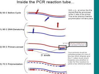
- 1. PCR EN TIEMPO REAL (Real-Time PCR) (RT-PCR) Benjamín Paz Aliaga UCSM-2004 Mientras la PCR tradicional detecta la amplificación en el punto final de la reacción con RT-PCR se detecta la amplificación mientras ocurre la reacción
- 5. PCR
- 8. KARY B MULLIS , EL DESCUBRIDOR DE LA PCR PREMIO NOBEL EN QUIMICA EN 1993
- 9. PCR EN TIEMPO REAL (Real-Time PCR) (RT-PCR) Benjamín Paz Aliaga UCSM-2004 Mientras la PCR tradicional detecta la amplificación en el punto final de la reacción con RT-PCR se detecta la amplificación mientras ocurre la reacción
- 15. www.biorad.com 2a. excitation filters 2b. emission filters 1. halogen tungsten lamp 4. sample plate 3. intensifier 5. ccd detector 350,000 pixels
- 20. SERIES OF 10-FOLD DILUTIONS
Notas del editor
- Real time PCR is a kinetic approach, where you look at the reaction in the early stages while it is still linear. There are many real time machines available. This is the one we use (the BioRad Icycler IQ real time PCR instrument). The lid slides back and then we put samples in a 96-well plate format inside, so one can look at a lot of samples simultaneously. The machine contains a sensitive camera which monitors the fluorescence in each well of the 96-well plate at frequent intervals during the PCR reaction. In our case, as DNA is synthesized, more SYBR green will bind and the fluorescence will increase.
- The real-time machine is connected to a computer and software on the computer is needed to run the real time PCR machine in real-time mode.
- So how do we measure differences in concentration of DNA or cDNA? This graph shows a series of 10-fold dilutions of a sample. As one dilutes the sample, it takes more cycles before the amplification is detectable. The blue line here is the same sample we have been following all along. Note that although the reactions show an orderly series of curves in order of dilution as they cross the orange line, if one looks at the upper parts of the curve, they are rather variable. Thus, if we stopped all these reactions at eg 33 cycles and ran a gel, it would indicate that the blue, red and purple reactions had the same amount of amplification, even though the reactions shown by the purple and red lines differ by a factor of 100 in the amount of dna. Reactions which differed by a factor of 2 would expect to be 1 cycle apart (samples that differ by 10 would be ~3.3 cycles apart). Is there any way to check that the correct fragments were amplified? One way to do some checking on the products is to do a melting curve.