
Recomendados
Más contenido relacionado
La actualidad más candente
La actualidad más candente (20)
Similar a RUSH ECO 117.pptx
Similar a RUSH ECO 117.pptx (20)
Más de franciscocarrera28
Más de franciscocarrera28 (20)
Último
Último (20)
RUSH ECO 117.pptx
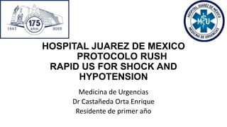
- 1. HOSPITAL JUAREZ DE MEXICO PROTOCOLO RUSH RAPID US FOR SHOCK AND HYPOTENSION Medicina de Urgencias Dr Castañeda Orta Enrique Residente de primer año
- 2. The RUSH Exam: Rapid Ultrasound in SHock in the Evaluation of the Critically lll Phillips Perera, MD, RDMS, FACEPa,*, Thomas Mailhot, MD, RDMSb, David Riley, MD, MS, RDMSa, Diku Mandavia, MD, FACEP,
- 3. Introducción Paciente chocado • Pre hospitalario • Intrahospitalario • Monitoreo The RUSH Exam: Rapid Ultrasound in SHock in the Evaluation of the Critically lll Phillips Perera, MD, RDMS, FACEPa,*, Thomas Mailhot, MD, RDMSb, David Riley, MD, MS, RDMSa, Diku Mandavia, MD, FACEP,
- 4. • Diagnósticos y tratamientos oportunos • Limitación en la disponibilidad horaria de algunos servicios de radiología •Diagnostico a pie de cama • Protocolo establecido • Repetible y Accesible • Operador dependiente • Requiere experiencia del operador • Equipo multiples transductores PROS CONTRAS
- 5. El Ultrasonido en pacientes criticos Cabecera Seguro No invasivo Bajo costo Rapido y en tiempo real The RUSH Exam: Rapid Ultrasound in SHock in the Evaluation of the Critically lll Phillips Perera, MD, RDMS, FACEPa,*, Thomas Mailhot, MD, RDMSb, David Riley, MD, MS, RDMSa, Diku Mandavia, MD, FACEP,
- 6. • En 2001, Rose et al. publico un protocolo de ultrasonido que habían creado para evaluar al paciente con hipotensión indiferenciada. • Weingart et al, en el 2007 creo el protocolo RUSH Rápido Este examen se realiza con equipo de ultrasonido estándar presente en muchos departamentos de emergencia. Que evaluamos? • La bomba • El tanque • Las tubería The RUSH Exam: Rapid Ultrasound in SHock in the Evaluation of the Critically lll Phillips Perera, MD, RDMS, FACEPa,*, Thomas Mailhot, MD, RDMSb, David Riley, MD, MS, RDMSa, Diku Mandavia, MD, FACEP,
- 7. Que se requiere • Equipos de ultrasonido estándar presentes en muchos departamentos de emergencia en la actualidad. • Transductor de matriz en fase (3,5 a 5 MHz) para permitir una exploración intercostal toracoabdominal adecuada. • Transductor de matriz lineal (7,5 a 10 MHz) para los exámenes venosos requeridos y para la evaluación del neumotórax. The RUSH Exam: Rapid Ultrasound in SHock in the Evaluation of the Critically lll Phillips Perera, MD, RDMS, FACEPa,*, Thomas Mailhot, MD, RDMSb, David Riley, MD, MS, RDMSa, Diku Mandavia, MD, FACEP,
- 8. Hipovolemico DISTRIBUTIVO CARDIOGENICO OBSTRUCTIVO • AAA • Hemorragia digestiva • Ectópico roto • Trauma grave • Deshidratación • Sepsis • Anafilaxia • Shock neurogénico • IAM • ICC • Valvulopatías severas • TEP masivo • Disección Ao ascendente • Taponamiento cardiaco • Neumotórax a tensión The RUSH Exam: Rapid Ultrasound in SHock in the Evaluation of the Critically lll Phillips Perera, MD, RDMS, FACEPa,*, Thomas Mailhot, MD, RDMSb, David Riley, MD, MS, RDMSa, Diku Mandavia, MD, FACEP,
- 9. ¿ORIGEN CARDIACO? ¿ORIGEN PULMONAR? ¿HIPOVOLEMIA? IAM ICC Taponamiento TEP Neumotórax AAA Sangrado digestivo Sepsis The RUSH Exam: Rapid Ultrasound in SHock in the Evaluation of the Critically lll Phillips Perera, MD, RDMS, FACEPa,*, Thomas Mailhot, MD, RDMSb, David Riley, MD, MS, RDMSa, Diku Mandavia, MD, FACEP,
- 10. Paso 1 protocolo RUSH // la bomba • El examen de eco se enfoca en buscar 3 hallazgos principales. 1. Saco pericárdico—Derrame pericardico o tamponade 2. El ventrículo izquierdo–Contractilidad global y tamaño de las cavidades 3. Tamaño relativo del ventrículo izquierdo con respecto al ventrículo derecho 4. Paraesternal eje corto subxifoideo The RUSH Exam: Rapid Ultrasound in SHock in the Evaluation of the Critically lll Phillips Perera, MD, RDMS, FACEPa,*, Thomas Mailhot, MD, RDMSb, David Riley, MD, MS, RDMSa, Diku Mandavia, MD, FACEP, SECTORIAL CONVEXO
- 11. • A) A la izquierda del esternón en el espacio intercostal 3 o 4 1. Paraesternal eje largo-–muesca hombro derecha 2. Paraesternal eje corto—muesca hombro izquierdo • B) Sonda dirigida hacia el hombro izquierdo desde una posición justo debajo de la punta subxifoidea del esternón • C) Decúbito lateral izquierdo (posición de pachon) y colocando la sonda justo debajo de la línea del pezón en el punto de máximo impulso del corazón--quinto espacio intercostal linea axilar anterior The RUSH Exam: Rapid Ultrasound in SHock in the Evaluation of the Critically lll Phillips Perera, MD, RDMS, FACEPa,*, Thomas Mailhot, MD, RDMSb, David Riley, MD, MS, RDMSa, Diku Mandavia, MD, FACEP,
- 13. The RUSH Exam: Rapid Ultrasound in SHock in the Evaluation of the Critically lll Phillips Perera, MD, RDMS, FACEPa,*, Thomas Mailhot, MD, RDMSb, David Riley, MD, MS, RDMSa, Diku Mandavia, MD, FACEP, • Sectorial apical • Subsifoido 4 camaras • Normal
- 14. The RUSH Exam: Rapid Ultrasound in SHock in the Evaluation of the Critically lll Phillips Perera, MD, RDMS, FACEPa,*, Thomas Mailhot, MD, RDMSb, David Riley, MD, MS, RDMSa, Diku Mandavia, MD, FACEP, • Paraesternal eje largo • Derrame pericardico
- 15. The RUSH Exam: Rapid Ultrasound in SHock in the Evaluation of the Critically lll Phillips Perera, MD, RDMS, FACEPa,*, Thomas Mailhot, MD, RDMSb, David Riley, MD, MS, RDMSa, Diku Mandavia, MD, FACEP, • Sectorial apical • Subsifoido 4 camaras • Derrame • Colapso ventriculo derecho en sistole • Tamponade
- 16. The RUSH Exam: Rapid Ultrasound in SHock in the Evaluation of the Critically lll Phillips Perera, MD, RDMS, FACEPa,*, Thomas Mailhot, MD, RDMSb, David Riley, MD, MS, RDMSa, Diku Mandavia, MD, FACEP, • Paraesternal eje largo • Dilatacion venticulo derecho
- 17. The RUSH Exam: Rapid Ultrasound in SHock in the Evaluation of the Critically lll Phillips Perera, MD, RDMS, FACEPa,*, Thomas Mailhot, MD, RDMSb, David Riley, MD, MS, RDMSa, Diku Mandavia, MD, FACEP, • Apical 4 camaras • Sectorial • trombo
- 18. Paso 2 protocolo RUSH // el tanque • Determinación del estado del volumen intravascular efectivo. • ¿Qué tan lleno esta el tanque? • Evaluación de las venas cava inferior y yugular en cuanto a tamaño y colapso con la inspiración. • Áreas donde el volumen intravascular podría estar comprometido The RUSH Exam: Rapid Ultrasound in SHock in the Evaluation of the Critically lll Phillips Perera, MD, RDMS, FACEPa,*, Thomas Mailhot, MD, RDMSb, David Riley, MD, MS, RDMSa, Diku Mandavia, MD, FACEP,
- 19. Estimación del volumen intravascular de forma no invasiva observando inicialmente la VCI Colocarse en el área epigástrica en una configuración de eje largo a lo largo de la IVC a medida que se extiende desde el abdomen hasta el corazón. Primero el corazón en el plano de 4 cámaras subxifoides y luego mover la sonda al plano de 2 cámaras subxifoides La aorta a menudo se verá rápidamente desde este plano como una estructura más profunda y de paredes más gruesas. Mover la sonda a la derecha del paciente traerá a la vista la IVC, discurriendo longitudinalmente junto a la aorta. La VCI debe examinarse en la unión de la aurícula derecha y la cava y seguirse de 2 a 3 cm caudalmente a lo largo del vaso A medida que el paciente respira, la VCI tendrá un patrón normal de colapso durante la inspiración The RUSH Exam: Rapid Ultrasound in SHock in the Evaluation of the Critically lll Phillips Perera, MD, RDMS, FACEPa,*, Thomas Mailhot, MD, RDMSb, David Riley, MD, MS, RDMSa, Diku Mandavia, MD, FACEP,
- 20. The RUSH Exam: Rapid Ultrasound in SHock in the Evaluation of the Critically lll Phillips Perera, MD, RDMS, FACEPa,*, Thomas Mailhot, MD, RDMSb, David Riley, MD, MS, RDMSa, Diku Mandavia, MD, FACEP, • Sectorial apical • Subsifoido 4 camaras • Normal
- 21. Correlación entre el tamaño y el porcentaje de cambio de la VCI con la variación respiratoria de la presión venosa central (PVC) Una VCI de menor calibre (<2 cm de diámetro) con colapso inspiratorio, más del 50% se correlaciona aproximadamente con una CVP de menos de 10 cm de agua. Una VCI de mayor tamaño (> 2 cm de diámetro) que colapsa menos del 50 % con la inspiración se correlaciona con una PVC de más de 10 cm de agua The RUSH Exam: Rapid Ultrasound in SHock in the Evaluation of the Critically lll Phillips Perera, MD, RDMS, FACEPa,*, Thomas Mailhot, MD, RDMSb, David Riley, MD, MS, RDMSa, Diku Mandavia, MD, FACEP,
- 22. Exepciónes a la regla • Tratamiento con vasodilatadores y/o diuréticos previo a la evaluación ecográfica en los que la VCI puede ser más pequeña que antes del tratamiento, alterando el estado fisiológico inicial. • La segunda advertencia existe en pacientes intubados que reciben ventilación con presión positiva, en los que se invierte la dinámica respiratoria de la VCI. The RUSH Exam: Rapid Ultrasound in SHock in the Evaluation of the Critically lll Phillips Perera, MD, RDMS, FACEPa,*, Thomas Mailhot, MD, RDMSb, David Riley, MD, MS, RDMSa, Diku Mandavia, MD, FACEP,
- 23. The RUSH Exam: Rapid Ultrasound in SHock in the Evaluation of the Critically lll Phillips Perera, MD, RDMS, FACEPa,*, Thomas Mailhot, MD, RDMSb, David Riley, MD, MS, RDMSa, Diku Mandavia, MD, FACEP, • Sectorial • Grosos de VCI
- 24. The RUSH Exam: Rapid Ultrasound in SHock in the Evaluation of the Critically lll Phillips Perera, MD, RDMS, FACEPa,*, Thomas Mailhot, MD, RDMSb, David Riley, MD, MS, RDMSa, Diku Mandavia, MD, FACEP, • Sectorial • Grosos de VCI
- 25. The RUSH Exam: Rapid Ultrasound in SHock in the Evaluation of the Critically lll Phillips Perera, MD, RDMS, FACEPa,*, Thomas Mailhot, MD, RDMSb, David Riley, MD, MS, RDMSa, Diku Mandavia, MD, FACEP, • Modo M • Grosos de VCI • Actualmente no recomendable
- 26. Filtración del tanque y examen FAST • "fugas anormales del tanque" • El examen FAST detecta colecciones de líquido intraperitoneal tan pequeñas como 100 ml, con un rango de 250 a 620 ml comúnmente citado • La sensibilidad y la especificidad generales del examen FAST son aproximadamente del 79 % y el 99 %, respectivamente. The RUSH Exam: Rapid Ultrasound in SHock in the Evaluation of the Critically lll Phillips Perera, MD, RDMS, FACEPa,*, Thomas Mailhot, MD, RDMSb, David Riley, MD, MS, RDMSa, Diku Mandavia, MD, FACEP,
- 27. El término de ecografía FAST (Focused Abdominal Sonography for Trauma) fue acuñado por Grace Rozycki y colaboradores en 1995. En 1997 el curso ATLS incluyó la ecografía fast como un método alternativo al lavado peritoneal diagnóstico en la evaluación de pacientes con trauma abdominal cerrado La motivación inicial para realizar el examen Focused Assessment with Sonography for Trauma (FAST) es la alta prevalencia de traumatismo abdominal, el cual es la tercera causa de muerte por trauma en el mundo, superada solo por el traumatismo craneo encefálico (TCE) y el trauma de tórax Además, es la principal causa de muerte evitable en trauma si se reconoce a tiempo. The RUSH Exam: Rapid Ultrasound in SHock in the Evaluation of the Critically lll Phillips Perera, MD, RDMS, FACEPa,*, Thomas Mailhot, MD, RDMSb, David Riley, MD, MS, RDMSa, Diku Mandavia, MD, FACEP,
- 28. El término FAST extendido (FAST-E) se refiere a la adición del examen torácico, ofreciendo así un espectro más amplio para el diagnóstico de posibles lesiones potencialmente detectables. Actualmente el FAST y FAST-E son un punto clave dentro de la atención de pacientes con trauma. The RUSH Exam: Rapid Ultrasound in SHock in the Evaluation of the Critically lll Phillips Perera, MD, RDMS, FACEPa,*, Thomas Mailhot, MD, RDMSb, David Riley, MD, MS, RDMSa, Diku Mandavia, MD, FACEP,
- 29. Actualmente hay evidencia suficiente para recomendar la realización de este procedimiento, mostrando una sensibilidad y una especificidad que varían entre el 73 al 99%. The RUSH Exam: Rapid Ultrasound in SHock in the Evaluation of the Critically lll Phillips Perera, MD, RDMS, FACEPa,*, Thomas Mailhot, MD, RDMSb, David Riley, MD, MS, RDMSa, Diku Mandavia, MD, FACEP,
- 30. Técnica Se requiere un transductor de baja frecuencia (3- 5 MHz) para evaluar el abdomen y la ventana subxifoidea. Un transductor de alta frecuencia para el examen torácico El no tener este último no hace inviable la adecuada realización del examen, aunque sí limita parcialmente la visualización de la pleura y del pulmón. The RUSH Exam: Rapid Ultrasound in SHock in the Evaluation of the Critically lll Phillips Perera, MD, RDMS, FACEPa,*, Thomas Mailhot, MD, RDMSb, David Riley, MD, MS, RDMSa, Diku Mandavia, MD, FACEP,
- 31. Al iniciar el FAST se busca responder inicialmente si hay hemoperitoneo; luego, al extender el examen al tórax se cuestiona si se encuentran lesiones torácicas como neumotórax, hemotórax o derrame pericacardico. The RUSH Exam: Rapid Ultrasound in SHock in the Evaluation of the Critically lll Phillips Perera, MD, RDMS, FACEPa,*, Thomas Mailhot, MD, RDMSb, David Riley, MD, MS, RDMSa, Diku Mandavia, MD, FACEP,
- 32. En el examen FAST se definen 3 ventanas. 1.- Hepatorrenal, que se ubica a nivel del cuadrante superior derecho 2.- Esplenorrenal, que está a nivel del cuadrante superior izquierdo 3.- Pélvica, que se visualiza por encima de la sínfisis del pubis 1 2 3 The RUSH Exam: Rapid Ultrasound in SHock in the Evaluation of the Critically lll Phillips Perera, MD, RDMS, FACEPa,*, Thomas Mailhot, MD, RDMSb, David Riley, MD, MS, RDMSa, Diku Mandavia, MD, FACEP,
- 33. Al realizar el examen FAST-E se adiciona: 1.- Ventana subxifoidea con el fin de evaluar la presencia de taponamiento cardiaco 2.- Ventanas pulmonares en ápices para descartar hemotórax o neumotórax. 1 2 2 2 2 2 2 2 2 The RUSH Exam: Rapid Ultrasound in SHock in the Evaluation of the Critically lll Phillips Perera, MD, RDMS, FACEPa,*, Thomas Mailhot, MD, RDMSb, David Riley, MD, MS, RDMSa, Diku Mandavia, MD, FACEP,
- 34. 1 2 3 4 5 6 The RUSH Exam: Rapid Ultrasound in SHock in the Evaluation of the Critically lll Phillips Perera, MD, RDMS, FACEPa,*, Thomas Mailhot, MD, RDMSb, David Riley, MD, MS, RDMSa, Diku Mandavia, MD, FACEP,
- 35. Cuando se habla de detectar hemoperitoneo se asume que el sangrado inicialmente proviene del hígado o del bazo. El FAST carece de sensibilidad para detectar lesiones de vísceras huecas y sangrado retroperitoneal. La presencia de hemoperitoneo en una ventana NO es específica de sangrado en ese sitio. The RUSH Exam: Rapid Ultrasound in SHock in the Evaluation of the Critically lll Phillips Perera, MD, RDMS, FACEPa,*, Thomas Mailhot, MD, RDMSb, David Riley, MD, MS, RDMSa, Diku Mandavia, MD, FACEP,
- 36. En las imágenes ecográficas el líquido libre abdominal se visualiza como una imagen anecoica que aparece en cualquiera de las ventanas descritas. The RUSH Exam: Rapid Ultrasound in SHock in the Evaluation of the Critically lll Phillips Perera, MD, RDMS, FACEPa,*, Thomas Mailhot, MD, RDMSb, David Riley, MD, MS, RDMSa, Diku Mandavia, MD, FACEP,
- 37. 1.- Ventana subxifoidea Evalúa el pericardio para detectar taponamiento cardiaco. El transductor se ubica a nivel subxifoideo; se utiliza el hígado como ventana acústica para obtener mejor resolución, y el indicador se ubica hacia la derecha del paciente. The RUSH Exam: Rapid Ultrasound in SHock in the Evaluation of the Critically lll Phillips Perera, MD, RDMS, FACEPa,*, Thomas Mailhot, MD, RDMSb, David Riley, MD, MS, RDMSa, Diku Mandavia, MD, FACEP,
- 38. Ventana subxifoidea The RUSH Exam: Rapid Ultrasound in SHock in the Evaluation of the Critically lll Phillips Perera, MD, RDMS, FACEPa,*, Thomas Mailhot, MD, RDMSb, David Riley, MD, MS, RDMSa, Diku Mandavia, MD, FACEP,
- 39. The RUSH Exam: Rapid Ultrasound in SHock in the Evaluation of the Critically lll Phillips Perera, MD, RDMS, FACEPa,*, Thomas Mailhot, MD, RDMSb, David Riley, MD, MS, RDMSa, Diku Mandavia, MD, FACEP,
- 40. Ventana subxifoidea normal. The RUSH Exam: Rapid Ultrasound in SHock in the Evaluation of the Critically lll Phillips Perera, MD, RDMS, FACEPa,*, Thomas Mailhot, MD, RDMSb, David Riley, MD, MS, RDMSa, Diku Mandavia, MD, FACEP,
- 41. En ocasiones no es posible obtener esta imagen (dolor, distensión, mala ventana), y entonces debe realizarse por medio del eje paraesternal izquierdo The RUSH Exam: Rapid Ultrasound in SHock in the Evaluation of the Critically lll Phillips Perera, MD, RDMS, FACEPa,*, Thomas Mailhot, MD, RDMSb, David Riley, MD, MS, RDMSa, Diku Mandavia, MD, FACEP,
- 42. Taponamiento cardiaco, líquido pericardiaco y colapso del ventrículo derecho. The RUSH Exam: Rapid Ultrasound in SHock in the Evaluation of the Critically lll Phillips Perera, MD, RDMS, FACEPa,*, Thomas Mailhot, MD, RDMSb, David Riley, MD, MS, RDMSa, Diku Mandavia, MD, FACEP,
- 43. Ventana hepatorrenal Evalúa el espacio hepatorrenal, el espacio de Morrison y la base pulmonar derecha. Es la más sensible para el diagnóstico de hemoperitoneo y la más fácil de obtener técnicamente. 4 The RUSH Exam: Rapid Ultrasound in SHock in the Evaluation of the Critically lll Phillips Perera, MD, RDMS, FACEPa,*, Thomas Mailhot, MD, RDMSb, David Riley, MD, MS, RDMSa, Diku Mandavia, MD, FACEP,
- 44. Se consigue ubicando el transductor un poco más adelante de la línea media axilar derecha entre el 7.° y 8.° arcos costales. El indicador de posición debe estar ubicado hacia la cabeza del paciente. Es importante visualizar el diafragma para definir la presencia de un posible hemotórax. Para la adecuada evaluación de esta ventana es necesario mover el transductor hacia arriba o hacia abajo en el torso paciente The RUSH Exam: Rapid Ultrasound in SHock in the Evaluation of the Critically lll Phillips Perera, MD, RDMS, FACEPa,*, Thomas Mailhot, MD, RDMSb, David Riley, MD, MS, RDMSa, Diku Mandavia, MD, FACEP,
- 45. The RUSH Exam: Rapid Ultrasound in SHock in the Evaluation of the Critically lll Phillips Perera, MD, RDMS, FACEPa,*, Thomas Mailhot, MD, RDMSb, David Riley, MD, MS, RDMSa, Diku Mandavia, MD, FACEP,
- 46. The RUSH Exam: Rapid Ultrasound in SHock in the Evaluation of the Critically lll Phillips Perera, MD, RDMS, FACEPa,*, Thomas Mailhot, MD, RDMSb, David Riley, MD, MS, RDMSa, Diku Mandavia, MD, FACEP,
- 47. The RUSH Exam: Rapid Ultrasound in SHock in the Evaluation of the Critically lll Phillips Perera, MD, RDMS, FACEPa,*, Thomas Mailhot, MD, RDMSb, David Riley, MD, MS, RDMSa, Diku Mandavia, MD, FACEP,
- 48. The RUSH Exam: Rapid Ultrasound in SHock in the Evaluation of the Critically lll Phillips Perera, MD, RDMS, FACEPa,*, Thomas Mailhot, MD, RDMSb, David Riley, MD, MS, RDMSa, Diku Mandavia, MD, FACEP,
- 49. The RUSH Exam: Rapid Ultrasound in SHock in the Evaluation of the Critically lll Phillips Perera, MD, RDMS, FACEPa,*, Thomas Mailhot, MD, RDMSb, David Riley, MD, MS, RDMSa, Diku Mandavia, MD, FACEP,
- 50. The RUSH Exam: Rapid Ultrasound in SHock in the Evaluation of the Critically lll Phillips Perera, MD, RDMS, FACEPa,*, Thomas Mailhot, MD, RDMSb, David Riley, MD, MS, RDMSa, Diku Mandavia, MD, FACEP,
- 51. The RUSH Exam: Rapid Ultrasound in SHock in the Evaluation of the Critically lll Phillips Perera, MD, RDMS, FACEPa,*, Thomas Mailhot, MD, RDMSb, David Riley, MD, MS, RDMSa, Diku Mandavia, MD, FACEP,
- 52. Ventana esplenorenal Se utiliza el bazo como ventana acústica, y también visualiza la base pulmonar izquierda. Se debe ubicar el diafragma, y para obtener mejores imágenes es necesario mover el transductor hacia arriba o abajo en el torso del paciente. Técnicamente es la ventana más difícil de visualizar. The RUSH Exam: Rapid Ultrasound in SHock in the Evaluation of the Critically lll Phillips Perera, MD, RDMS, FACEPa,*, Thomas Mailhot, MD, RDMSb, David Riley, MD, MS, RDMSa, Diku Mandavia, MD, FACEP,
- 53. Se consigue ubicando el transductor a nivel de la línea axilar posterior entre el 7.° y 8.° arco costal. El marcador de posición debe estar ubicado hacia la cabeza del paciente. The RUSH Exam: Rapid Ultrasound in SHock in the Evaluation of the Critically lll Phillips Perera, MD, RDMS, FACEPa,*, Thomas Mailhot, MD, RDMSb, David Riley, MD, MS, RDMSa, Diku Mandavia, MD, FACEP,
- 54. The RUSH Exam: Rapid Ultrasound in SHock in the Evaluation of the Critically lll Phillips Perera, MD, RDMS, FACEPa,*, Thomas Mailhot, MD, RDMSb, David Riley, MD, MS, RDMSa, Diku Mandavia, MD, FACEP,
- 55. The RUSH Exam: Rapid Ultrasound in SHock in the Evaluation of the Critically lll Phillips Perera, MD, RDMS, FACEPa,*, Thomas Mailhot, MD, RDMSb, David Riley, MD, MS, RDMSa, Diku Mandavia, MD, FACEP,
- 56. The RUSH Exam: Rapid Ultrasound in SHock in the Evaluation of the Critically lll Phillips Perera, MD, RDMS, FACEPa,*, Thomas Mailhot, MD, RDMSb, David Riley, MD, MS, RDMSa, Diku Mandavia, MD, FACEP,
- 57. The RUSH Exam: Rapid Ultrasound in SHock in the Evaluation of the Critically lll Phillips Perera, MD, RDMS, FACEPa,*, Thomas Mailhot, MD, RDMSb, David Riley, MD, MS, RDMSa, Diku Mandavia, MD, FACEP,
- 58. Líquido libre en base pulmonar izquierda; se visualizó mediante ventana esplenorrenal y corresponde a hemotórax traumático The RUSH Exam: Rapid Ultrasound in SHock in the Evaluation of the Critically lll Phillips Perera, MD, RDMS, FACEPa,*, Thomas Mailhot, MD, RDMSb, David Riley, MD, MS, RDMSa, Diku Mandavia, MD, FACEP,
- 59. The RUSH Exam: Rapid Ultrasound in SHock in the Evaluation of the Critically lll Phillips Perera, MD, RDMS, FACEPa,*, Thomas Mailhot, MD, RDMSb, David Riley, MD, MS, RDMSa, Diku Mandavia, MD, FACEP,
- 60. Ventana pélvica o suprapúbica Evaluar la presencia de líquido libre a nivel pélvico. NO descarta fracturas pélvicas porque este líquido puede provenir de lesión de víscera sólida. The RUSH Exam: Rapid Ultrasound in SHock in the Evaluation of the Critically lll Phillips Perera, MD, RDMS, FACEPa,*, Thomas Mailhot, MD, RDMSb, David Riley, MD, MS, RDMSa, Diku Mandavia, MD, FACEP,
- 61. En las mujeres puede haber líquido libre en esta ventana de manera fisiológica. La vejiga llena se utiliza como ventana ecográfica; la ausencia de orina hace que el resultado sea no confiable. The RUSH Exam: Rapid Ultrasound in SHock in the Evaluation of the Critically lll Phillips Perera, MD, RDMS, FACEPa,*, Thomas Mailhot, MD, RDMSb, David Riley, MD, MS, RDMSa, Diku Mandavia, MD, FACEP,
- 62. Se obtiene ubicando el transductor a nivel suprapúbico. Se debe visualizar en eje longitudinal y transversal (indicador de posición hacia la cabeza y luego hacia el lado derecho del paciente) The RUSH Exam: Rapid Ultrasound in SHock in the Evaluation of the Critically lll Phillips Perera, MD, RDMS, FACEPa,*, Thomas Mailhot, MD, RDMSb, David Riley, MD, MS, RDMSa, Diku Mandavia, MD, FACEP,
- 63. The RUSH Exam: Rapid Ultrasound in SHock in the Evaluation of the Critically lll Phillips Perera, MD, RDMS, FACEPa,*, Thomas Mailhot, MD, RDMSb, David Riley, MD, MS, RDMSa, Diku Mandavia, MD, FACEP,
- 64. The RUSH Exam: Rapid Ultrasound in SHock in the Evaluation of the Critically lll Phillips Perera, MD, RDMS, FACEPa,*, Thomas Mailhot, MD, RDMSb, David Riley, MD, MS, RDMSa, Diku Mandavia, MD, FACEP,
- 65. The RUSH Exam: Rapid Ultrasound in SHock in the Evaluation of the Critically lll Phillips Perera, MD, RDMS, FACEPa,*, Thomas Mailhot, MD, RDMSb, David Riley, MD, MS, RDMSa, Diku Mandavia, MD, FACEP,
- 66. The RUSH Exam: Rapid Ultrasound in SHock in the Evaluation of the Critically lll Phillips Perera, MD, RDMS, FACEPa,*, Thomas Mailhot, MD, RDMSb, David Riley, MD, MS, RDMSa, Diku Mandavia, MD, FACEP,
- 67. The RUSH Exam: Rapid Ultrasound in SHock in the Evaluation of the Critically lll Phillips Perera, MD, RDMS, FACEPa,*, Thomas Mailhot, MD, RDMSb, David Riley, MD, MS, RDMSa, Diku Mandavia, MD, FACEP,
- 68. Paso 3 protocolo RUSH // Las tuberias • ''Rotura de los conductos'': aneurisma de aorta y disección • Primero el lado arterial del sistema circulatorio y luego el lado venoso • Aneurismas aórticos abdominales (AAA) y las disecciones aórticas • Imagen de la aorta en la orientación longitudinal • Los aneurismas más pequeños pueden ser sintomáticos, aunque la ruptura es más común en los aneurismas que miden más de 5 cm The RUSH Exam: Rapid Ultrasound in SHock in the Evaluation of the Critically lll Phillips Perera, MD, RDMS, FACEPa,*, Thomas Mailhot, MD, RDMSb, David Riley, MD, MS, RDMSa, Diku Mandavia, MD, FACEP,
- 69. Paso 3 protocolo RUSH // Las tuberias • Hay que prestar especial atención por debajo de las arterias renales donde se localizan la mayoría de los AAA. • La disseccón aortica se presenta como la presencia de dilatación de la raíz aórtica y un colgajo de íntima aórtica. • TVP se observa Un coágulo de sangre agudo forma una masa en la luz de la vena y el hallazgo patognomónico de la TVP será una compresión incompleta de las paredes anterior y posterior de la vena. The RUSH Exam: Rapid Ultrasound in SHock in the Evaluation of the Critically lll Phillips Perera, MD, RDMS, FACEPa,*, Thomas Mailhot, MD, RDMSb, David Riley, MD, MS, RDMSa, Diku Mandavia, MD, FACEP,
- 70. The RUSH Exam: Rapid Ultrasound in SHock in the Evaluation of the Critically lll Phillips Perera, MD, RDMS, FACEPa,*, Thomas Mailhot, MD, RDMSb, David Riley, MD, MS, RDMSa, Diku Mandavia, MD, FACEP,
- 71. The RUSH Exam: Rapid Ultrasound in SHock in the Evaluation of the Critically lll Phillips Perera, MD, RDMS, FACEPa,*, Thomas Mailhot, MD, RDMSb, David Riley, MD, MS, RDMSa, Diku Mandavia, MD, FACEP,
- 72. The RUSH Exam: Rapid Ultrasound in SHock in the Evaluation of the Critically lll Phillips Perera, MD, RDMS, FACEPa,*, Thomas Mailhot, MD, RDMSb, David Riley, MD, MS, RDMSa, Diku Mandavia, MD, FACEP,
- 73. The RUSH Exam: Rapid Ultrasound in SHock in the Evaluation of the Critically lll Phillips Perera, MD, RDMS, FACEPa,*, Thomas Mailhot, MD, RDMSb, David Riley, MD, MS, RDMSa, Diku Mandavia, MD, FACEP, Patron de reconocimiento