Descargar como PDF, PPTX


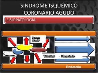


Este documento presenta información sobre el síndrome isquémico coronario agudo. Define el síndrome, describe su epidemiología y factores de riesgo. Explica la fisiopatología que involucra disfunción endotelial, inflamación y aterotrombosis. Finalmente, cubre temas como clasificaciones de infarto, electrocardiogramas, y biomarcadores utilizados para el diagnóstico.


